ACETABULAR FRACTURE AND MANAGEMENT
•Download as PPTX, PDF•
2 likes•438 views

This document discusses acetabulum fractures, which involve the articular surface of the hip joint. It covers the anatomy, incidence, etiology, classification, evaluation, treatment, surgical approaches, and complications of these fractures. The key points are: 1) Acetabulum fractures can involve one or both columns of the hip joint and have a bimodal distribution, occurring most often in younger patients due to high-energy trauma and elderly patients due to low-energy falls. 2) Treatment depends on the fracture pattern and degree of displacement, with nonoperative management for minimally displaced or stable fractures and operative treatment for displaced or unstable fractures. 3) Surgical approaches include anterior, posterior, and

Recommended
More Related Content
What's hot
What's hot (20)
Similar to ACETABULAR FRACTURE AND MANAGEMENT
Similar to ACETABULAR FRACTURE AND MANAGEMENT (20)
Recently uploaded
Recently uploaded (20)
ACETABULAR FRACTURE AND MANAGEMENT
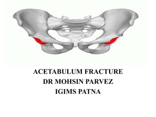
- 1. ACETABULUM FRACTURE DR MOHSIN PARVEZ IGIMS PATNA
- 2. ANATOMY : incomplete hemispherical socket with horshoeshaped Articular surface surrounding the medial nonarticular cotyloid fossa partial ball n socket joint ,six components :
- 4. Posterior column : quadrilateral surface,posterior wall and dome,ischial tuberosity greater/lesser sciatic notches Anterior column : anterior ilium (gluteus medius tubercle),anterior wall and dome iliopectineal eminence,lateral superior pubic ramus
- 5. Anatomic reconstruction of the dome/roof with the concentric reduction of the femoral head beneath this dome is the goal of both the operative and nonoperative treatment.
- 6. Acetabulum fractures are pelvis fractures that involve the articular surface of the hip joint and may involve one or two columns, one or two walls, or the roof within the pelvis Incidence ~ 4 per 100,000 per year Demographics fractures occur in a bimodal distribution high energy trauma in younger patients (e.g., motor vehicle accidents) low energy trauma in elderly patients (e.g., fall from standing height) Etiology Pathoanatomy fracture pattern predominately determined by force vector position of femoral head at time of injury bone quality (e.g., age) Associated conditions orthopaedic manifestations lower extremity injury (36%) ,nerve palsy (13%) most commonly seen in transverse + posterior wall fracture patterns
- 7. Radiographic evaluation: AP pelvic view , Judet view –iliac and obturator Iliac oblique view –posterior column and anterior wall Obturator oblique---anterior column and posterior wall
- 9. If any of the roof arc measurement in a displaced fracture are less than 45 degrees operative treatment should be considered roof arc angle : angle between vertical line through femoral head and line through fracture stable- if the fracture line exits outside the weight bearing dome of the acetabulum not applicable for associated both column or posterior wall pattern because no intact portion of the acetabulum to measure
- 10. On CT : transverse and anterior and posterior wall fracture are in sagittal plane; anterior and posterior column fractures extend through the quadrilateral surace and into obturator foramen with a more coronal orientation
- 12. Optional views inlet/outlet if concerned for pelvic ring involvement examination under anesthesia (EUA) used to assess posterior wall stability hip positioned in flexion, adduction and axial load obtain obturator oblique view opening of the medial clear space suggests instability of the posterior wall fracture
- 14. Axial CT scans showing the superior 10 mm of the acetabular roof to be intact have been shown to correspond to radiographic roof arc measurements of 45 degrees. gull sign :represents impaction of superomedial roof seen on iliac oblique view,pathognomic for posterior wall fractures spur sign :represents most caudal part of intact ilium due to medialization of articular components,seen on obturator oblique view,pathognomic for ABC fractures
- 15. Judet and Letournel classification system classifed as 5 elementary and 5 associated fracture patterns most common fracture patterns Younger:posterior wall,transverse fracture "family" Elderly:anterior column (e.g., quadrilateral plate fractures) anterior column, posterior hemitransverse assoicated both column fractures
- 27. TREATMENT: Follow ATLS protocol urgent operative intervention within 1-24 hours if it is a part of open fracture or associated with an irreducible dislocation of the hip Nonoperative protected weight bearing for 6-8 weeks Indications: patient factors high operative risk (e.g., elderly patients, presence of DVT) morbid obesity ,open contaminated wound late presenting > 3 weeks fracture characteristics-minimally displaced fracture (< 2 mm) < 20% posterior wall fractures treatment based on size of posterior wall is controversial recommend an exam under anesthesia (EUA) using fluoroscopy best method to test stability -femoral head congruency with weight bearing roof (out of traction) both column fracture pattern with secondary congruence (out of traction) displaced fracture with roof arcs > 45° in AP and Judet views or >10 mm on axial CT cuts
- 28. Operative treatment open reduction and internal fixation indications < 3 weeks from date of injury,physiologically stable adequate soft-tissue envelope,no local infection pregnancy is not contraindication to surgical fixation displacement of roof (> 2 mm) unstable fracture pattern (e.g. posterior wall fracture involving > 40- 50%) marginal impaction,intra-articular loose bodies irreducible fracture-dislocation Approaches Anterior-ilioinguinal,iliofemoral,modified stoppa Posterior-Kocher-Langenbach Combined-extended iliofemoral
- 29. total hip arthroplasty indications usually elderly patients with significant osteopenia and/or significant comminution pre-existing arthritis post-traumatic arthritis in all ages column fixation strategies reconstruction bridging plate and screws percutaneous column screws cable fixation wall fixation strategies bridge plate and screws lag screw and neutralization plate spring (butress) plate
- 30. MEDICAL CONTRAINDICATIONS TO SURGERY On occasion, the severity of the medical condition mandates that operative intervention be delayed. If deemed needed, the articular cartilage of the hip should be protected during these delays with the patient in skeletal traction. On occasion, severe head trauma with a tenuous, evolving spectrum of injury may preclude a surgical procedure.
- 31. Approaches : Anterior approaches – Ilioinguinal in Anterior wall and anterior column Both column fracture Posterior hemitransverse fracture Posterior Kocher Langenbach - Posterior wall and posterior column fx Most transverse and T-shaped Combination of above fracture Extended ilioinguinal approach - Only single approach that allows direct visualization of both columns Associated fracture pattern 21 days after injury Some transverse fxs and T types Some both column fxs (if posterior comminution is present) Modified Stoppa - Access to quadrilateral plate to buttress comminuted medial wall fractures
- 40. Post-traumatic DJD most common complication Heterotopic ossification highest incidence with extensile approach treat with lowest incidence with anterior ilioinguinal approach Osteonecrosis DVT and PE Infection Bleeding Neurovascular injury risk factor highest incidence with transverse + posterior wall fractures Intraarticular hardware placement Abductor muscle weakness