
Recommended
More Related Content
What's hot
What's hot (20)
Similar to Aortic Valve Disease: Causes, Symptoms, and Treatment
Similar to Aortic Valve Disease: Causes, Symptoms, and Treatment (20)
Recently uploaded
Recently uploaded (20)
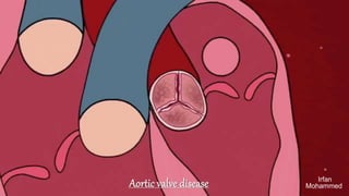
Aortic Valve Disease: Causes, Symptoms, and Treatment
- 2. Aortic valve 3 cusps/ leaflets. Separated by 3 commissures. Commissures supported by a fibrous annulus. Cordae tendinae and papillary muscles are absent. Normal aortic valve surface area: 3 -4 cm2. each Cusp is crescent shaped and capable of opening fully to allow unimpeded forward flow, then closing tightly to prevent regurgitation Aortic valve stenosis Aortic valve regurgitation.
- 3. Aortic sclerosis Aortic valve sclerosis is defined as calcification and thickening of a tri leaflet aortic valve in the absence of obstruction of ventricular outflow. Aortic sclerosis is very common in the elderly It is related to atherosclerosis. Aortic sclerosis means thickening, degeneration and calcification. Occurs with aging or on a diseased valve or anatomically abnormal valve. Incidence of aortic sclerosis increases with age, male gender, smoking, hypertension, high lipoprotein (Lp) (a), high low-density lipoprotein (LDL), and diabetes mellitus. Pathophysiology : Due to inflammation, atherosclerosis, osteoblastic transformation. Age Aortic sclerosis Aortic stenosis 65 – 75 yrs 75-85 yrs >85 yrs 20 % 35% 48% 1.3% 2.4% 4%
- 4. Aortic valve stenosis : Aortic valve stenosis — or aortic stenosis — occurs when the heart's aortic valve narrows. The valve doesn't open fully, which reduces or blocks blood flow from your heart into the main artery to your body (aorta) and to the rest of your body. 80% patients are males. Degenerative calcification of the aortic cusp on a substrate of bicuspid aortic valve or rheumatic valve. valve myofibroblast differentiates into osteoblast :deposition of calcium hydroxyapatite. 30% of elderly have valve sclerosis not severe to cause obstruction. mc cause of aortic stenosis in elderly : Aortic sclerosis. mc cause of aortic stenosis in young : Bicuspid aortic valve.
- 5. Classification of aortic stenosis : Left ventricle outflow issue. To maintain cardiac output - it undergoes concentric LV hypertrophy. • Increased gradient across the valve. • Increased peak flow velocity across valve. • Decreased valve surface area due to stenosis. 40 / 4 / I Rule : (For severe aortic stenosis) mean transvalvular gradient across valve : > 40 mmHg. Peak flow velocity across valve : > 4 m/s valve surface area is < I cm2
- 6. Bicuspid aortic valve : Bicuspid aortic valve is a type of abnormality in the aortic valve in the heart. In bicuspid aortic valve, the valve has only two small parts, called leaflets, instead of the normal three. This condition is present from birth. It can occur with other heart defects. most common congenital valve defect. male > female Autosomal dominant NOTCH I defect or endothelial nitric oxide synthase defect. medial degeneration with ascending aortic aneurysm/dissection. 75% AS VS 20% AR
- 7. Supravalvular aortic stenosis Sub-valvular aortic stenosis associated with Hypertrophic cardiomyopathy due to sub-valvular obstruction. (membrane) SVAS occurs in 1 in 20,000 newborns worldwide. Supravalvular aortic stenosis (SVAS) is a heart defect that develops before birth. This defect is a narrowing (stenosis) of the large blood vessel that carries blood from the heart to the rest of the body (the aorta) Associated with Williams syndrome: Hypercalcemia, growth failure, mental retardation Mutations in the ELN gene cause SVAS. Pathophysiology : Concentric LVH + increased flow gradient + increased velocity LV systolic cardiac output maintained LV filling pressure is high - diastolic dysfunction. After disease progresses, contractile function lost. This leads to systolic failure.
- 8. Symptomatology of aortic stenosis median survival based on symptoms : A 5 (Angina) - 5 yrs S 3 (syncope) - 3 yrs D 2 (dyspnoea) - 2 yrs unexplained sudden cardiac death can occur at any point(<1 %). Difficulty walking short distances Swollen ankles or feet Difficulty sleeping or needing to sleep sitting up Decline in activity level or reduced ability to do normal activities Fatigue upon exertion, Failure to gain weight, Poor or inadequate feeding, Breathing problems
- 9. symptoms . HEYDES syndrome Aortic stenosis Angiodysplasia Of Ascending colon. Angina Dyspnoea syncope Angina is a type of chest pain Left ventricular hypertrophy. Increases o2 demand Decreased diastolic perfusion time. Decreased capillary density with respect to wall thickness. Atherosclerosis. Shortness of breath Fixed cardiac output state. Increased LV filling pressure Due to diastolic failure Increased LV filling pressure Increased LA pressure Increased pulmonary capillary wedge pressure Dyspnoea Syncope is another word for fainting or passing out. due to fixed cardiac output state
- 10. Clinical findings Pulse - normal. In severe AS : Pulsus Tardus (slow-rising pulse) In very severe AS (with heart failure): pulsus parvus ET tardus. (low amplitude) JVP - normal BP - normal In very severe AS - Narrow pulse pressure. Signs of cardiac failure : Left heart failure due to increased LV filling pressure. Inspection :Sustained heaving apex laterally displaced Signs of pulmonary HTN are rare here.
- 11. Heart sounds It is best heard along the right sternal border in the 2nd intercostal space. S1 normal, aortic ejection click may be heard. S2 : Soft S2 , In degenerated sclerosed valve : Soft S2, In bicuspid aortic valve stenosis : Normal/ loud S2 , Severe AS: reverse split. P2&P1 S3: only in very severe AS (in LV failure) S4 : Heard in aortic stenosis.
- 12. murmur Ejection systolic murmurs (esm) identified by late systolic peaking. Harsh, medium pitched murmur. Best heard in sitting and leaning forward position and at the end of expiration. Best heard in aortic area. Radiating to the carotid/apex Late systolic accentuation. High pitch (musical quality) radiating to apex : Gallavardin phenomenon. Low pitch (rough quality) radiated to carotid Longer duration of murmur indicates severity. Severe aortic stenosis Very severe aortic stenosis Pulsus tardus, sustained heaving apex. presence of S4, longer duration of murmur paradoxical reverse split Pulsus parvus et tardus Narrow pulse pressure S3 heard. ECG : look for LVH V1 or V2s + v5 or v6r is > 35mm
- 13. management I. Transthoracic Echo Sclerosed valve without stenosis (< 25 mmHg) 2. Dobutamine stress echo To differentiate true and pseudo severe AS. AS causing heart Failure - true severe AS. Heart failure with mild AS - Pseudo severe AS. Treatment: Symptomatic patient Aortic valve replacement. Has best success in Aortic Stenosis. In Asymptomatic patient: IP ejection fraction < 50% Progression of yearly ECHO dobutamine stress test – positive bicuspid valve undergoing any other procedure. Trans catheter aortic valve replacement. Aortic valvotomy, if patient unfit for aortic replacement. https://www.youtube.com/watch?v=VZt8fYdXsnI .
- 14. Aortic valve regurgitation Aortic valve regurgitation — or aortic regurgitation — is a condition that occurs when your heart's aortic valve doesn't close tightly. As a result, some of the blood pumped out of your heart's main pumping chamber (left ventricle) leaks backward. Aortic valve regurgitation can develop suddenly or over decades. We have 2 types of aortic regurgitation 1.Chronic aortic regurgitation. 2.Acute aortic regurgitation.
- 15. Chronic Aortic regurgitation It is of 2 types 1.Valvular rheumatic 2.aortic root disease (mc) Aortic dissection, Syphilis, Ankylosing spondylitis/ Reiters syndrome, marfan syndrome – (most important),Osteogenesis imperfecta. Left ventricle undergoes eccentric dilatation/hypertrophy. Elevation of end diastolic volume (EDv) - Cardiac output is normal/increased. Increased sv and maintained EDP due to compliance. Filling pressures are normal. No pulmonary edema. LV dilatation increases the LV systolic tension requires to develop any level of systolic pressure thus Both Preload and afterload are high. So Patient would remain asymptomatic for years. (LV failure)
- 16. symptoms Asymptomatic for decades. Palpations due to increased LV size Exertional dyspnea only when there is LV systolic dysfunction In severe AR Increase in stroke volume causes distension of peripheral arteries. Increases in SBP In diastole, regurgitation of blood back in LV: Quick collapse of vessel Diastolic BP reduces wide pulse pressure Nocturnal angina due to fall in arterial diastolic pressures and increased regurgitant volume reducing coronary perfusion.
- 17. Clinical findings in AR (severe AR) Corrigan's pulse : A rapid and forceful distension of the arterial pulse with a quick collapse. De musset sign : Bobbing of head with each heart beat. Becker sign : Visible pulsations on retinal arterioles. Landolfe's sign : Systolic contraction and diastolic dilation of pupil. Rosenbachs sign : Hepatic pulsations. Gerhardt's sign : Pulsation of the spleen in presence of splenomegaly. muller's sign: visible pulsations of the uvula Traube's sign : pistol shot sounds over femoral artery. Duroziez sign : Systolic murmur over the femoral artery when compressed proximally and diastolic murmur when compressed distally. quincke sign : Capillary pulsations seen on light compression of the nail bed. Lincoln sign : Pulsations in popliteal artery. Hill sign : popliteal cuff systolic pressure exceeding brachial cuff systolic pressure by more than 20 mmHg
- 18. other findings and heart sounds : Pulse : Corrigans pulse (severe AR) : A pulse that is forceful and then suddenly collapses Pulsus bisferiens (sever AR or mild severe AR with mild AS ) BP: wide pulse pressure. JVP : normal Palpation : Hyperdynamic apex with cardiomegaly Heart sounds: S1 : Soft due to premature closure. S2 : Soft in valvular disease and loud in root disease. S3 : Heard only in LV failure S4 : Not heard
- 19. Murmur Patient in sitting and leaning forward, end expiratory. High pitched murmur De crescendo murmur. Blowing murmur best heard with diaphragm of stethoscope. No radiation Best area : Neo aortic area 3rd left intercostal space. In root disease - right stemal border. Short in acute AR / severe chronic AR with LV dysfunction. musical murmur in perforation of leaflet. Ejection systolic flow murmur - heard in aortic Austin flint murmur :Regurgitation in diastole causes anterior leaflet displacement. Low pitched mid diastolic murmur heard at apex
- 20. management of AR : Aortic valve replacement. AS>AR>MR Indications for surgery : Symptomatic : surgery Asymptomatic with severe AR in echo 50/50 rule Asymptomatic with LV EF < 5O% or Lv end systolic diameter > 50 mm.
- 21. Acute aortic regurgitation medical emergency. Inability of LV to quickly adapt to the abrupt increase in end diastolic volume LV fiiling pressure increases Pulmonary venous pressure increase & cardiac output decreases. Patient presents with Acute pulmonary edema or cardiogenic shock. No signs of wide pulse pressure S3 heard, Shorter duration murmur (low pitched). Requires immediate surgery. Causes : Aortic dissection Infective endocarditis Trauma
- 22. reference https://www.mayoclinic.org/diseases-conditions/aortic-stenosis/symptoms- causes/syc- 20353139#:~:text=Aortic%20valve%20stenosis%20%E2%80%94%20or%20aortic,the%20 severity%20of%20your%20condition. https://www.ncbi.nlm.nih.gov/books/NBK470446/ https://www.mayoclinic.org/diseases-conditions/angina/symptoms-causes/syc- 20369373#:~:text=Angina%20(an%2DJIE%2Dnuh,or%20pain%20in%20the%20chest. https://www.healio.com/cardiology/learn-the-heart/cardiology-review/topic- reviews/aortic- stenosis/diagnosis#:~:text=The%20ECG%20in%20patients%20with,in%20aortic%20ste nosis%20is%20concentric. https://www.youtube.com/watch?v=pgDWz1JybzE https://www.leehealth.org/our-services/cardiothoracic-surgery/heart-valve- surgical-repair
- 23. Thank you
Editor's Notes
- Lp protine deposition raas activation.
- A double pulse noticed during systole in the peripheral pulse