SlideShare a Scribd company logo
1 of 58
Download to read offline
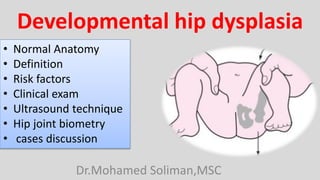
Developmental hip dysplasia
Dr.Mohamed Soliman,MSC
• Normal Anatomy
• Definition
• Risk factors
• Clinical exam
• Ultrasound technique
• Hip joint biometry
• cases discussion
• Hip joint is Ball and socket joint with wide
range of motion 2nd only to glenohumeral
joint
• 2/3 of sphere
• Covered by articular cartilage
• Cartilage thickest superiorly
• Cartilage thins at head/neck junction
• Ossification centre of femoral head appears
on ultrasound before it appears
radiographically (6th-8th week)
Femoral Head
Triradiate cartilage
(growth cartilage)
• formed by portions of ilium, ischium,
and pubis
• unite at triradiate cartilage (growth
cartilage)
• Oriented anterior, inferior, lateral
• Covers > 50% femoral head
• Anterior and posterior rims: Osseous
margins of acetabulum
• Medial wall: Quadrilateral plate ilium
• Echogenic fibrocartilage at edge of
cartilaginous roof is the true acetabular
labrum
Acetabulum
• Laxity of joint capsule
• Inadequate contact between
acetabulum and femoral head
• Acetabulum is particularly susceptible
to remodeling during first 10 postnatal
weeks
• Contact between femoral head and
acetabulum is necessary for normal
acetabular development
Definition
Developmental dysplasia of hip (DDH) & Congenital dysplasia of hip (CDH)
Underdevelopment of acetabular component of hip joint
Shallow acetabulum
± subluxation of femoral head
± Secondary hypoplasia of femoral head
Ranges from shallow acetabulum to complete dislocation with false acetabulum
Dysplasia: 0.8% newborn (0.3% boys, 1.4% girls)
• M:F = 1:4
• female hormone relaxin, which may contribute to ↑ ligamentous laxity
• Left hip is 3x more commonly affected than right hip : Possibly related
to left occiput anterior position of most non breech babies in utero , In this
position, left hip lies against maternal spine, which may limit abduction
Risk factors
1. First child
2. Female
3. Positive family history
4. breech presentation
5. Oligohydramnios
6. Torticollis
7. foot deformity
3% of births are breech
8% of girls with breech birth have developmental hip dysplasia
Due of hip flexed, knee extended position of breech babies in utero
Ortolani maneuver
▪ Hip is flexed to 90° and gently abducted while thigh is lifted anteriorly
▪ "Clunk" is felt as dislocated femoral head reduces
Barlow maneuver
▪ Hip is flexed to 90° and gently adducted while thigh is pushed posteriorly
▪ "Clunk" is felt as femoral head dislocates
• Most useful between birth and 6 months
• Both static and dynamic evaluation
Longitudinal
coronal
Transverse
Static Dynamic
Longitudinal coronal plane
Transverse plane
Longitudinal Coronal view ("egg in spoon" view)
• Most important .
• lateral decubitus position
• the hip flexed to only 20° .
• transducer 10-15° obliquely
(posteriorly) from coronal plane to
obtain straight iliac line
• Position too anterior → false-positive
result
• Gas can be found within normal hip
Longitudinal Coronal view
"egg in spoon" view
Non stress scan
single image showing
8
structures
1. straight iliac contour
2. acetabular labrum
3. Osseous acetabular roof
4. cartilaginous acetabular roof
5. triradiate cartilage
6. Ischium
7. center of femoral head
8. Ossified femoral metaphysis
Coronal view
"egg in spoon" view
Non stress scan
straight iliac contour
cartilaginous acetabular roof
triradiate cartilage
Ischium
femoral head
Ossified
femoral
metaphysis
Egg in spoon view
Osseous
acetabular roof
cartilaginous
acetabular roof
acetabular labrum
Triradiate
cartilage
Ischium
Normal Longitudinal coronal , non stress test
Ossified femoral
metaphysis
femoral head
Ilium
• lateral decubitus position.
• transducer in coronal position.
• Barlow maneuver (i.e. posterior
force on adducted and flexed hip)
• Attempt to displace femoral head
from acetabulum during real-time
imaging
• Lack of infant relaxation could
hinder or prevent acquisition of
optimal stress view and can
produce false-negative examination
• Nonstress views provide more
information than stress views
Coronal view
"egg in spoon" view
stress scan
Stress test produces an axial section of the femoral head. The femoral head
should appear round in this section.
Normal Longitudinal coronal , stress test
Transverse view
golf ball on tee
Ice cream cone
• Shows femoral head overlying
triradiate cartilage
• Useful to continue screening hip
during active movement if child
is restless
• Will give indication as to stability
of hip during normal movement
Transverse view
golf ball on tee
Non stress scan
single image showing
7
structures
1. Acetabular roof
2. acetabular labrum
3. Triradiate cartilage
4. Femoral head
5. Femoral metaphysis
6. Gluteal muscles
7. Ischium
Transverse view
golf ball on tee
Ice cream cone
Non stress scan
Femoral head
Acetabular roof
acetabular labrum
Gluteal muscles
Femoral
metaphysis
Triradiate
cartilage
ischium
golf ball on tee
ice cream cone
Normal transverse
Femoral head
Acetabular roof
acetabular labrum
Gluteal muscles
Femoral
metaphysis
Triradiate
cartilage
ischium
ice cream cone
golf ball on tee
Hip joint Biometry
Ultrasound X ray
1. Graf staging
2. Bony coverage index
Acetabular roof line
• Line along osseous
acetabular roof line
• α angle is measured at
intersection between iliac
line and line along osseous
acetabular roof
• α angle is acetabular angle
Reflects
1. steepness of acetabular roof
2. Portion of femoral head
contained within bony
acetabulum
3. Acetabular bony coverage
4. Center of femoral head relative
to iliac straight line
Iliac line
Line along straight
contour of ilium
Labrum line
• Line from promontory to tip of
labrum
• β angle is measured at
intersection between iliac line
and line from acetabular
promontory to tip of Labrum
• Reflects elevation of
acetabular labrum
Ultrasound 3 lines
straight iliac contour
triradiate cartilage
Ischium
femoral head Ossified
femoral
metaphysis
Hip joint Biometry
iliac line
α
β
β angle
a angle
Normal values
α angle > 60°
β angle < 55° .
α angle is more critical than β angle
Measurements outside of these ranges are suggestive of developmental hip dysplasia.
Coronal scan shows the hip of a 4-month-old child with the hip in extension. The center of
the femoral head has begun to ossify, showing up as an echogenic focus with posterior
acoustic shadowing.
Acetabulum immaturity
Immature
roundish acetabular promontory
mature
Angular acetabular promontory
Modified Graf staging
grade a angle Β angle Hip status
Type Ia > 60° < 55° mature hip, angular bony promontory
Type Ib > 60° < 55° mature hip, roundish bony promontory
Type IIa 50-60° 55-77° physiologic immaturity < 3 months
Type IIb 50-60° 55-77° Delayed maturity > 3 months
Type IIc 43-49° >77° critical hip, subluxation, severe dysplasia
Type III α < 43° dislocated hip
Type IV < 43° dislocated hip, inverted labrum
Pathological cases
• Shallow acetabulum .
• The α angle is 44°
• Moderate femoral head
subluxation.
• The acetabular labrum is
everted.
• The femoral head is relatively
small.
Moderately dysplastic hip
type IIc
• The α angle is 25 °
• dislocated femoral head .
• The labrum is interposed
between the bony acetabulum
and the femoral head.
severely hypoplastic acetabulum
type IV DDH.
• rounded shallow acetabulum .
• femoral head is severely
subluxed
• the acetabular labrum is
located between the
acetabulum and the femoral
head.
severely dysplastic hip
Type III DDH.
• reduction in α angle (34°)
• increase in β angle (55°)
type III DDH.
same patient
measurements
• lateral and cephalad displacement
of the femoral head.
• The labrum is interposed between
the femoral head and acetabulum
and is thickened.
• The pulvinar is also thickened.
severely dysplastic hip
type IV DDH.
• severe dysplastic acetabulum
with a dislocated hip.
• The bony acetabulum is not
measurable,
• the labrum cannot be well
delineated.
Type IV DDH.
* Should have > 50% of femoral head covered by bony acetabulum
* Important and easy to comprehend measurement
bony coverage index
straight iliac contour
triradiate cartilage
Ischium
femoral
head
Ossified
femoral
metaphysis
Hip joint Biometry
Bony coverage index
iliac line
d
D
Longitudinal coronal ultrasound
normal hip with
a well-covered
femoral head
The portion of the
femoral head
diameter (D) covered
by bone acetabulum
(d) is > 50%
(D/d > 50%).
Iliac line
Important terminology according to bony coverage index
Capsular laxity
 50% bony coverage femoral head at rest
 < 50% bone coverage on stress examination or active movement
Subluxation
< 50% bony coverage at rest
o Some centers define subluxation as < 33% bony coverage and 33-50% as
indeterminate
Dislocation
Femoral head lies completely outside of bony acetabulum at rest
• Immature acetabulum with a
roundish acetabular promontory
• good bony coverage of the
femoral head.
Previously roundish bony
acetabular promontory has now
become less rounded .
It is expected finding with normal
maturation of the acetabulum.
Normal acetabular labrum .
same patient
Follow up 2 months later
roundish acetabular promontory
indicative of acetabular immaturity.
The acetabular roof is still deep with
good bony coverage of the femoral
head.
Longitudinal coronal ultrasound
slightly roundish acetabular
promontory . However, the
acetabular bony roof is well
formed and deep.
same patient
contralateral hip
• moderate capsular laxity
• < 50% coverage of the femoral
head by the bony acetabulum as
indicated by the iliac line
• Treatment is indicated
• with follow-up ultrasound in 4
weeks.
Same patient
during stress scan
• femoral head is now not subluxed
during stress maneuver,
• index > 50%.
• The acetabulum promontory is
sharper.
Early diagnosis and proper treatment
can lead to dramatic improvement.
Same patient
Stress scan
4 weeks later
• immature acetabulum
• roundish acetabular
promontory .
• The majority of the unossified
femoral head lies below the
iliac line .
• good bony coverage.
Longitudinal ultrasound
• Most of the femoral head lies
above the iliac line , indicating
capsular laxity and is common
in newborns and premature
infants.
• Gas in the hip joint (normal finding)
Same patient during stress test
Radiographic findings ( after 6 months)
Hip dysplasia associated with
• relatively shallow acetabulum
• slanting roof
• relatively small superolaterally placed femoral head
Acetabular index
Perkin line
Hilgenreiner line
HPA
Hilgenreiner line
Perkin line
Perkin line
Acetabular index
Hilgenreiner line
• links tops of both triradiate
cartilages
• Femoral metaphysis lie below it
Perkin line
• perpendicular to Hilgenreiner line
through lateral margin of bony
acetabulum
• Femoral capital epiphysis should
lie within inner lower quadrant of
intersection between Perkin and
Hilgenreiner lines
Acetabular index
• angle between acetabular roof
and Hilgenreiner line
• 30-32° in newborn; ↓ in older
children
left-sided acetabular dysplasia
• dislocation of the femoral head
• delayed ossification of the left
femoral capital epiphysis.
Hilgenreiner line
Perkins Perkins
Acetabular
index
angle
1. Ultrasound is useful up to 6 months of age
2. Limited at 6-9 months and not useful thereafter
3. Ensure that definitions of capsular laxity, subluxation, and dislocation are
clear to those interpreting the ultrasound result
4. Useful to have these definitions written on ultrasound report
5. On ultrasound examination, incorrect orientation can make deep hip appear
shallow , Not possible to make shallow hip appear deep
6. Prognosis is excellent if dysplasia is diagnosed and treated early, even for
severe degree of DDH
7. Key to diagnosis rests on obtaining correct coronal (longitudinal) view
8. If in doubt, repeat ultrasound in 2-4 weeks
Prognosis
• Physiological hip joint laxity of newborn without acetabular dysplasia resolves
spontaneously
• Early treatment of DDH: Excellent result with harness or splint
• Delayed treatment of DDH: Irreversible dysplasia
1. Limited range of movement, adductor spasm
2. Limb shortening, osteoarthrosis
Treatment
•Conservative
o Pavlik harness ± closed reduction (if dislocated)
•Surgical for dislocation or subluxation failing to respond to conservative
treatment
1. Open reduction + spica cast
2. Adductor tenotomy + release of iliopsoas
3. Varus (derotational) vs. reconstructive osteotomy
Clinical photograph shows a baby
wearing a Pavlik harness to treat
DDH.
The harness helps to keep the hips
in an abducted flexed position.
This position ensures that the
femoral heads remain within the
acetabular fossae.
Ultrasound of Developmental dysplasia of hip Joint ..Dr.Mohamed Soliman

More Related Content

What's hot

Pelvis acetabulum - anatomy , imaging , classification
Pelvis acetabulum   - anatomy , imaging , classificationPelvis acetabulum   - anatomy , imaging , classification
Pelvis acetabulum - anatomy , imaging , classificationjatinder12345
 
Imaging in hip disorders
Imaging in hip disordersImaging in hip disorders
Imaging in hip disordersVikram Patil
 
RADIOGRAPHIC VIEWS FOR HIP JOINT
RADIOGRAPHIC VIEWS FOR HIP JOINTRADIOGRAPHIC VIEWS FOR HIP JOINT
RADIOGRAPHIC VIEWS FOR HIP JOINTGanesan Yogananthem
 
Ankle joint radiography
Ankle joint radiographyAnkle joint radiography
Ankle joint radiographyNikhil Murkey
 
Shoulder labral tears MRI
Shoulder labral tears MRIShoulder labral tears MRI
Shoulder labral tears MRIDr. Mohit Goel
 
Approach to skeletal dysplasia
Approach to skeletal dysplasiaApproach to skeletal dysplasia
Approach to skeletal dysplasiaNavni Garg
 
Presentation1.pptx, ultrasound examination of the hip joint
Presentation1.pptx, ultrasound examination of the hip jointPresentation1.pptx, ultrasound examination of the hip joint
Presentation1.pptx, ultrasound examination of the hip jointAbdellah Nazeer
 
Radiographic evaluation of shoulder
Radiographic evaluation of shoulderRadiographic evaluation of shoulder
Radiographic evaluation of shouldermrinal joshi
 
developemental dysplasia of hip
developemental dysplasia of hipdevelopemental dysplasia of hip
developemental dysplasia of hipHardik Pawar
 
Presentation1.pptx, interpretation of x ray on bone tumour.
Presentation1.pptx, interpretation of x ray on bone tumour.Presentation1.pptx, interpretation of x ray on bone tumour.
Presentation1.pptx, interpretation of x ray on bone tumour.Abdellah Nazeer
 
Hip ultrasonography in infants and children
Hip ultrasonography in infants  and  childrenHip ultrasonography in infants  and  children
Hip ultrasonography in infants and childrenvishwanath0908
 
Developmental Dysplasia of Hip Radiological findings
Developmental Dysplasia of Hip Radiological findingsDevelopmental Dysplasia of Hip Radiological findings
Developmental Dysplasia of Hip Radiological findingsDr Gandhi Kota
 
Presentation1, radiological imaging of shoulder dislocation.
Presentation1, radiological imaging of shoulder dislocation.Presentation1, radiological imaging of shoulder dislocation.
Presentation1, radiological imaging of shoulder dislocation.Abdellah Nazeer
 
Legg calve perthes
Legg calve perthesLegg calve perthes
Legg calve perthesairwave12
 
Achondroplasia, pseudoachondroplasia, hypochondroplasia
Achondroplasia, pseudoachondroplasia, hypochondroplasiaAchondroplasia, pseudoachondroplasia, hypochondroplasia
Achondroplasia, pseudoachondroplasia, hypochondroplasiaNikhil Murkey
 
X-RAY PELVIS PROJECTIONS
X-RAY PELVIS PROJECTIONSX-RAY PELVIS PROJECTIONS
X-RAY PELVIS PROJECTIONSJai Kumar
 

What's hot (20)

Pelvis acetabulum - anatomy , imaging , classification
Pelvis acetabulum   - anatomy , imaging , classificationPelvis acetabulum   - anatomy , imaging , classification
Pelvis acetabulum - anatomy , imaging , classification
 
Hip joint
Hip jointHip joint
Hip joint
 
Imaging in hip disorders
Imaging in hip disordersImaging in hip disorders
Imaging in hip disorders
 
RADIOGRAPHIC VIEWS FOR HIP JOINT
RADIOGRAPHIC VIEWS FOR HIP JOINTRADIOGRAPHIC VIEWS FOR HIP JOINT
RADIOGRAPHIC VIEWS FOR HIP JOINT
 
Ankle joint radiography
Ankle joint radiographyAnkle joint radiography
Ankle joint radiography
 
Shoulder labral tears MRI
Shoulder labral tears MRIShoulder labral tears MRI
Shoulder labral tears MRI
 
Approach to skeletal dysplasia
Approach to skeletal dysplasiaApproach to skeletal dysplasia
Approach to skeletal dysplasia
 
Presentation1.pptx, ultrasound examination of the hip joint
Presentation1.pptx, ultrasound examination of the hip jointPresentation1.pptx, ultrasound examination of the hip joint
Presentation1.pptx, ultrasound examination of the hip joint
 
Radiographic evaluation of shoulder
Radiographic evaluation of shoulderRadiographic evaluation of shoulder
Radiographic evaluation of shoulder
 
developemental dysplasia of hip
developemental dysplasia of hipdevelopemental dysplasia of hip
developemental dysplasia of hip
 
Presentation1.pptx, interpretation of x ray on bone tumour.
Presentation1.pptx, interpretation of x ray on bone tumour.Presentation1.pptx, interpretation of x ray on bone tumour.
Presentation1.pptx, interpretation of x ray on bone tumour.
 
Hip ultrasonography in infants and children
Hip ultrasonography in infants  and  childrenHip ultrasonography in infants  and  children
Hip ultrasonography in infants and children
 
Spine radiography
Spine radiographySpine radiography
Spine radiography
 
Developmental Dysplasia of Hip Radiological findings
Developmental Dysplasia of Hip Radiological findingsDevelopmental Dysplasia of Hip Radiological findings
Developmental Dysplasia of Hip Radiological findings
 
Presentation1, radiological imaging of shoulder dislocation.
Presentation1, radiological imaging of shoulder dislocation.Presentation1, radiological imaging of shoulder dislocation.
Presentation1, radiological imaging of shoulder dislocation.
 
Scoliosis
ScoliosisScoliosis
Scoliosis
 
X ray of wrist and hand
X ray of wrist and handX ray of wrist and hand
X ray of wrist and hand
 
Legg calve perthes
Legg calve perthesLegg calve perthes
Legg calve perthes
 
Achondroplasia, pseudoachondroplasia, hypochondroplasia
Achondroplasia, pseudoachondroplasia, hypochondroplasiaAchondroplasia, pseudoachondroplasia, hypochondroplasia
Achondroplasia, pseudoachondroplasia, hypochondroplasia
 
X-RAY PELVIS PROJECTIONS
X-RAY PELVIS PROJECTIONSX-RAY PELVIS PROJECTIONS
X-RAY PELVIS PROJECTIONS
 

Similar to Ultrasound of Developmental dysplasia of hip Joint ..Dr.Mohamed Soliman

Developmental dysplasia of hip
Developmental dysplasia of hipDevelopmental dysplasia of hip
Developmental dysplasia of hipHarsha Nandini
 
Developmental dysplasia of hip Ddh
Developmental dysplasia of hip  DdhDevelopmental dysplasia of hip  Ddh
Developmental dysplasia of hip DdhRziUllah
 
congenitalhipdislocation-181019181229.pptx
congenitalhipdislocation-181019181229.pptxcongenitalhipdislocation-181019181229.pptx
congenitalhipdislocation-181019181229.pptxssuser3d2170
 
Developmental dysplasia of hip
Developmental dysplasia of hipDevelopmental dysplasia of hip
Developmental dysplasia of hipArun Sivaram
 
DEVELOPMENT DYSPLASIA OF THE HIP.pptx
DEVELOPMENT DYSPLASIA OF THE HIP.pptxDEVELOPMENT DYSPLASIA OF THE HIP.pptx
DEVELOPMENT DYSPLASIA OF THE HIP.pptxDr Abhishek Rastogi
 
Developmental Dysplasia of Hip final.pptx
Developmental Dysplasia of Hip final.pptxDevelopmental Dysplasia of Hip final.pptx
Developmental Dysplasia of Hip final.pptxsudarshan731
 
Slipped capital femoral epiphysis
Slipped capital femoral epiphysisSlipped capital femoral epiphysis
Slipped capital femoral epiphysisDr. Ditesh Jain
 
Developmental displasia of hip
Developmental displasia of hipDevelopmental displasia of hip
Developmental displasia of hipPriyesh Jaiswal
 
Adult hip dysplasia
Adult hip dysplasiaAdult hip dysplasia
Adult hip dysplasiaPebySingh
 
Case discussion of perthes disease-Dr. Siddharth Deshwal PG Orthopaedics
Case discussion of perthes disease-Dr. Siddharth Deshwal PG OrthopaedicsCase discussion of perthes disease-Dr. Siddharth Deshwal PG Orthopaedics
Case discussion of perthes disease-Dr. Siddharth Deshwal PG OrthopaedicsSIDDHARTHDESHWAL3
 
DEVELOPMENTAL DYSPLASIA OF HIP.pptx
DEVELOPMENTAL  DYSPLASIA OF HIP.pptxDEVELOPMENTAL  DYSPLASIA OF HIP.pptx
DEVELOPMENTAL DYSPLASIA OF HIP.pptxJo Martin Kuncheria
 
Cervical spine anatomy.pptx
Cervical spine anatomy.pptxCervical spine anatomy.pptx
Cervical spine anatomy.pptxHMU
 

Similar to Ultrasound of Developmental dysplasia of hip Joint ..Dr.Mohamed Soliman (20)

DDH .pptx
 DDH .pptx DDH .pptx
DDH .pptx
 
Developmental dysplasia of hip
Developmental dysplasia of hipDevelopmental dysplasia of hip
Developmental dysplasia of hip
 
DDH
DDHDDH
DDH
 
Developmental dysplasia of hip Ddh
Developmental dysplasia of hip  DdhDevelopmental dysplasia of hip  Ddh
Developmental dysplasia of hip Ddh
 
Dr charan ddh
Dr charan ddhDr charan ddh
Dr charan ddh
 
congenitalhipdislocation-181019181229.pptx
congenitalhipdislocation-181019181229.pptxcongenitalhipdislocation-181019181229.pptx
congenitalhipdislocation-181019181229.pptx
 
DDH
DDHDDH
DDH
 
DDH
DDHDDH
DDH
 
CDH AND DDH
CDH AND DDHCDH AND DDH
CDH AND DDH
 
Developmental dysplasia of hip
Developmental dysplasia of hipDevelopmental dysplasia of hip
Developmental dysplasia of hip
 
DEVELOPMENT DYSPLASIA OF THE HIP.pptx
DEVELOPMENT DYSPLASIA OF THE HIP.pptxDEVELOPMENT DYSPLASIA OF THE HIP.pptx
DEVELOPMENT DYSPLASIA OF THE HIP.pptx
 
Developmental Dysplasia of Hip final.pptx
Developmental Dysplasia of Hip final.pptxDevelopmental Dysplasia of Hip final.pptx
Developmental Dysplasia of Hip final.pptx
 
Slipped capital femoral epiphysis
Slipped capital femoral epiphysisSlipped capital femoral epiphysis
Slipped capital femoral epiphysis
 
Developmental displasia of hip
Developmental displasia of hipDevelopmental displasia of hip
Developmental displasia of hip
 
Skull radiography
Skull radiography Skull radiography
Skull radiography
 
Adult hip dysplasia
Adult hip dysplasiaAdult hip dysplasia
Adult hip dysplasia
 
Scfe seminar
Scfe seminarScfe seminar
Scfe seminar
 
Case discussion of perthes disease-Dr. Siddharth Deshwal PG Orthopaedics
Case discussion of perthes disease-Dr. Siddharth Deshwal PG OrthopaedicsCase discussion of perthes disease-Dr. Siddharth Deshwal PG Orthopaedics
Case discussion of perthes disease-Dr. Siddharth Deshwal PG Orthopaedics
 
DEVELOPMENTAL DYSPLASIA OF HIP.pptx
DEVELOPMENTAL  DYSPLASIA OF HIP.pptxDEVELOPMENTAL  DYSPLASIA OF HIP.pptx
DEVELOPMENTAL DYSPLASIA OF HIP.pptx
 
Cervical spine anatomy.pptx
Cervical spine anatomy.pptxCervical spine anatomy.pptx
Cervical spine anatomy.pptx
 

Recently uploaded

Russian Call Girls in Pune Riya 9907093804 Short 1500 Night 6000 Best call gi...
Russian Call Girls in Pune Riya 9907093804 Short 1500 Night 6000 Best call gi...Russian Call Girls in Pune Riya 9907093804 Short 1500 Night 6000 Best call gi...
Russian Call Girls in Pune Riya 9907093804 Short 1500 Night 6000 Best call gi...Miss joya
 
Call Girls Electronic City Just Call 7001305949 Top Class Call Girl Service A...
Call Girls Electronic City Just Call 7001305949 Top Class Call Girl Service A...Call Girls Electronic City Just Call 7001305949 Top Class Call Girl Service A...
Call Girls Electronic City Just Call 7001305949 Top Class Call Girl Service A...narwatsonia7
 
Call Girl Coimbatore Prisha☎️ 8250192130 Independent Escort Service Coimbatore
Call Girl Coimbatore Prisha☎️  8250192130 Independent Escort Service CoimbatoreCall Girl Coimbatore Prisha☎️  8250192130 Independent Escort Service Coimbatore
Call Girl Coimbatore Prisha☎️ 8250192130 Independent Escort Service Coimbatorenarwatsonia7
 
Call Girl Lucknow Mallika 7001305949 Independent Escort Service Lucknow
Call Girl Lucknow Mallika 7001305949 Independent Escort Service LucknowCall Girl Lucknow Mallika 7001305949 Independent Escort Service Lucknow
Call Girl Lucknow Mallika 7001305949 Independent Escort Service Lucknownarwatsonia7
 
VIP Call Girls Pune Vrinda 9907093804 Short 1500 Night 6000 Best call girls S...
VIP Call Girls Pune Vrinda 9907093804 Short 1500 Night 6000 Best call girls S...VIP Call Girls Pune Vrinda 9907093804 Short 1500 Night 6000 Best call girls S...
VIP Call Girls Pune Vrinda 9907093804 Short 1500 Night 6000 Best call girls S...Miss joya
 
Bangalore Call Girls Marathahalli 📞 9907093804 High Profile Service 100% Safe
Bangalore Call Girls Marathahalli 📞 9907093804 High Profile Service 100% SafeBangalore Call Girls Marathahalli 📞 9907093804 High Profile Service 100% Safe
Bangalore Call Girls Marathahalli 📞 9907093804 High Profile Service 100% Safenarwatsonia7
 
Hi,Fi Call Girl In Mysore Road - 7001305949 | 24x7 Service Available Near Me
Hi,Fi Call Girl In Mysore Road - 7001305949 | 24x7 Service Available Near MeHi,Fi Call Girl In Mysore Road - 7001305949 | 24x7 Service Available Near Me
Hi,Fi Call Girl In Mysore Road - 7001305949 | 24x7 Service Available Near Menarwatsonia7
 
Call Girl Bangalore Nandini 7001305949 Independent Escort Service Bangalore
Call Girl Bangalore Nandini 7001305949 Independent Escort Service BangaloreCall Girl Bangalore Nandini 7001305949 Independent Escort Service Bangalore
Call Girl Bangalore Nandini 7001305949 Independent Escort Service Bangalorenarwatsonia7
 
Call Girls In Andheri East Call 9920874524 Book Hot And Sexy Girls
Call Girls In Andheri East Call 9920874524 Book Hot And Sexy GirlsCall Girls In Andheri East Call 9920874524 Book Hot And Sexy Girls
Call Girls In Andheri East Call 9920874524 Book Hot And Sexy Girlsnehamumbai
 
Kesar Bagh Call Girl Price 9548273370 , Lucknow Call Girls Service
Kesar Bagh Call Girl Price 9548273370 , Lucknow Call Girls ServiceKesar Bagh Call Girl Price 9548273370 , Lucknow Call Girls Service
Kesar Bagh Call Girl Price 9548273370 , Lucknow Call Girls Servicemakika9823
 
Mumbai Call Girls Service 9910780858 Real Russian Girls Looking Models
Mumbai Call Girls Service 9910780858 Real Russian Girls Looking ModelsMumbai Call Girls Service 9910780858 Real Russian Girls Looking Models
Mumbai Call Girls Service 9910780858 Real Russian Girls Looking Modelssonalikaur4
 
Ahmedabad Call Girls CG Road 🔝9907093804 Short 1500 💋 Night 6000
Ahmedabad Call Girls CG Road 🔝9907093804  Short 1500  💋 Night 6000Ahmedabad Call Girls CG Road 🔝9907093804  Short 1500  💋 Night 6000
Ahmedabad Call Girls CG Road 🔝9907093804 Short 1500 💋 Night 6000aliya bhat
 
Call Girls ITPL Just Call 7001305949 Top Class Call Girl Service Available
Call Girls ITPL Just Call 7001305949 Top Class Call Girl Service AvailableCall Girls ITPL Just Call 7001305949 Top Class Call Girl Service Available
Call Girls ITPL Just Call 7001305949 Top Class Call Girl Service Availablenarwatsonia7
 
Asthma Review - GINA guidelines summary 2024
Asthma Review - GINA guidelines summary 2024Asthma Review - GINA guidelines summary 2024
Asthma Review - GINA guidelines summary 2024Gabriel Guevara MD
 
Aspirin presentation slides by Dr. Rewas Ali
Aspirin presentation slides by Dr. Rewas AliAspirin presentation slides by Dr. Rewas Ali
Aspirin presentation slides by Dr. Rewas AliRewAs ALI
 
Sonagachi Call Girls Services 9907093804 @24x7 High Class Babes Here Call Now
Sonagachi Call Girls Services 9907093804 @24x7 High Class Babes Here Call NowSonagachi Call Girls Services 9907093804 @24x7 High Class Babes Here Call Now
Sonagachi Call Girls Services 9907093804 @24x7 High Class Babes Here Call NowRiya Pathan
 
Call Girls Service In Shyam Nagar Whatsapp 8445551418 Independent Escort Service
Call Girls Service In Shyam Nagar Whatsapp 8445551418 Independent Escort ServiceCall Girls Service In Shyam Nagar Whatsapp 8445551418 Independent Escort Service
Call Girls Service In Shyam Nagar Whatsapp 8445551418 Independent Escort Serviceparulsinha
 
Call Girls Service Surat Samaira ❤️🍑 8250192130 👄 Independent Escort Service ...
Call Girls Service Surat Samaira ❤️🍑 8250192130 👄 Independent Escort Service ...Call Girls Service Surat Samaira ❤️🍑 8250192130 👄 Independent Escort Service ...
Call Girls Service Surat Samaira ❤️🍑 8250192130 👄 Independent Escort Service ...CALL GIRLS
 
Call Girls Jayanagar Just Call 7001305949 Top Class Call Girl Service Available
Call Girls Jayanagar Just Call 7001305949 Top Class Call Girl Service AvailableCall Girls Jayanagar Just Call 7001305949 Top Class Call Girl Service Available
Call Girls Jayanagar Just Call 7001305949 Top Class Call Girl Service Availablenarwatsonia7
 

Recently uploaded (20)

Russian Call Girls in Pune Riya 9907093804 Short 1500 Night 6000 Best call gi...
Russian Call Girls in Pune Riya 9907093804 Short 1500 Night 6000 Best call gi...Russian Call Girls in Pune Riya 9907093804 Short 1500 Night 6000 Best call gi...
Russian Call Girls in Pune Riya 9907093804 Short 1500 Night 6000 Best call gi...
 
Call Girls Electronic City Just Call 7001305949 Top Class Call Girl Service A...
Call Girls Electronic City Just Call 7001305949 Top Class Call Girl Service A...Call Girls Electronic City Just Call 7001305949 Top Class Call Girl Service A...
Call Girls Electronic City Just Call 7001305949 Top Class Call Girl Service A...
 
sauth delhi call girls in Bhajanpura 🔝 9953056974 🔝 escort Service
sauth delhi call girls in Bhajanpura 🔝 9953056974 🔝 escort Servicesauth delhi call girls in Bhajanpura 🔝 9953056974 🔝 escort Service
sauth delhi call girls in Bhajanpura 🔝 9953056974 🔝 escort Service
 
Call Girl Coimbatore Prisha☎️ 8250192130 Independent Escort Service Coimbatore
Call Girl Coimbatore Prisha☎️  8250192130 Independent Escort Service CoimbatoreCall Girl Coimbatore Prisha☎️  8250192130 Independent Escort Service Coimbatore
Call Girl Coimbatore Prisha☎️ 8250192130 Independent Escort Service Coimbatore
 
Call Girl Lucknow Mallika 7001305949 Independent Escort Service Lucknow
Call Girl Lucknow Mallika 7001305949 Independent Escort Service LucknowCall Girl Lucknow Mallika 7001305949 Independent Escort Service Lucknow
Call Girl Lucknow Mallika 7001305949 Independent Escort Service Lucknow
 
VIP Call Girls Pune Vrinda 9907093804 Short 1500 Night 6000 Best call girls S...
VIP Call Girls Pune Vrinda 9907093804 Short 1500 Night 6000 Best call girls S...VIP Call Girls Pune Vrinda 9907093804 Short 1500 Night 6000 Best call girls S...
VIP Call Girls Pune Vrinda 9907093804 Short 1500 Night 6000 Best call girls S...
 
Bangalore Call Girls Marathahalli 📞 9907093804 High Profile Service 100% Safe
Bangalore Call Girls Marathahalli 📞 9907093804 High Profile Service 100% SafeBangalore Call Girls Marathahalli 📞 9907093804 High Profile Service 100% Safe
Bangalore Call Girls Marathahalli 📞 9907093804 High Profile Service 100% Safe
 
Hi,Fi Call Girl In Mysore Road - 7001305949 | 24x7 Service Available Near Me
Hi,Fi Call Girl In Mysore Road - 7001305949 | 24x7 Service Available Near MeHi,Fi Call Girl In Mysore Road - 7001305949 | 24x7 Service Available Near Me
Hi,Fi Call Girl In Mysore Road - 7001305949 | 24x7 Service Available Near Me
 
Call Girl Bangalore Nandini 7001305949 Independent Escort Service Bangalore
Call Girl Bangalore Nandini 7001305949 Independent Escort Service BangaloreCall Girl Bangalore Nandini 7001305949 Independent Escort Service Bangalore
Call Girl Bangalore Nandini 7001305949 Independent Escort Service Bangalore
 
Call Girls In Andheri East Call 9920874524 Book Hot And Sexy Girls
Call Girls In Andheri East Call 9920874524 Book Hot And Sexy GirlsCall Girls In Andheri East Call 9920874524 Book Hot And Sexy Girls
Call Girls In Andheri East Call 9920874524 Book Hot And Sexy Girls
 
Kesar Bagh Call Girl Price 9548273370 , Lucknow Call Girls Service
Kesar Bagh Call Girl Price 9548273370 , Lucknow Call Girls ServiceKesar Bagh Call Girl Price 9548273370 , Lucknow Call Girls Service
Kesar Bagh Call Girl Price 9548273370 , Lucknow Call Girls Service
 
Mumbai Call Girls Service 9910780858 Real Russian Girls Looking Models
Mumbai Call Girls Service 9910780858 Real Russian Girls Looking ModelsMumbai Call Girls Service 9910780858 Real Russian Girls Looking Models
Mumbai Call Girls Service 9910780858 Real Russian Girls Looking Models
 
Ahmedabad Call Girls CG Road 🔝9907093804 Short 1500 💋 Night 6000
Ahmedabad Call Girls CG Road 🔝9907093804  Short 1500  💋 Night 6000Ahmedabad Call Girls CG Road 🔝9907093804  Short 1500  💋 Night 6000
Ahmedabad Call Girls CG Road 🔝9907093804 Short 1500 💋 Night 6000
 
Call Girls ITPL Just Call 7001305949 Top Class Call Girl Service Available
Call Girls ITPL Just Call 7001305949 Top Class Call Girl Service AvailableCall Girls ITPL Just Call 7001305949 Top Class Call Girl Service Available
Call Girls ITPL Just Call 7001305949 Top Class Call Girl Service Available
 
Asthma Review - GINA guidelines summary 2024
Asthma Review - GINA guidelines summary 2024Asthma Review - GINA guidelines summary 2024
Asthma Review - GINA guidelines summary 2024
 
Aspirin presentation slides by Dr. Rewas Ali
Aspirin presentation slides by Dr. Rewas AliAspirin presentation slides by Dr. Rewas Ali
Aspirin presentation slides by Dr. Rewas Ali
 
Sonagachi Call Girls Services 9907093804 @24x7 High Class Babes Here Call Now
Sonagachi Call Girls Services 9907093804 @24x7 High Class Babes Here Call NowSonagachi Call Girls Services 9907093804 @24x7 High Class Babes Here Call Now
Sonagachi Call Girls Services 9907093804 @24x7 High Class Babes Here Call Now
 
Call Girls Service In Shyam Nagar Whatsapp 8445551418 Independent Escort Service
Call Girls Service In Shyam Nagar Whatsapp 8445551418 Independent Escort ServiceCall Girls Service In Shyam Nagar Whatsapp 8445551418 Independent Escort Service
Call Girls Service In Shyam Nagar Whatsapp 8445551418 Independent Escort Service
 
Call Girls Service Surat Samaira ❤️🍑 8250192130 👄 Independent Escort Service ...
Call Girls Service Surat Samaira ❤️🍑 8250192130 👄 Independent Escort Service ...Call Girls Service Surat Samaira ❤️🍑 8250192130 👄 Independent Escort Service ...
Call Girls Service Surat Samaira ❤️🍑 8250192130 👄 Independent Escort Service ...
 
Call Girls Jayanagar Just Call 7001305949 Top Class Call Girl Service Available
Call Girls Jayanagar Just Call 7001305949 Top Class Call Girl Service AvailableCall Girls Jayanagar Just Call 7001305949 Top Class Call Girl Service Available
Call Girls Jayanagar Just Call 7001305949 Top Class Call Girl Service Available
 

Ultrasound of Developmental dysplasia of hip Joint ..Dr.Mohamed Soliman

  • 1. Developmental hip dysplasia Dr.Mohamed Soliman,MSC • Normal Anatomy • Definition • Risk factors • Clinical exam • Ultrasound technique • Hip joint biometry • cases discussion
  • 2. • Hip joint is Ball and socket joint with wide range of motion 2nd only to glenohumeral joint • 2/3 of sphere • Covered by articular cartilage • Cartilage thickest superiorly • Cartilage thins at head/neck junction • Ossification centre of femoral head appears on ultrasound before it appears radiographically (6th-8th week) Femoral Head
  • 3. Triradiate cartilage (growth cartilage) • formed by portions of ilium, ischium, and pubis • unite at triradiate cartilage (growth cartilage) • Oriented anterior, inferior, lateral • Covers > 50% femoral head • Anterior and posterior rims: Osseous margins of acetabulum • Medial wall: Quadrilateral plate ilium • Echogenic fibrocartilage at edge of cartilaginous roof is the true acetabular labrum Acetabulum
  • 4. • Laxity of joint capsule • Inadequate contact between acetabulum and femoral head • Acetabulum is particularly susceptible to remodeling during first 10 postnatal weeks • Contact between femoral head and acetabulum is necessary for normal acetabular development
  • 5.
  • 6. Definition Developmental dysplasia of hip (DDH) & Congenital dysplasia of hip (CDH) Underdevelopment of acetabular component of hip joint Shallow acetabulum ± subluxation of femoral head ± Secondary hypoplasia of femoral head Ranges from shallow acetabulum to complete dislocation with false acetabulum Dysplasia: 0.8% newborn (0.3% boys, 1.4% girls)
  • 7. • M:F = 1:4 • female hormone relaxin, which may contribute to ↑ ligamentous laxity • Left hip is 3x more commonly affected than right hip : Possibly related to left occiput anterior position of most non breech babies in utero , In this position, left hip lies against maternal spine, which may limit abduction Risk factors 1. First child 2. Female 3. Positive family history 4. breech presentation 5. Oligohydramnios 6. Torticollis 7. foot deformity 3% of births are breech 8% of girls with breech birth have developmental hip dysplasia Due of hip flexed, knee extended position of breech babies in utero
  • 8. Ortolani maneuver ▪ Hip is flexed to 90° and gently abducted while thigh is lifted anteriorly ▪ "Clunk" is felt as dislocated femoral head reduces
  • 9. Barlow maneuver ▪ Hip is flexed to 90° and gently adducted while thigh is pushed posteriorly ▪ "Clunk" is felt as femoral head dislocates
  • 10. • Most useful between birth and 6 months • Both static and dynamic evaluation Longitudinal coronal Transverse Static Dynamic
  • 12. Longitudinal Coronal view ("egg in spoon" view)
  • 13. • Most important . • lateral decubitus position • the hip flexed to only 20° . • transducer 10-15° obliquely (posteriorly) from coronal plane to obtain straight iliac line • Position too anterior → false-positive result • Gas can be found within normal hip Longitudinal Coronal view "egg in spoon" view Non stress scan
  • 14. single image showing 8 structures 1. straight iliac contour 2. acetabular labrum 3. Osseous acetabular roof 4. cartilaginous acetabular roof 5. triradiate cartilage 6. Ischium 7. center of femoral head 8. Ossified femoral metaphysis Coronal view "egg in spoon" view Non stress scan
  • 15. straight iliac contour cartilaginous acetabular roof triradiate cartilage Ischium femoral head Ossified femoral metaphysis Egg in spoon view
  • 16. Osseous acetabular roof cartilaginous acetabular roof acetabular labrum Triradiate cartilage Ischium Normal Longitudinal coronal , non stress test Ossified femoral metaphysis femoral head Ilium
  • 17. • lateral decubitus position. • transducer in coronal position. • Barlow maneuver (i.e. posterior force on adducted and flexed hip) • Attempt to displace femoral head from acetabulum during real-time imaging • Lack of infant relaxation could hinder or prevent acquisition of optimal stress view and can produce false-negative examination • Nonstress views provide more information than stress views Coronal view "egg in spoon" view stress scan
  • 18. Stress test produces an axial section of the femoral head. The femoral head should appear round in this section. Normal Longitudinal coronal , stress test
  • 19. Transverse view golf ball on tee Ice cream cone
  • 20. • Shows femoral head overlying triradiate cartilage • Useful to continue screening hip during active movement if child is restless • Will give indication as to stability of hip during normal movement Transverse view golf ball on tee Non stress scan
  • 21. single image showing 7 structures 1. Acetabular roof 2. acetabular labrum 3. Triradiate cartilage 4. Femoral head 5. Femoral metaphysis 6. Gluteal muscles 7. Ischium Transverse view golf ball on tee Ice cream cone Non stress scan
  • 22. Femoral head Acetabular roof acetabular labrum Gluteal muscles Femoral metaphysis Triradiate cartilage ischium golf ball on tee ice cream cone
  • 23. Normal transverse Femoral head Acetabular roof acetabular labrum Gluteal muscles Femoral metaphysis Triradiate cartilage ischium ice cream cone
  • 25. Hip joint Biometry Ultrasound X ray 1. Graf staging 2. Bony coverage index
  • 26. Acetabular roof line • Line along osseous acetabular roof line • α angle is measured at intersection between iliac line and line along osseous acetabular roof • α angle is acetabular angle Reflects 1. steepness of acetabular roof 2. Portion of femoral head contained within bony acetabulum 3. Acetabular bony coverage 4. Center of femoral head relative to iliac straight line Iliac line Line along straight contour of ilium Labrum line • Line from promontory to tip of labrum • β angle is measured at intersection between iliac line and line from acetabular promontory to tip of Labrum • Reflects elevation of acetabular labrum Ultrasound 3 lines
  • 27. straight iliac contour triradiate cartilage Ischium femoral head Ossified femoral metaphysis Hip joint Biometry iliac line α β
  • 29. Normal values α angle > 60° β angle < 55° . α angle is more critical than β angle Measurements outside of these ranges are suggestive of developmental hip dysplasia.
  • 30. Coronal scan shows the hip of a 4-month-old child with the hip in extension. The center of the femoral head has begun to ossify, showing up as an echogenic focus with posterior acoustic shadowing.
  • 31. Acetabulum immaturity Immature roundish acetabular promontory mature Angular acetabular promontory
  • 32. Modified Graf staging grade a angle Β angle Hip status Type Ia > 60° < 55° mature hip, angular bony promontory Type Ib > 60° < 55° mature hip, roundish bony promontory Type IIa 50-60° 55-77° physiologic immaturity < 3 months Type IIb 50-60° 55-77° Delayed maturity > 3 months Type IIc 43-49° >77° critical hip, subluxation, severe dysplasia Type III α < 43° dislocated hip Type IV < 43° dislocated hip, inverted labrum
  • 34. • Shallow acetabulum . • The α angle is 44° • Moderate femoral head subluxation. • The acetabular labrum is everted. • The femoral head is relatively small. Moderately dysplastic hip type IIc
  • 35. • The α angle is 25 ° • dislocated femoral head . • The labrum is interposed between the bony acetabulum and the femoral head. severely hypoplastic acetabulum type IV DDH.
  • 36. • rounded shallow acetabulum . • femoral head is severely subluxed • the acetabular labrum is located between the acetabulum and the femoral head. severely dysplastic hip Type III DDH.
  • 37. • reduction in α angle (34°) • increase in β angle (55°) type III DDH. same patient measurements
  • 38. • lateral and cephalad displacement of the femoral head. • The labrum is interposed between the femoral head and acetabulum and is thickened. • The pulvinar is also thickened. severely dysplastic hip type IV DDH.
  • 39. • severe dysplastic acetabulum with a dislocated hip. • The bony acetabulum is not measurable, • the labrum cannot be well delineated. Type IV DDH.
  • 40. * Should have > 50% of femoral head covered by bony acetabulum * Important and easy to comprehend measurement bony coverage index
  • 41. straight iliac contour triradiate cartilage Ischium femoral head Ossified femoral metaphysis Hip joint Biometry Bony coverage index iliac line d D
  • 42. Longitudinal coronal ultrasound normal hip with a well-covered femoral head The portion of the femoral head diameter (D) covered by bone acetabulum (d) is > 50% (D/d > 50%). Iliac line
  • 43. Important terminology according to bony coverage index Capsular laxity  50% bony coverage femoral head at rest  < 50% bone coverage on stress examination or active movement Subluxation < 50% bony coverage at rest o Some centers define subluxation as < 33% bony coverage and 33-50% as indeterminate Dislocation Femoral head lies completely outside of bony acetabulum at rest
  • 44. • Immature acetabulum with a roundish acetabular promontory • good bony coverage of the femoral head.
  • 45. Previously roundish bony acetabular promontory has now become less rounded . It is expected finding with normal maturation of the acetabulum. Normal acetabular labrum . same patient Follow up 2 months later
  • 46. roundish acetabular promontory indicative of acetabular immaturity. The acetabular roof is still deep with good bony coverage of the femoral head. Longitudinal coronal ultrasound
  • 47. slightly roundish acetabular promontory . However, the acetabular bony roof is well formed and deep. same patient contralateral hip
  • 48. • moderate capsular laxity • < 50% coverage of the femoral head by the bony acetabulum as indicated by the iliac line • Treatment is indicated • with follow-up ultrasound in 4 weeks. Same patient during stress scan
  • 49. • femoral head is now not subluxed during stress maneuver, • index > 50%. • The acetabulum promontory is sharper. Early diagnosis and proper treatment can lead to dramatic improvement. Same patient Stress scan 4 weeks later
  • 50. • immature acetabulum • roundish acetabular promontory . • The majority of the unossified femoral head lies below the iliac line . • good bony coverage. Longitudinal ultrasound
  • 51. • Most of the femoral head lies above the iliac line , indicating capsular laxity and is common in newborns and premature infants. • Gas in the hip joint (normal finding) Same patient during stress test
  • 52. Radiographic findings ( after 6 months) Hip dysplasia associated with • relatively shallow acetabulum • slanting roof • relatively small superolaterally placed femoral head Acetabular index Perkin line Hilgenreiner line HPA
  • 53. Hilgenreiner line Perkin line Perkin line Acetabular index Hilgenreiner line • links tops of both triradiate cartilages • Femoral metaphysis lie below it Perkin line • perpendicular to Hilgenreiner line through lateral margin of bony acetabulum • Femoral capital epiphysis should lie within inner lower quadrant of intersection between Perkin and Hilgenreiner lines Acetabular index • angle between acetabular roof and Hilgenreiner line • 30-32° in newborn; ↓ in older children
  • 54. left-sided acetabular dysplasia • dislocation of the femoral head • delayed ossification of the left femoral capital epiphysis. Hilgenreiner line Perkins Perkins Acetabular index angle
  • 55. 1. Ultrasound is useful up to 6 months of age 2. Limited at 6-9 months and not useful thereafter 3. Ensure that definitions of capsular laxity, subluxation, and dislocation are clear to those interpreting the ultrasound result 4. Useful to have these definitions written on ultrasound report 5. On ultrasound examination, incorrect orientation can make deep hip appear shallow , Not possible to make shallow hip appear deep 6. Prognosis is excellent if dysplasia is diagnosed and treated early, even for severe degree of DDH 7. Key to diagnosis rests on obtaining correct coronal (longitudinal) view 8. If in doubt, repeat ultrasound in 2-4 weeks
  • 56. Prognosis • Physiological hip joint laxity of newborn without acetabular dysplasia resolves spontaneously • Early treatment of DDH: Excellent result with harness or splint • Delayed treatment of DDH: Irreversible dysplasia 1. Limited range of movement, adductor spasm 2. Limb shortening, osteoarthrosis Treatment •Conservative o Pavlik harness ± closed reduction (if dislocated) •Surgical for dislocation or subluxation failing to respond to conservative treatment 1. Open reduction + spica cast 2. Adductor tenotomy + release of iliopsoas 3. Varus (derotational) vs. reconstructive osteotomy
  • 57. Clinical photograph shows a baby wearing a Pavlik harness to treat DDH. The harness helps to keep the hips in an abducted flexed position. This position ensures that the femoral heads remain within the acetabular fossae.