
Recommended
More Related Content
Similar to physiology26-respiratory system1.pdf
Similar to physiology26-respiratory system1.pdf (20)
More from metti007
Recently uploaded
Recently uploaded (20)
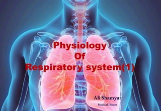
physiology26-respiratory system1.pdf
- 2. contents 1)Physiological anatomy of respiratory system 2)Mechanism of respiration 3)Pulmonary volume, capacities and function tests 4)Transport of Gases 5)Exchange of Gases 6)Regulation of respiration 7)Applied aspects
- 3. 1)Physiological anatomy of respiratory system Anatomically, respiratory tract is divided into : 1-upper, which consist of organs outside thorax ( nasal cavity, pharynx pharynx and larynx) 2-lower respiratory tract which consist of organs within thorax ( trachea, bronchi, bronchioles, alveolar duct and alveoli). The discussion is mainly concentrated on the lower respiratory tract and the related physiology.
- 4. Respieatory unit Respiratory unit is the terminal portion of the respiratory tract. It includes: 1-respiratory bronchioles 2-alveolar ducts 3-alveolar atrium 4-alveolar sac 5-alveolus A typical pair of human lungs contain about 375-800 million alveoli, producing 70 m2 (750 sq ft) of surface area. Each alveolus is wrapped in a fine mesh of capillaries covering about 70% of its area. The diameter of an alveolus is between 200 and 500 μm.
- 5. Water surface tension The surface tension of water is 7.2 Pa at 25°C . The surface tension arises from the polar nature of the water molecule
- 9. Tytpes of respirationn: 1-external respiration: -exchange of O2 &CO2 between lungs and blood 2-internal respirationn: -exchange of O2 and CO2 between blood and tissues discussion
- 10. 2)Mechanism of ventilation Major respiratory muscles:
- 12. Mechanics of ventilation Boyle’s law states that the volume of gas is inversely proportional to pressure (when temperature is constant). Therefore: 1-When the volume of the thoracic cavity increases – the volume of the lungs increases and the pressure within the lungs decreases. 2-When the volume of the thoracic cavity decreases – the volume of the lungs decreases and the pressure within the lungs increases. Air ouside lungs Air ouside lungs
- 14. Mechanics of ventilation When the plastic wrap is pulled, the balloon inflates. When the plastic wrap is pushed, the balloon deflates.
- 15. Mechanics of ventilation The space between the outer surface of the lungs and inner thoracic wall is known as the pleural space. This is usually filled with pleural fluid, forming a seal which holds the lungs against the thoracic wall. This seal ensures that when the thoracic cavity expands or reduces, the lungs undergo expansion or reduction in size accordingly. During breathing, the contraction and relaxation of muscles acts to change the volume of the thoracic cavity – thereby altering the volume of the lungs, and changing the pressure inside the lungs.
- 16. Basic Concepts Elastic recoil of the chest wall, tries to pull the chest outward Elastic recoil of lung, creates an inward pull Intra-alveolar pressure P < atmospheric P Inspiration Intra-alveolar pressure P > atmospheric P Expiration
- 17. Transmural( or alveolar distending Pressure )Gradient
- 18. Mechanics of ventilation Inspiration is the phase of ventilation in which air enters the lungs. It is initiated by contraction of the inspiratory muscles: 1-Diaphragm: - flattens, extending the superior/inferior dimension of the thoracic cavity. 2-External intercostal muscles : - elevates the ribs and sternum, extending the anterior/posterior dimension of the thoracic cavity.
- 19. Mechanics of ventilation The action of the inspiratory muscles results in an increase in the volume of the thoracic cavity. As the lungs are held against the inner thoracic wall by the pleural seal, they also undergo an increase in volume.
- 20. Mechanics of ventilation As per Boyle’s law, an increase in lung volume results in a decrease in the pressure within the lungs. The pressure outside the lungs is now higher; – and so air rushes into the lungs, moving down the pressure gradient.
- 21. Mechanics of ventilation Expiration is the phase of ventilation in which air is expelled from the lungs. It is initiated by relaxation of the inspiratory muscles: 1-Diaphragm : - returns to resting position, reducing the superior/inferior dimension of the thoracic cavity. 2-External intercostal muscles : - relaxes to depress the ribs and sternum, reducing the anterior/posterior dimension of the thoracic cavity.
- 22. Mechanics of ventilation The relaxation of the inspiratory muscles results in a decrease in the volume of the thoracic cavity. The elastic recoil of the previously expanded lung tissue allows them to return to their original size.
- 23. Mechanics of ventilation As per Boyle’s law, a decrease in lung volume results in an increase in the pressure within the lungs. The pressure inside the lungs is now higher than outside – and so air moves out of the lungs, down the pressure gradient.
- 25. Forced breathing Forced breathing is an active mode of breathing which utilizes additional muscles to rapidly expand and contract the thoracic cavity volume. It most commonly occurs during exercise.
- 26. Forced breathing Active Inspiration Active inspiration involves the contraction of the accessory muscles of breathing (in addition to the diaphragm and external intercostals). All these muscles act to increase the volume of the thoracic cavity: 1-Scalenes – elevates the upper ribs. 2-Sternocleidomastoid – elevates the sternum. 3-Pectoralis major and minor – pulls ribs outwards. 4-Serratus anterior – elevates the ribs (when the scapulae are fixed). 5-Latissimus dorsi – elevates the lower ribs.
- 27. Forced breathing Active Expiration: Active expiration utilises the contraction of several thoracic and abdominal muscles. These muscles act the decrease the volume of the thoracic cavity: 1_Anterolateral abdominal wall – increases the intra- abdominal pressure, pushing the diaphragm upwards into the thoracic cavity. 2-Internal intercostal – depresses the ribs. 3-Innermost intercostal – depresses the ribs.
- 28. 3)Pulmonary volume, capacities and function tests
- 29. 500 ml
- 30. 2000-3000 ml
- 31. 800-1100 ml
- 32. Residual Volume(RV) It is the volume of air remaining in the lungs after maximal exhalation.It is indirectly measured from summation of FRC and ERV and cannot be measured by spirometry. In obstructive lung diseases with features of incomplete emptying of the lungs and air trapping, RV may be significantly high. 1000-1200 ml
- 33. Inspiratory capacity(IC) It is the maximum volume of air that can be inhaled following a resting state. It is calculated from the sum of inspiratory reserve volume and tidal volume. IC = IRV+TV 2500-3500 ml
- 34. Vital Capacity(VC) It is the total amount of air exhaled after maximal inhalation. The value varies according to age and body size. It is calculated by summing tidal volume, inspiratory reserve volume, and expiratory reserve volume. VC = TV+IRV+ERV. 3300-4700 ml
- 35. Function Residual Capacity(FRC) : It is the amount of air remaining in the lungs at the end of a normal exhalation. It is calculated by adding together residual and expiratory reserve volumes. FRC = RV+ERV. 1800-2200 ml
- 36. Total Lung Capacity(TLC) It is the maximum volume of air the lungs can accommodate or sum of all volume compartments or volume of air in lungs after maximum inspiration. TLC is calculated by summation of the four primary lung volumes. (TV, IRV, ERV, RV) 4000-6000 ml
- 37. Dead space
- 38. Dead space represents the volume of ventilated air that does not participate in gas exchange. The two types of dead space are anatomical dead space and physiologic dead space: 1- Anatomical dead space is represented by the volume of air that fills the conducting zone of respiration made up by the nose, trachea, and bronchi. This volume is considered to be 30% of normal tidal volume (500 mL ); (the value of anatomic dead space is 150 mL ). 2-Alveolar dead space is the volume of air in the respiratory zone that does not take part in gas exchange. Physiologic or total dead space is equal to anatomic plus alveolar dead space .
- 39. spirometer