SlideShare a Scribd company logo
1 of 47
Download to read offline
General Properties of Viruses
Viruses are the smallest infectious agents (ranging from about 20 nm to 300 (in some cases 1000) nm in diameter) and contain
only one kind of nucleic acid (RNA or DNA) as their genome.
Viruses are obligatory intracellular parasites, it means they absolutely require living host cells in order to multiply.
The nucleic acid is encased in a protein shell, which may be surrounded by a lipid-containing membrane.
Virion is the physical particle in extracellular phase which is able to spread to new host cells, complete intact virus partical .
Viruses are inert in the extracellular environment; they replicate only in living cells, being parasites at the genetic level.
VIRUS - the Latin word for poison, to describe filterable infectious agents.
• 1892, tobacco mosaic disease (lMO), the Russian bacteriologist Dimitri Iwanowski
• The first human disease associated with a filterable agent was yellow fever.
• Advances in the molecular biological techniques in the 1980s and 1990s led to the recognition of several new viruses,
including human immunodeficiency virus (HIV) and SARS associated coronavirus. COVID 19 in our days.
The viral nucleic acid contains information necessary for programming the infected host cell to synthesize virus-specific
macromolecules required for the production of viral progeny. During the replicative cycle, numerous copies of viral nucleic acid
and coat proteins are produced.
The coat proteins assemble together to form the capsid, which encases and stabilizes the viral nucleic acid against the
extracellular environment and facilitates the attachment and penetration by the virus upon contact with new susceptible cells.
Viruses have few or no enzymes of their own for metabolism; for example, they lack enzymes for protein synthesis and ATP
generation.
To multiply, viruses must take over the metabolic machinery of the host cell.
The virus infection may have little or no effect on the host cell or may result in cell damage or death.
The universe of viruses is rich in diversity. varying in size, shape, and genetic content; only some types possess a lipid
envelope.
Viruses vary greatly in structure, genome organization and expression, and strategies of replication and transmission.
The host range for a given virus may be broad or extremely limited.
Viruses are known to infect unicellular organisms such as mycoplasmas, bacteria, and algae and all higher plants and animals.
TERMS AND DEFINITIONS IN VIROLOGY
Capsid: The protein shell, or coat, that encloses the nucleic acid
genome.
Capsomeres: Morphologic units seen in the electron microscope on
the surface of icosahedral virus particles. Capsomeres represent
clusters of polypeptides, but the morphologic units do not necessarily
correspond to the chemically defined structural units.
Defective virus: A virus particle that is functionally deficient in some
aspect of replication.
Envelope: A lipid-containing membrane that surrounds some virus
particles. It is acquired during viral maturation by a budding process
through a cellular membrane. Virus encoded glycoproteins are
exposed on the surface of the envelope. These projections are called
peplomers.
Nucleocapsid: The protein–nucleic acid complex representing the
packaged form of the viral genome. The term is commonly used in
cases in which the nucleocapsid is a substructure of a more complex
virus particle.
Structural units: The basic protein building blocks of the coat. They
are usually a collection of more than one nonidentical protein subunit.
The structural unit is often referred to as a protomer.
Subunit: A single folded viral polypeptide chain.
Enveloped virus with icosahedral symmetry.
Not all icosahedral viruses have envelopes.
Virus with helical symmetry
Host Range
The host range of a virus is the spectrum of host cells the virus can infect.
There are viruses that infect invertebrates, vertebrates, plants, protists, fungi, and bacteria. However, most viruses are able to infect
specific types of cells of only one host species. In rare cases, viruses cross the host-range barrier, thus expanding their host range.
Viruses that infect bacteria are called bacteriophages, or phages.
The particular host range of a virus is determined by the virus's requirements for its specific attachment to the host cell and the
availability within the potential host of cellular factors required for viral multiplication.
For the virus to infect the host cell, the outer surface of the virus must chemically interact with specific receptor sites on the
surface of the cell.
For some bacteriophages, the receptor site is part of the cell wall of the host; in other cases, it is part of the fimbriae
or flagella.
For animal viruses, the receptor sites are on the plasma membranes of the host cells.
CLASSIFICATION OF ANIMAL VIRUSES
The following properties have been used as a basis for the classification of viruses.
Virion morphology, including
• size,
• shape,
• type of symmetry,
• presence or absence of peplomers, and
• presence or absence of membranes.
Virus genome properties, including
• type of nucleic acid (DNA or RNA),
• size of genome in kilobases (kb) or kilobase pairs (kbp),
• strandedness (single or double), whether linear or circular,
• sense (positive, negative),
• segments (number, size),
• nucleotide sequence, G + C content, and
• presence of special features (repetitive elements, isomerization)
Genome organization and replication, including
• gene order,
• number and position of open reading frames,
• strategy of replication (patterns of transcription, translation),
• cellular sites (accumulation of proteins, virion assembly, virion
release).
Virus protein properties, including
• number,
• size,
• functional activities of structural and nonstructural proteins,
• amino acid sequence,
• modifications (glycosylation, phosphorylation, myristylation),
• special functional activities (transcriptase, reverse transcriptase, neuraminidase,
fusion activities).
Physicochemical properties of the virion, including
• molecular mass,
• pH stability,
• thermal stability, and
• susceptibility to physical and chemical agents
Biologic properties, including
• natural host range,
• mode of transmission,
• vector relationships,
• pathogenicity,
• tissue tropisms,
• pathology.
Antigenic properties
Viral Sizes and shapes
Direct observation in the electron microscope is the most widely
used method for estimating particle size.
Another method that can be used is sedimentation in the
ultracentrifuge. The relationship between the size and shape of a
particle and its rate of sedimentation permits determination of
particle size.
Different viruses vary considerably in size. Although most are quite
a bit smaller than bacteria, some of the larger viruses (such as the
vaccinia virus) are about the same size as some very small bacteria
(such as the mycoplasmas, rickettsias, and chlamydias).
Viruses range from 20 to 1000 nm in length.
Universal System of Virus Taxonomy
Viruses are separated into major groupings—called families—on the basis of virion morphology, genome structure, and strategies
of replication. Virus family names have the suffix -viridae.
Within each family, subdivisions called genera are usually based on biological, genomic, physicochemical, or serologic
differences.
Criteria used to define genera vary from family to family. Genus names carry the suffix -virus.
By 2000, the International Committee on Taxonomy of Viruses had organized more than 4000 animal and plant viruses into 56
families and 233 genera, with hundreds of viruses still unassigned. Of these, 24 families contained viruses that infect humans and
animals.
Families of Animal Viruses that Contain Members Able to Infect Humans
Capsid and Envelope
The nucleic acid of a virus is protected by a protein coat called the capsid. The structure
of the capsid is ultimately determined by the viral nucleic acid and accounts for most of
the mass of a virus, especially of small ones. Each capsid is composed of protein
subunits called capsomeres.
In some viruses, the proteins composing the capsomeres are of a single type; in other
viruses, several types of protein may be present.
The arrangement of capsomeres is characteristic of a particular type of virus.
In some viruses, the capsid is covered by an envelope which usually consists of some
combination of lipids, proteins, and carbohydrates. Some animal viruses are released
from the host cell by an extrusion process that coats the virus with a layer of the host
cell 's plasma membrane; that layer becomes the viral envelope.
In many cases, the envelope contains proteins determined by the viral nucleic acid and
materials derived from normal host cell components.
Depending on the virus, envelopes may or may not be covered by spikes, which are
carbohydrate-protein complexes that project from the surface of the envelope. Some
viruses attach to host cells by means of spikes. Spikes are such a reliable characteristic
of some viruses that they can be used as a means of identification.
Viruses whose capsids are not covered by an envelope are known as nonenveloped
viruses. The capsid of a nonenveloped virus protects the nucleic acid from nuclease
enzymes in biological fluids and promotes the virus's attachment to susceptible host
cells.
PRINCIPLES OF VIRUS STRUCTURE
Electron microscopy, cryoelectron microscopy, and x-ray diffraction techniques have made it possible to resolve fine
differences in the basic morphology of viruses.
Cubic Symmetry (Polyhedral viruses)
Many animal, plant, and bacterial viruses are polyhedral, or manysided, viruses.
All cubic symmetry observed with animal viruses is of the icosahedral pattern, a
regular polyhedron with 20 triangular faces and 12 corners (vertices) and
fivefold, threefold, and twofold axes of rotational symmetry. The vertex units
have five neighbors (pentavalent), and all others have six (hexavalent).
There are exactly 60 identical subunits on the surface of an icosahedron. To
build a particle size adequate to encapsidate viral genomes, viral shells are
composed of multiples of 60 structural units.
An example of a polyhedral virus in the shape of an icosahedron are
adenoviruses and polioviruses.
Viral architecture can be grouped into three types based on the
arrangement of morphologic subunits:
(1) cubic symmetry (eg, adenoviruses),
(2) helical symmetry (eg, orthomyxoviruses), and
(3) complex structures (eg, poxviruses).
Helical Symmetry
In cases of helical symmetry, protein subunits are bound in a periodic way to the viral nucleic
acid, winding it into a helix. The filamentous viral nucleic acid–protein complex
(nucleocapsid) is then coiled inside a lipid-containing envelope. Thus, there is a regular,
periodic interaction between capsid protein and nucleic acid in viruses with helical symmetry.
All known examples of animal viruses with helical symmetry contain RNA genomes and,
with the exception of rhabdoviruses, have flexible nucleocapsids that are wound into a ball
inside envelopes.
Helical viruses resemble long rods that may be rigid or flexible. The viruses that cause rabies
and Ebola hemorrhagic fever are helical viruses.
Enveloped viruses are roughly sphericaL When helical or polyhedral viruses are enclosed by
envelopes, they are called enveloped helical or enveloped polyhedral viruses. An example of
an enveloped helical virus is the influenza virus.
An example of an enveloped polyhedral (icosahedral) virus is the herpes simplex virus.
Complex Structures
Some virus particles do not exhibit simple cubic or helical
symmetry but are more complicated in structure.
Some viruses, particularly bacterial viruses, have complicated
structures and are called complex viruses. One example of a
complex virus is a bacteriophage. Some bacteriophages have
capsids to which additional structures are attached (Figure
13.5a).
The capsid (head) is polyhedral and that the tail sheath is
helicaL The head contains the nucleic acid.
Another example of complex viruses are poxviruses, which
do not contain clearly identifiable capsids but have several
coats around the nucleic acid.
CHEMICAL COMPOSITION OF VIRUSES
Viral Protein
The structural proteins of viruses have several important functions.
Their major purpose is to facilitate transfer of the viral nucleic acid from one host cell to another.
They serve to protect the viral genome against inactivation by nucleases, participate in the attachment of the virus
particle to a susceptible cell.
They provide the structural symmetry of the virus particle.
The proteins determine the antigenic characteristics of the virus.
Some surface proteins may also exhibit specific activities (eg, influenza virus hemagglutinin agglutinates red blood
cells).
Some viruses carry enzymes (which are proteins) inside the virions. The enzymes are present in very small amounts
and are probably not important in the structure of the virus particles; however, they are essential for the initiation of
the viral replicative cycle when the virion enters a host cell.
Examples include an RNA polymerase carried by viruses with negative-sense RNA genomes (eg, orthomyxoviruses, rhabdoviruses) that is
needed to copy the first mRNAs, and reverse transcriptase, an enzyme in retroviruses that makes a DNA copy of the viral RNA, an essential
step in replication and transformation.
At the extreme in this respect are the poxviruses, the cores of which contain a transcriptional system; many different enzymes are packaged in
poxvirus particles.
Lyzoceam - bacteriaphage
Viral Nucleic Acid
Viruses contain a single kind of nucleic acid—either DNA or RNA—that
encodes the genetic information necessary for replication of the virus.
The genome may be single or double stranded, circular or linear, and
segmented or nonsegmented. The type of nucleic acid, its strandedness, and its
size are major characteristics used for classifying viruses into families.
The size of the viral DNA genome ranges from 3.2 kbp (hepadnaviruses) to
375 kbp (poxviruses).
The size of the viral RNA genome ranges from about 4 kb (picobirnaviruses)
to 32 kb (coronaviruses).
All major DNA viral groups have genomes that are single molecules of DNA
and have a linear or circular configuration.
Viral RNAs exist in several forms.
The RNA may be a single linear molecule (eg, picornaviruses).
For other viruses (eg, orthomyxoviruses), the genome consists of several segments
of RNA that may be loosely associated within the virion.
The isolated RNA of viruses with positive-sense genomes (ie, picornaviruses,
togaviruses) is infectious, and the molecule functions as an mRNA within the
infected cell.
The isolated RNA of the negative-sense RNA viruses, such as rhabdoviruses and
orthomyxoviruses, is not infectious. For these viral families, the virions carry an
RNA polymerase that in the cell transcribes the genomic RNA molecules into
several complementary RNA molecules, each of which may serve as an mRNA.
Viral nucleic acid may be characterized by its G + C content.
Viral Lipid Envelopes
A number of different viruses contain lipid envelopes as part of
their structure.
The lipid is acquired when the viral nucleocapsid buds through a
cellular membrane in the course of maturation. Budding occurs
only at sites where virus-specific proteins have been inserted into
the host cell membrane. The budding process varies markedly
depending on the replication strategy of the virus and the structure
of the nucleocapsid.
The specific phospholipid composition of a virion envelope is
determined by the specific type of cell membrane involved in the
budding process. For example, herpesviruses bud through the
nuclear membrane of the host cell, and the phospholipid
composition of the purified virus reflects the lipids of the nuclear
membrane.
Lipid-containing viruses are sensitive to treatment with
desinfectants indicating that disruption or loss of lipid results in
loss of infectivity.
Nonlipid-containing viruses are generally resistant to ether.
Viral Glycoproteins
Viral envelopes contain glycoproteins. In contrast to the
lipids in viral membranes, which are derived from the
host cell, the envelope glycoproteins are virus encoded.
However, the sugars added to viral glycoproteins often
reflect the host cell in which the virus is grown.
The surface glycoproteins of an enveloped virus attach
the virus particle to a target cell by interacting with a
cellular receptor.
They are also often involved in the membrane fusion
step of infection.
The glycoproteins are also important viral antigens.
Influenza virus membrane glycoproteins are
hemagglutinin, neuraminidase.
REACTION TO PHYSICALAND CHEMICALAGENTS
Heat and Cold
There is great variability in the heat stability of different viruses.
Icosahedral viruses are heat stable, but enveloped viruses are much more heat labile.
Viral infectivity is generally destroyed by heating at 50–60°C for 30 minutes, although there are some notable exceptions (eg,
hepatitis B virus, polyomaviruses).
Viruses can be preserved by storage at subfreezing temperatures, and some may withstand lyophilization and can thus be
preserved in the dry state at 4°C or even at room temperature.
Viruses that withstand lyophilization are more heat resistant when heated in the dry state. Enveloped viruses tend to lose
infectivity after prolonged storage even at –90°C and are particularly sensitive to repeated freezing and thawing.
Stabilization of Viruses by Salts
Many viruses can be stabilized by salts in concentrations of 1 mol/L. The mechanism by which the salts stabilize viral
preparations is not known.
Viruses are preferentially stabilized by certain salts. MgCl2, 1 mol/L, stabilizes picornaviruses and reoviruses; MgSO4, 1
mol/L, stabilizes orthomyxoviruses and paramyxoviruses; and Na2SO4, 1 mol/L, stabilizes herpesviruses.
The stability of viruses is important in the preparation of vaccines. The ordinary nonstabilized oral polio vaccine must be stored
at freezing temperatures to preserve its potency.
However, with the addition of salts for stabilization of the virus, potency can be maintained for weeks at ambient temperatures
even in the high temperatures of the tropics.
pH
Viruses are usually stable between pH values of 5.0 and 9.0.
Some viruses (eg, enteroviruses) are resistant to acidic conditions. All viruses are destroyed by alkaline conditions.
Radiation
Ultraviolet, x-ray, and high-energy particles inactivate viruses. The dose varies for different viruses. Infectivity is the most
radiosensitive property because replication requires expression of the entire genetic contents. Irradiated particles that are
unable to replicate may still be able to express some specific functions in host cells.
Ether Susceptibility
Ether susceptibility can be used to distinguish viruses that possess an envelope from those that do not.
Detergents
Nonionic detergents (eg, Nonidet P40 and Triton X-100) solubilize lipid constituents of viral membranes. The viral
proteins in the envelope are released (undenatured). Anionic detergents (eg, sodium dodecyl sulfate) also solubilize viral
envelopes; in addition, they disrupt capsids into separated polypeptides.
Formaldehyde
Formaldehyde destroys viral infectivity by reacting with nucleic acid. Viruses with single-stranded genomes are inactivated
much more readily than those with double-stranded genomes. Formaldehyde has minimal adverse effects on the
antigenicity of proteins and therefore has been used frequently in the production of inactivated viral vaccines.
Photodynamic Inactivation
Viruses are penetrable to a varying degree by vital dyes such as toluidine blue, neutral red, and proflavine.
These dyes bind to the viral nucleic acid, and the virus then becomes susceptible to inactivation by visible light.
Antibiotics and Other Antibacterial Agents
Antibacterial antibiotics and sulfonamides, organic iodine compounds, quaternary ammonium compounds are not effective
against viruses. However some antiviral drugs are available. Larger concentrations of chlorine are required to destroy viruses than
to kill bacteria, especially in the presence of extraneous proteins.
Alcohols, such as isopropanol and ethanol, are relatively ineffective against certain viruses, especially picornaviruses.
Common Methods of Inactivating Viruses for Various Purposes
Viruses may be inactivated for various reasons, such as to sterilize laboratory supplies and equipment, disinfect surfaces or skin,
make drinking water safe, and produce inactivated virus vaccines.
Different methods and chemicals are used for these purposes.
Sterilization may be accomplished by steam under pressure, dry heat, ethylene oxide, and γ-irradiation.
Surface disinfectants include sodium hypochlorite, glutaraldehyde, formaldehyde, and peracetic acid.
Skin disinfectants include chlorhexidine, 70% ethanol, and iodophores.
Vaccine production may involve the use of formaldehyde, β-propiolactone, psoralen + ultraviolet irradiation, or detergents
(subunit vaccines) to inactivate the vaccine virus.
Multiplication of Bacteriophages
Bacteriophages
The lytic cycle And lysogenic cycle
Homework for practical course
Growing Bacteriophages in the Laboratory
Growing Animal Viruses in the laboratory
In the laboratory, three methods are commonly used for culturing animal viruses.
These methods involve using
• living animals,
• embryonated eggs, or
• cell cultures.
Viral Identification
• Electron microscope.
• Serological methods, such as Western blotting, are the most commonly used means of identification. In these tests, the
virus is detected and identified by its reaction with antibodies.
• Observation of cytopathic effects is also useful for the identification of a virus.
• Restriction fragment length polymorphisms (RFLPs)
• Polymerase chain reaction (PCR) was used to amplify viral RNA (West Nile virus in 1999 in the United Statesand the
SARS-associated corona virus in China in 2002 and COVID19 these days
Cells grown in vitro are central to the cultivation and characterization of viruses.
A. Detection of Virus-Infected Cells
Multiplication of a virus can be monitored in a variety of ways:
1. Development of cytopathic effects (ie, morphologic changes in the
cells). Types of virus-induced cytopathic effects include cell lysis or
necrosis, inclusion formation, giant cell formation, and cytoplasmic
vacuolization
2. Appearance of a virus-encoded protein, such as the hemagglutinin of
influenza virus. Specific antisera can be used to detect the synthesis of
viral proteins in infected cells.
3. Detection of virus-specific nucleic acid. Molecular-based assays such as
polymerase chain reaction provide rapid, sensitive, and specific methods of
detection.
4. Adsorption of erythrocytes to infected cells, called hemadsorption, caused
by the presence of virus-encoded hemagglutinin
(parainfluenza, influenza) in cellular membranes. This reaction becomes
positive before cytopathic changes are visible and in some cases occurs in
the absence of cytopathic effects
5. Viral growth in an embryonated chick egg may result in death of the
embryo (eg, encephalitis viruses), production of pocks or plaques on the
chorioallantoic membrane (eg, herpes, smallpox, vaccinia), or development
of hemagglutinins in the embryonic fluids or tissues (eg, influenza).
B. Inclusion Body Formation
In the course of viral multiplication within cells, virusspecific structures called inclusion bodies may be
produced. They become far larger than the individual virus particle and often have an affinity for acid
dyes (eg, eosin). They may be situated in the nucleus (herpesvirus), in the cytoplasm (poxvirus), or in
both (measles virus).
The presence of inclusion bodies may be of considerable diagnostic aid. The intracytoplasmic inclusion
in nerve cells—the Negri body—is pathognomonic for rabies.
Quantitation of Viruses
A. Physical Methods
• Quantitative polymerase chain reaction
• Hemagglutination assays - are an easy and rapid method of quantitating these types of viruses. Certain viruses contain a
protein (hemagglutinin) that has the ability to agglutinate red blood cells of humans or some animal.
• Radioimmunoassays and enzyme-linked immunosorbent assays. These tests do not distinguish infectious from noninfectious
particles and sometimes detect viral proteins not assembled into particles.
• electron microscope (infectious virus particles cannot be distinguished from noninfectious ones).
B. Biologic Methods
Plaque assay - A widely used assay for infectious virus growing well in tissue culture. Monolayers of host cells are inoculated
with suitable dilutions of virus and after adsorption are overlaid with medium containing agar or carboxymethylcellulose to
prevent virus spreading throughout the culture. After several days, the cells initially infected have produced virus that spreads
only to surrounding cells. Multiple cycles of replication and cell killing produce a small area of infection, or plaque. Under
controlled conditions, a single plaque can arise from a single infectious virus particle, termed a plaqueforming unit. The
cytopathic effect of infected cells within the plaque can be distinguished from uninfected cells of the monolayer with or without
suitable staining, and plaques can usually be counted macroscopically.
A more rapid method of assay is based on determination of the number of infected cells producing a viral antigen, such as by
immunofluorescence. Certain viruses (eg, herpes and vaccinia) form pocks when inoculated onto the chorioallantoic membrane of
an embryonated egg. Such viruses can be quantitated by relating the number of pocks counted to the viral dilution inoculated.
REPLICATION OF VIRUSES
Viruses multiply only in living cells. The host cell must provide the energy and synthetic machinery and the lowmolecular-
weight precursors for the synthesis of viral proteins and nucleic acids. The viral nucleic acid carries the genetic specificity to
code for all of the virus-specific macromolecules in a highly organized fashion.
The unique feature of viral multiplication is that soon after interaction with a host cell the infecting virion is
disrupted and its measurable infectivity is lost. This phase of the growth cycle is called the eclipse period.
After the synthesis of viral nucleic acid and viral proteins, the components assemble to form new infectious
virions. The yield of infectious virus per cell ranges widely, from modest numbers to more than 100,000 particles.
The duration of the virus replication cycle also varies widely, from 6 to 8 hours (picornaviruses) to more than 40
hours (some herpesviruses).
Not all infections lead to new progeny virus.
• Productive infections occur in permissive cells and result in the production of infectious virus.
• Abortive infections fail to produce infectious progeny, either because the cell may be nonpermissive and
unable to support the expression of all viral genes or because the infecting virus may be defective, lacking
some functional viral gene.
• A latent infection may ensue, with the persistence of viral genomes, the expression of no or a few viral genes,
and the survival of the infected cell.
General Steps in Viral Replication Cycles
Steps in viral replication include
• attachment to a cell via binding to specific receptors on the cell surface,
• entry into the cell,
• uncoating of the viral genome,
• regulated expression of viral transcripts,
• synthesis of viral proteins,
• replication of viral genomic nucleic acid,
• assembly of new progeny viruses, and
• release of new virions from the cell.
The duration of replication cycles varies widely among different virus types.
The infected cells may be killed or may survive with little damage.
Not all infections lead to new progeny virus.
Attachment, Penetration, and Uncoating
The first step in viral infection is attachment,
interaction of a virion with a specific receptor site on the
surface of a cell. Receptor molecules differ for different
viruses but are generally glycoproteins. Successful
infection of a cell by a virus may involve interaction
with more than one type of receptor.
The presence or absence of receptors plays an important
determining role in cell tropism and viral pathogenesis.
The attachment step may initiate irreversible structural
changes in the virion. After binding, the virus particle is
taken up inside the cell. This step is referred to as
penetration or engulfment.
Uncoating occurs shortly after penetration. It is the
physical separation of the viral nucleic acid from the
outer structural components of the virion so that it can
function.
The genome may be released as free nucleic acid
(picornaviruses) or as a nucleocapsid (reoviruses). The
nucleocapsids usually contain polymerases. Uncoating
may require acidic pH in the endosome.
Expression of Viral Genomes and Synthesis of Viral Components
The essential theme in viral replication is that specific mRNAs must be transcribed from the viral nucleic acid for successful
expression and duplication of genetic information. After this is accomplished, viruses use cell components to translate the
mRNA. Various classes of viruses use different pathways to synthesize the mRNAs depending on the structure of the viral
nucleic acid.
Negative-strand (negativesense) RNA viruses (eg, rhabdoviruses) carry RNA polymerases to synthesize mRNAs. Their
single-strand RNA genome is complementary to mRNA, which is conventionally designated positive strand (positive sense).
The negative-strand viruses must supply their own RNA polymerase because eukaryotic cells lack enzymes able to synthesize
mRNA off an RNA template.
Positive strand (positive sense) RNA viruses contain RNA which can act as mRNA.
In the course of viral replication, all of the virus specified macromolecules are synthesized in a highly organized sequence.
In some viral infections, notably those involving double-stranded DNA-containing viruses, early viral proteins are
synthesized soon after infection and late proteins are made only late in infection after viral DNA synthesis begins.
DNA viruses that replicate in the nucleus generally use host cell DNA and RNA polymerases and processing enzymes. The
larger viruses (herpesviruses, poxviruses) are more independent of cellular functions than are the smaller viruses. This is one
reason the larger viruses are more susceptible to antiviral chemotherapy because more virus-specific processes are available
as targets for drug action.
Viral DNA is usually replicated in the nucleus. Viral genomic RNA is generally duplicated in the cell cytoplasm, although
there are exceptions.
Morphogenesis and Release
Newly synthesized viral genomes and capsid polypeptides assemble together to form progeny viruses.
In general, nonenveloped viruses accumulate in infected cells, and the cells eventually lyse and release the virus particles.
Enveloped viruses mature by a budding process. Virusspecific envelope glycoproteins are inserted into cellular membranes; viral
nucleocapsids then bud through the membrane at these modified sites and in so doing acquire an envelope.
Budding frequently occurs at the plasma membrane but may involve other membranes in the cell. Enveloped viruses are not
infectious until they have acquired their envelopes.
Viral components may accumulate and be involved in the formation of inclusion bodies in the cell. As a result of the profound
deleterious effects of viral replication, cellular cytopathic effects eventually develop and the cell dies.
The growth cycle of a nonenveloped, double-
stranded DNA virus
(1) After penetrating the host cell, viral DNA is
uncoated and enters the nucleus.
(2) Viral genes are transcribed.
(3) The mRNAs are translated in the cytoplasm.
Newly synthesized proteins enter the nucleus.
(4) Viral DNA is replicated in the nucleus,
sometimes with the help of newly synthesized
viral replication proteins.
(5) Viral DNA and viral structural proteins assemble
in the nucleus to produce new progeny virions.
(6) On rare occasions, viral DNA may be
incorporated into cellular DNA as a side-effect
of infection.
The growth cycle of a positive-sense, single-
stranded RNA virus
(1) The virus enters the cell and the viral
RNA genome is uncoated.
(2) As a positive-sense, single-stranded
genome, the RNA is directly translated,
producing viral proteins.
(3) A negative-sense RNA copy of the
positive template is synthesized.
(4) It is used to produce many positive-sense
copies.
(5) The newly synthesized positive-sense
RNA molecules are assembled with viral
structural proteins to produce new
progeny virions.
Interactions Among Viruses
When two or more virus particles infect the same host cell, they may interact in a variety of ways.
A. Recombination
Recombination results in the production of progeny virus (recombinant) that carries traits not found together in either parent.
The classic mechanism is that the nucleic acid strands break, and part of the genome of one parent is joined to part of the
genome of the second parent. The recombinant virus is genetically stable, yielding progeny similar to itself upon replication.
Viruses vary widely in the frequency with which they undergo recombination. In the case of viruses with segmented
genomes (eg, influenza virus) the formation of recombinants is caused by reassortment of individual genome fragments
rather than by an actual crossover event.
B. Complementation
This refers to the interaction of viral gene products in cells infected with two viruses, one or both of which may be defective.
It results in the replication of one or both under conditions in which replication would not ordinarily occur. The basis for
complementation is that one virus provides a gene product in which the second is defective, allowing the second virus to
grow. The genotypes of the two viruses remain unchanged.
If both mutants are defective in the same gene product, they will not be able to complement each other’s growth.
C. Phenotypic Mixing
A special case of complementation is phenotypic mixing, or the association of a genotype with a heterologous phenotype.
This occurs when the genome of one virus becomes randomly incorporated within capsid proteins specified by a different virus or
a capsid consisting of components of both viruses. If the genome is encased in a completely heterologous protein coat, this
extreme example of phenotypic mixing may be called “phenotypic masking” or “transcapsidation.” Such mixing is not a stable
genetic change because, upon replication, the phenotypically mixed parent will yield progeny encased in capsids homologous to
the genotype. Phenotypic mixing usually occurs between different members of the same virus family; the intermixed capsid
proteins must be able to interact correctly to form a structurally intact capsid. However, phenotypic mixing also can occur between
enveloped viruses, and in this case, the viruses do not have to be closely related.
D. Interference
Infection of either cell cultures or whole animals with two viruses often leads to an inhibition of multiplication of one of the
viruses, an effect called interference. Interference in animals is distinct from specific immunity. Furthermore, interference does not
occur with all viral combinations; two viruses may infect and multiply within the same cell as efficiently as in single infections.
Several mechanisms have been elucidated as causes of interference:
(1) One virus may inhibit the ability of the second to adsorb to the cell, either by blocking its receptors (retroviruses,
enteroviruses) or by destroying its receptors (orthomyxoviruses).
(2) One virus may compete with the second for components of the replication apparatus (eg, polymerase, translation initiation
factor).
(3) The first virus may cause the infected cell to produce an inhibitor that prevents replication of the second virus.
MODES OF TRANSMISSION OF VIRUSES
Viruses may be transmitted in the following ways:
1. Direct transmission from person to person by contact. The major means of transmission include
• droplet or aerosol infection (eg, influenza, measles, smallpox);
• by sexual contact (eg, papillomavirus, hepatitis B, herpes simplex type 2, human immunodeficiency virus);
• by hand– mouth, hand–eye, or mouth–mouth contact (eg, herpes simplex, rhinovirus, Epstein-Barr virus);
• by exchange of contaminated blood (eg, hepatitis B, human immunodeficiency virus).
2. Indirect transmission
• by the fecal–oral route (eg, enteroviruses, rotaviruses, infectious hepatitis A)
• by fomites (eg, Norwalk virus, rhinovirus).
3. Transmission from animal to animal, with humans an accidental host. Spread may be
• by bite (rabies) or by droplet or aerosol infection from rodent-contaminated quarters (eg, arenaviruses, hantaviruses).
4. Transmission by means of an arthropod vector (eg, arboviruses, now classified primarily as togaviruses, flaviviruses, and
bunyaviruses).
Emerging Viral Diseases
Owing to wide-reaching changes in social attitudes, technology, and the environment—plus the decreased effectiveness of
previous approaches to disease control—the spectrum of infectious diseases is expanding today. New agents appear, and diseases
once thought to be under control are increasing in incidence as pathogens evolve and spread. The term “emerging infectious
diseases” denotes these phenomena.
Viral diseases emerge following one of three general patterns:
• recognition of a new agent,
• abrupt increase in illnesses caused by an endemic agent, and
• invasion of a new host population.
Combinations of factors contribute to disease emergence. Factors include
(1) environmental changes (deforestation, damming or other changes in water ecosystems, flood or drought, famine),
(2) human behavior (sexual behavior, drug use, outdoor recreation),
(3) socioeconomic and demographic phenomena (war, poverty, population growth and migration, urban decay),
(4) travel and commerce (highways, international air travel),
(5) food production (globalization of food supplies, changes in methods of food processing and packaging),
(6) health care (new medical devices, blood transfusions, organ and tissue transplantation, drugs causing immunosuppression,
widespread use of antibiotics),
(7) microbial adaptation (changes in virulence, development of drug resistance, cofactors in chronic diseases), and
(8) public health measures (inadequate sanitation and vector control measures, curtailment of prevention programs, lack of
trained personnel in sufficient numbers).
Examples of emerging viral infections in different regions of the world include
• Ebola virus,
• Nipah virus,
• hantavirus pulmonary disease,
• human immunodeficiency virus infection,
• dengue hemorrhagic fever,
• West Nile virus,
• Rift Valley fever,
• COVID19
• bovine spongiform encephalopathy (the latter a prion disease).
Of potential concern also is the possible use of animal organs as xenografts in humans (xenotransplantation).
Concerns exist about the potential accidental introduction of new viral pathogens from the donor species into humans.
Bioterrorism Agents
Bioterrorism agents are microorganisms (or toxins) that could be used to produce death and disease in humans, animals, or plants
for terrorist purposes.
Such microorganisms could be genetically modified to increase their virulence, make them resistant to drugs or vaccines, or
enhance their ability to be disseminated in the environment. Potential bioterrorism agents are classified into risk categories based
on the ease of dissemination or transmission from person to person, mortality rates, ability to cause public panic, and requirement
for public health preparedness.
Viral agents in the highest risk category are smallpox and the viral hemorrhagic fevers;
Highest risk bacteria include the agents of anthrax, botulism, plague, and tularemia.
Viroids
Viroids are small infectious agents that cause diseases of plants.
Viroids are agents that do not fit the definition of classic viruses. They are nucleic
acid molecules without a protein coat.
Plant viroids are single-stranded, covalently closed circular RNA molecules
consisting of about 360 nucleotides and with a highly base-paired rod like structure.
Viroids replicate by an entirely novel mechanism. Viroid RNA does not encode any
protein products; the devastating plant diseases induced by viroids occur by an
unknown mechanism.
Plant viruses cause many diseases of economically important crops, including beans
(bean mosaic virus), corn and sugarcane (wound tumor virus), and potatoes (potato
yellow dwarf virus).
Viruses can cause color change, deformed growth, wilting, and stunted growth in
their plant hosts. Some hosts, however, remain symptomless and only serve as
reservoirs of infection.
Plant cells are generally protected from disease by an impermeable cell wall. Viruses
must enter through wounds or be assisted by other plant parasites, including
nematodes, fungi, and, most often, insects that suck the plant's sap. Once one plant is
infected, it can spread infection to other plants in its pollen and seeds.
In laboratories, plant viruses are cultured in protoplasts (plant cells with the cell
walls removed) and in insect cell cuitures.
Prions
A few infectious diseases are caused by prions. In 1982, American
neurobiologist Stanley Prusiner proposed that infectious proteins
caused a neurological disease in sheep called scrapie. The infectivity
of scrapie-infected brain tissue is reduced by treatment with proteases
but not by treatment with radiation, suggesting that the infectious
agent is pure protein.
Prusiner coined the name prion for proteinaceous infectious particle.
They are highly resistant to inactivation by heat, formaldehyde, and
ultraviolet light that inactivate viruses.
Prion diseases, called “transmissible spongiform encephalopathies,”
include scrapie in sheep, mad cow disease in cattle, and kuru, fatal
familial insomnia and Creutzfeldt-Jakob disease in humans.
These diseases are caused by the conversion of a normal host
glycoprotein called Prpc (for cellular prion protein) into an infectious
form called PrpSc (for scrapie protein). The gene for Prpc is located
on chromosome 20 in humans. Recent evidence suggests that Prpc is
involved in regulating cell death. The actual cause of cell damage is
not known. Fragments of PrpSc molecules accumulate in the brain,
forming plaques; these plaques are used for postmortem diagnosis,
but they do not appear to be the cause of cell damage.
General Characteristics of Viruses
I. Depending on one's viewpoint, viruses may be regarded as exceptionally complex aggregations of nonliving
chemicals or as exceptionally simple living microbes.
2. Viruses contain a single type of nucleic acid (DNA or RNA) and a protein coat, sometimes enclosed by an
envelope composed of lipids, proteins, and carbohydrates.
3. Viruses are obligatory intracellular parasites. They multiply by using the host cell's synthesizing machinery to
cause the synthesis of specialized elements that can transfer the viral nucleic acid to other cells.
Host Range
1. Host rauge refers to the spectrum of host cells in which a virus can multiply.
2. Most viruses infect only specific types of cells in one host species.
3. Host range is determined by the specific attachment site on the host cell's surface and the availability of
host cellular factors.
Viral Size
1. Viral size is ascertained by electron microscopy.
2. Viruses range from 20 to 1000 nm in length.
Viral Structure
A virion is a complete, fully developed viral particle composed of nucleic acid surrounded by a coat.
Nucleic Acid
1. Viruses contain either DNA or RNA, never both, and the nucleic acid may be single- or double-stranded, linear
or circular, or divided into several separate molecules.
2. The proportion of nucleic acid in relation to protein in viruses ranges from about 1 % to about 50%.
Capsid and Envelope
1. The protein coat surrounding the nucleic acid of a virus is called the capsid.
2. The capsid is composed of subunits, capsomeres, which can be a single type of protein or several types.
3. The capsid of some viruses is enclosed by an envelope consisting of lipids, proteins, and carbohydrates.
4. Some envelopes are covered with carbohydrate-protein complexes called spikes.
General Morphology
1. Helical viruses (for example, Ebola virus) resemble long rods, and their capsids are hollow cylinders
surrounding the nucleic acid.
2. Polyhedral viruses (for example, adenovirus) are many-sided. Usually the capsid is an icosahedron.
3. Enveloped viruses are covered by an envelope and are roughly spherical but highly pleomorphic. There are also
enveloped helical viruses (for example, influenza virus) and enveloped polyhedral viruses (for example,
Simpiexvirus).
4. Complex viruses have complex structures. For example, many bacteriophages have a polyhedral capsid with a
helical tail attached
Isolation, Cultivation, and Identification of Viruses
1. Viruses must be grown in living cells.
2. The easiest viruses to grow are bacteriophages.
Growing Bacteriophages in the laboratory
1. The plaque method mixes bacteriophages with host bacteria and nutrient agar.
2. After several viral multiplication cycles, the bacteria in the area surrounding the original virus are destroyed; the
area of lysis is called a plaque.
3. Each plaque originates with a single viral particle; the concentration of viruses is given as plaque-forming units.
Growing Animal Viruses in the Laboratory
1. Cultivation of some animal viruses requires whole animals.
2. Some animal viruses can be cultivated in embryonated eggs.
3. Cell cultures are cells growing in culture media in the laboratory.
4. Primary cell lines and embryonic diploid cell lines grow for a short time in vitro.
5. Continuous cell lines can be maintained in vitro indefinitely.
6. Viral growth can cause cytopathic effects in the cell culture,
Viral Identification
1. Serological tests are used most often to identify viruses.
2. Viruses may be identified by RFLPs and PCR.
Viral Multiplication
1. Viruses do not contain enzymes for energy production or protein synthesis.
2. For a virus to multiply, it must invade a host cell and direct the host's metabolic machinery to produce viral enzymes and
components.
Multiplication of Bacteriophages
1. During the lytic cycle, a phage causes the lysis and death of a host cell.
2. Some viruses can either cause lysis or have their DNA incorporated as a prophage into the DNA of the host cell. The latter
situation is called lysogeny.
3. During the attachment phase of the lytic cycle, sites on the phage's tail fibers attach to complementary receptor sites on the
bacterial cell.
4. In penetration, phage lysozyme opens a portion of the bacterial cell wall, the tail sheath contracts to force the tail core through
the cell wall, and phage DNA enters the bacterial cell. The capsid remains outside.
5. In biosynthesis, transcription of phage DNA produces mRNA coding for proteins necessary for phage multiplication. Phage
DNA is replicated, and capsid proteins are produced. During the eclipse period, separate phage DNA and protein can be
found.
6. During maturation, phage DNA and capsids are assembled into complete viruses.
7. During release, phage lysozyme breaks down the bacterial cell wall, and the new phages are released.
8. During the lysogenic cycle, prophage genes are regulated by a repressor coded for by the prophage. The prophage is replicated
each time the cell divides.
9. Exposure to certain mutagens can lead to excision of the prophage and initiation of the lytic cycle.
10. Because of lysogeny, lysogenic cells become immune to reinfection with the same phage and may undergo phage conversion.
11. A lysogenic phage can transfer bacterial genes from one cell to another through transduction. Any genes can be transferred in
generalized transduction, and specific genes can be transferred in specialized transduction.
Multiplication of Animal Viruses
1. Animal viruses attach to the plasma membrane of the host cell.
2. Entry occurs by endocytosis or fusion.
3. Animal viruses are uncoated by viral or host cell enzymes.
4. The DNA of most DNA viruses is released into the nucleus of the host cell. Transcription of viral DNA and
translation produce viral DNA and, later, capsid proteins. Capsid proteins are synthesized in the cytoplasm of the
host cell.
5. DNA viruses include members of the families Adenoviridae, Poxviridae, Herpesviridae, Papovaviridae, and
Hepadnaviridae.
6. Multiplication of RNA viruses occurs in the cytoplasm of the host cell. RNA-dependent RNA polymerase
synthesizes a doublestranded RNA.
7. Picornaviridae + strand RNA acts as mRNA and directs the synthesis of RNA-dependent RNA polymerase.
8. Togaviridae + strand RNA acts as a template for RNA-dependent RNA polymerase, and mRNA is transcribed
from a new - RNA strand.
9. Rhabdoviridae - strand RNA is a template for viral RNA-dependent RNA polymerase, which transcribes mRNA.
10. Reoviridae are digested in host cell cytoplasm to release mRNA for viral biosynthesis.
11. Retroviridae reverse transcriptase (RNA-dependent DNA polymerase) transcribes DNA from RNA.
12. After maturation, viruses are released, One method of release (and envelope formation) is budding.
Nonenveloped viruses are released through ruptures in the host cell membrane.
Viruses and Cancer
I. The earliest relationship between cancer and viruses was demonstrated in the early 1905, when chicken leukemia
and chicken sarcoma were transferred to healthy animals by cell -free filtrates.
The Transformation of Normal Cells Into Tumor Cells
1. When activated, oncogenes transform normal cells into cancerous cells.
2. Viruses capable of producing tumors are called oncogenic viruses.
3. Several DNA viruses and retroviruses are oncogenic.
4. The genetic material of oncogenic viruses becomes integrated into the host cell 's DNA.
5. Transformed cells lose contact inhibition, contain virus-specific antigens (TSTA and T antigen), exhibit
chromosome abnormalities, and can produce tumors when injected into susceptible animals.
DNA Oncogenic Viruses
1. Oncogenic viruses are found among the Adenoviridae, Herpesviridae, Poxviridae, and Papovaviridae.
2. The EB virus, a herpesvirus, causes Burkin's lymphoma and nasopharyngeal carcinoma. Hepadllavirus causes
liver cancer.
RNA Oncogenic Viruses
1. Among the RNA viruses, only retroviruses seem to be oncogenic.
2. HTLV-I and HTLV-2 have been associated with human leukemia and lymphoma.
3. The virus's ability to produce tumors is related to the production o f reverse transcriptase. The DNA synthesized
from the viral RNA becomes incorporated as a provirus into the host cell's DNA.
4. A provirus can remain latent, can produce viruses, or can transform the host cell
latent Viral Infections
I. A la tent viral infection is one in which the virus remains in the host cell for long periods without producing an
infection.
2. Examples are cold sores and shingles.
Persistent Viral Infections
I. Persistent viral infections are disease processes that occur over a long period and are generally fatal.
2. Persistent viral infections are caused by conventional viruses; viruses accumulate over a long period .
Prions
I. Prions are infectious proteins first discovered in the 1980s.
2. Prion diseases, such as CJD and mad cow disease, all involve the degeneration of brain tissue.
3. Prion diseases are the result of an altered protein; the cause can be a mutation in the normal gene for Prpc or
contact with an altered protein (PrpSc) .
Plant Viruses and Viroids
I. Plant viruses must enter plant hosts through wounds or with invasive parasites, such as insects.
2. Some plant viruses also multiply in insect (vector) cells.
3. Viroids are infectious pieces of RNA that cause some plant diseases, such as potato spindle tuber disease.

More Related Content

Similar to 7.-Virology.pdf

Presentation 0n virus (09 12-2014)
Presentation 0n virus (09 12-2014)Presentation 0n virus (09 12-2014)
Presentation 0n virus (09 12-2014)Asraful Islam Rayhan
 
Viruses – Morphology & Classification.pptx
Viruses – Morphology & Classification.pptxViruses – Morphology & Classification.pptx
Viruses – Morphology & Classification.pptxSanket Ratnaparkhi
 
Morphology, Classification, Cultivation and Replication of Virus
Morphology, Classification, Cultivation and Replication of VirusMorphology, Classification, Cultivation and Replication of Virus
Morphology, Classification, Cultivation and Replication of VirusKrutika Pardeshi
 
Baltimore classification virus
Baltimore classification  virusBaltimore classification  virus
Baltimore classification virusSuganyaPaulraj
 
Virology. Structure of Viruses. Methods of cultivation
Virology. Structure of Viruses. Methods of cultivationVirology. Structure of Viruses. Methods of cultivation
Virology. Structure of Viruses. Methods of cultivationEneutron
 
Bsshusdhbsbsjsbsnsbbdhdndndndbdbbdbdbsbsvsvsvs
BsshusdhbsbsjsbsnsbbdhdndndndbdbbdbdbsbsvsvsvsBsshusdhbsbsjsbsnsbbdhdndndndbdbbdbdbsbsvsvsvs
Bsshusdhbsbsjsbsnsbbdhdndndndbdbbdbdbsbsvsvsvshsaif4231
 
1 lecture virology 2021
1 lecture virology 20211 lecture virology 2021
1 lecture virology 2021ssuser958c39
 
Viruses : characteristics, structure, reproduction , transmission and economi...
Viruses : characteristics, structure, reproduction , transmission and economi...Viruses : characteristics, structure, reproduction , transmission and economi...
Viruses : characteristics, structure, reproduction , transmission and economi...nishakataria10
 
Bohomolets Microbiology Lecture #4
Bohomolets Microbiology Lecture #4Bohomolets Microbiology Lecture #4
Bohomolets Microbiology Lecture #4Dr. Rubz
 
Virus structure by Kainat Ramzan
Virus structure by Kainat RamzanVirus structure by Kainat Ramzan
Virus structure by Kainat RamzanKainatRamzan3
 
virology-201119022601 (1).pdf
virology-201119022601 (1).pdfvirology-201119022601 (1).pdf
virology-201119022601 (1).pdfJamesHelserMoola
 
morphologyyyy of VIRUSSSSSSSSSSSSSS.pptx
morphologyyyy of VIRUSSSSSSSSSSSSSS.pptxmorphologyyyy of VIRUSSSSSSSSSSSSSS.pptx
morphologyyyy of VIRUSSSSSSSSSSSSSS.pptxDiptiPriya6
 

Similar to 7.-Virology.pdf (20)

Presentation virus (09 12-2014)
Presentation virus (09 12-2014)Presentation virus (09 12-2014)
Presentation virus (09 12-2014)
 
Presentation 0n virus (09 12-2014)
Presentation 0n virus (09 12-2014)Presentation 0n virus (09 12-2014)
Presentation 0n virus (09 12-2014)
 
Viruses – Morphology & Classification.pptx
Viruses – Morphology & Classification.pptxViruses – Morphology & Classification.pptx
Viruses – Morphology & Classification.pptx
 
Morphology, Classification, Cultivation and Replication of Virus
Morphology, Classification, Cultivation and Replication of VirusMorphology, Classification, Cultivation and Replication of Virus
Morphology, Classification, Cultivation and Replication of Virus
 
Baltimore classification virus
Baltimore classification  virusBaltimore classification  virus
Baltimore classification virus
 
Virology. Structure of Viruses. Methods of cultivation
Virology. Structure of Viruses. Methods of cultivationVirology. Structure of Viruses. Methods of cultivation
Virology. Structure of Viruses. Methods of cultivation
 
Bsshusdhbsbsjsbsnsbbdhdndndndbdbbdbdbsbsvsvsvs
BsshusdhbsbsjsbsnsbbdhdndndndbdbbdbdbsbsvsvsvsBsshusdhbsbsjsbsnsbbdhdndndndbdbbdbdbsbsvsvsvs
Bsshusdhbsbsjsbsnsbbdhdndndndbdbbdbdbsbsvsvsvs
 
1 lecture virology 2021
1 lecture virology 20211 lecture virology 2021
1 lecture virology 2021
 
Viruses : characteristics, structure, reproduction , transmission and economi...
Viruses : characteristics, structure, reproduction , transmission and economi...Viruses : characteristics, structure, reproduction , transmission and economi...
Viruses : characteristics, structure, reproduction , transmission and economi...
 
Bohomolets Microbiology Lecture #4
Bohomolets Microbiology Lecture #4Bohomolets Microbiology Lecture #4
Bohomolets Microbiology Lecture #4
 
Study of viruses
Study of virusesStudy of viruses
Study of viruses
 
Virus structure by Kainat Ramzan
Virus structure by Kainat RamzanVirus structure by Kainat Ramzan
Virus structure by Kainat Ramzan
 
Virology
VirologyVirology
Virology
 
Virsus
VirsusVirsus
Virsus
 
Virus.ppt
Virus.pptVirus.ppt
Virus.ppt
 
Chapter 13 viruse
Chapter 13 viruseChapter 13 viruse
Chapter 13 viruse
 
Virology
VirologyVirology
Virology
 
virology-201119022601 (1).pdf
virology-201119022601 (1).pdfvirology-201119022601 (1).pdf
virology-201119022601 (1).pdf
 
morphologyyyy of VIRUSSSSSSSSSSSSSS.pptx
morphologyyyy of VIRUSSSSSSSSSSSSSS.pptxmorphologyyyy of VIRUSSSSSSSSSSSSSS.pptx
morphologyyyy of VIRUSSSSSSSSSSSSSS.pptx
 
Unit 1 virology
Unit 1 virologyUnit 1 virology
Unit 1 virology
 

More from metti007

1.-Microbiology-subject-objects-history-methods.pdf
1.-Microbiology-subject-objects-history-methods.pdf1.-Microbiology-subject-objects-history-methods.pdf
1.-Microbiology-subject-objects-history-methods.pdfmetti007
 
2.-Cell-envelope.pdf
2.-Cell-envelope.pdf2.-Cell-envelope.pdf
2.-Cell-envelope.pdfmetti007
 
2.-Cell-structers.pdf
2.-Cell-structers.pdf2.-Cell-structers.pdf
2.-Cell-structers.pdfmetti007
 
physiology26-respiratory system1.pdf
physiology26-respiratory system1.pdfphysiology26-respiratory system1.pdf
physiology26-respiratory system1.pdfmetti007
 
EasySkeletalSystem-3.ppt
EasySkeletalSystem-3.pptEasySkeletalSystem-3.ppt
EasySkeletalSystem-3.pptmetti007
 
Diseases 2nd page.pdf
Diseases 2nd page.pdfDiseases 2nd page.pdf
Diseases 2nd page.pdfmetti007
 
HEALTH SERVICES.pptx
HEALTH SERVICES.pptxHEALTH SERVICES.pptx
HEALTH SERVICES.pptxmetti007
 

More from metti007 (7)

1.-Microbiology-subject-objects-history-methods.pdf
1.-Microbiology-subject-objects-history-methods.pdf1.-Microbiology-subject-objects-history-methods.pdf
1.-Microbiology-subject-objects-history-methods.pdf
 
2.-Cell-envelope.pdf
2.-Cell-envelope.pdf2.-Cell-envelope.pdf
2.-Cell-envelope.pdf
 
2.-Cell-structers.pdf
2.-Cell-structers.pdf2.-Cell-structers.pdf
2.-Cell-structers.pdf
 
physiology26-respiratory system1.pdf
physiology26-respiratory system1.pdfphysiology26-respiratory system1.pdf
physiology26-respiratory system1.pdf
 
EasySkeletalSystem-3.ppt
EasySkeletalSystem-3.pptEasySkeletalSystem-3.ppt
EasySkeletalSystem-3.ppt
 
Diseases 2nd page.pdf
Diseases 2nd page.pdfDiseases 2nd page.pdf
Diseases 2nd page.pdf
 
HEALTH SERVICES.pptx
HEALTH SERVICES.pptxHEALTH SERVICES.pptx
HEALTH SERVICES.pptx
 

Recently uploaded

VIP Call Girls Pune Vrinda 9907093804 Short 1500 Night 6000 Best call girls S...
VIP Call Girls Pune Vrinda 9907093804 Short 1500 Night 6000 Best call girls S...VIP Call Girls Pune Vrinda 9907093804 Short 1500 Night 6000 Best call girls S...
VIP Call Girls Pune Vrinda 9907093804 Short 1500 Night 6000 Best call girls S...Miss joya
 
High Profile Call Girls Jaipur Vani 8445551418 Independent Escort Service Jaipur
High Profile Call Girls Jaipur Vani 8445551418 Independent Escort Service JaipurHigh Profile Call Girls Jaipur Vani 8445551418 Independent Escort Service Jaipur
High Profile Call Girls Jaipur Vani 8445551418 Independent Escort Service Jaipurparulsinha
 
Call Girls Service Surat Samaira ❤️🍑 8250192130 👄 Independent Escort Service ...
Call Girls Service Surat Samaira ❤️🍑 8250192130 👄 Independent Escort Service ...Call Girls Service Surat Samaira ❤️🍑 8250192130 👄 Independent Escort Service ...
Call Girls Service Surat Samaira ❤️🍑 8250192130 👄 Independent Escort Service ...CALL GIRLS
 
Call Girl Koramangala | 7001305949 At Low Cost Cash Payment Booking
Call Girl Koramangala | 7001305949 At Low Cost Cash Payment BookingCall Girl Koramangala | 7001305949 At Low Cost Cash Payment Booking
Call Girl Koramangala | 7001305949 At Low Cost Cash Payment Bookingnarwatsonia7
 
Mumbai Call Girls Service 9910780858 Real Russian Girls Looking Models
Mumbai Call Girls Service 9910780858 Real Russian Girls Looking ModelsMumbai Call Girls Service 9910780858 Real Russian Girls Looking Models
Mumbai Call Girls Service 9910780858 Real Russian Girls Looking Modelssonalikaur4
 
Kolkata Call Girls Services 9907093804 @24x7 High Class Babes Here Call Now
Kolkata Call Girls Services 9907093804 @24x7 High Class Babes Here Call NowKolkata Call Girls Services 9907093804 @24x7 High Class Babes Here Call Now
Kolkata Call Girls Services 9907093804 @24x7 High Class Babes Here Call NowNehru place Escorts
 
VIP Mumbai Call Girls Hiranandani Gardens Just Call 9920874524 with A/C Room ...
VIP Mumbai Call Girls Hiranandani Gardens Just Call 9920874524 with A/C Room ...VIP Mumbai Call Girls Hiranandani Gardens Just Call 9920874524 with A/C Room ...
VIP Mumbai Call Girls Hiranandani Gardens Just Call 9920874524 with A/C Room ...Garima Khatri
 
Low Rate Call Girls Pune Esha 9907093804 Short 1500 Night 6000 Best call girl...
Low Rate Call Girls Pune Esha 9907093804 Short 1500 Night 6000 Best call girl...Low Rate Call Girls Pune Esha 9907093804 Short 1500 Night 6000 Best call girl...
Low Rate Call Girls Pune Esha 9907093804 Short 1500 Night 6000 Best call girl...Miss joya
 
Call Girls Jp Nagar Just Call 7001305949 Top Class Call Girl Service Available
Call Girls Jp Nagar Just Call 7001305949 Top Class Call Girl Service AvailableCall Girls Jp Nagar Just Call 7001305949 Top Class Call Girl Service Available
Call Girls Jp Nagar Just Call 7001305949 Top Class Call Girl Service Availablenarwatsonia7
 
Call Girl Indore Vrinda 9907093804 Independent Escort Service Indore
Call Girl Indore Vrinda 9907093804 Independent Escort Service IndoreCall Girl Indore Vrinda 9907093804 Independent Escort Service Indore
Call Girl Indore Vrinda 9907093804 Independent Escort Service IndoreRiya Pathan
 
Call Girls Whitefield Just Call 7001305949 Top Class Call Girl Service Available
Call Girls Whitefield Just Call 7001305949 Top Class Call Girl Service AvailableCall Girls Whitefield Just Call 7001305949 Top Class Call Girl Service Available
Call Girls Whitefield Just Call 7001305949 Top Class Call Girl Service Availablenarwatsonia7
 
College Call Girls Pune Mira 9907093804 Short 1500 Night 6000 Best call girls...
College Call Girls Pune Mira 9907093804 Short 1500 Night 6000 Best call girls...College Call Girls Pune Mira 9907093804 Short 1500 Night 6000 Best call girls...
College Call Girls Pune Mira 9907093804 Short 1500 Night 6000 Best call girls...Miss joya
 
Call Girls Frazer Town Just Call 7001305949 Top Class Call Girl Service Avail...
Call Girls Frazer Town Just Call 7001305949 Top Class Call Girl Service Avail...Call Girls Frazer Town Just Call 7001305949 Top Class Call Girl Service Avail...
Call Girls Frazer Town Just Call 7001305949 Top Class Call Girl Service Avail...narwatsonia7
 
Low Rate Call Girls Mumbai Suman 9910780858 Independent Escort Service Mumbai
Low Rate Call Girls Mumbai Suman 9910780858 Independent Escort Service MumbaiLow Rate Call Girls Mumbai Suman 9910780858 Independent Escort Service Mumbai
Low Rate Call Girls Mumbai Suman 9910780858 Independent Escort Service Mumbaisonalikaur4
 
Call Girls Service in Bommanahalli - 7001305949 with real photos and phone nu...
Call Girls Service in Bommanahalli - 7001305949 with real photos and phone nu...Call Girls Service in Bommanahalli - 7001305949 with real photos and phone nu...
Call Girls Service in Bommanahalli - 7001305949 with real photos and phone nu...narwatsonia7
 
Call Girls Electronic City Just Call 7001305949 Top Class Call Girl Service A...
Call Girls Electronic City Just Call 7001305949 Top Class Call Girl Service A...Call Girls Electronic City Just Call 7001305949 Top Class Call Girl Service A...
Call Girls Electronic City Just Call 7001305949 Top Class Call Girl Service A...narwatsonia7
 
Call Girls Service In Shyam Nagar Whatsapp 8445551418 Independent Escort Service
Call Girls Service In Shyam Nagar Whatsapp 8445551418 Independent Escort ServiceCall Girls Service In Shyam Nagar Whatsapp 8445551418 Independent Escort Service
Call Girls Service In Shyam Nagar Whatsapp 8445551418 Independent Escort Serviceparulsinha
 
Aspirin presentation slides by Dr. Rewas Ali
Aspirin presentation slides by Dr. Rewas AliAspirin presentation slides by Dr. Rewas Ali
Aspirin presentation slides by Dr. Rewas AliRewAs ALI
 
Bangalore Call Girls Majestic 📞 9907093804 High Profile Service 100% Safe
Bangalore Call Girls Majestic 📞 9907093804 High Profile Service 100% SafeBangalore Call Girls Majestic 📞 9907093804 High Profile Service 100% Safe
Bangalore Call Girls Majestic 📞 9907093804 High Profile Service 100% Safenarwatsonia7
 

Recently uploaded (20)

VIP Call Girls Pune Vrinda 9907093804 Short 1500 Night 6000 Best call girls S...
VIP Call Girls Pune Vrinda 9907093804 Short 1500 Night 6000 Best call girls S...VIP Call Girls Pune Vrinda 9907093804 Short 1500 Night 6000 Best call girls S...
VIP Call Girls Pune Vrinda 9907093804 Short 1500 Night 6000 Best call girls S...
 
High Profile Call Girls Jaipur Vani 8445551418 Independent Escort Service Jaipur
High Profile Call Girls Jaipur Vani 8445551418 Independent Escort Service JaipurHigh Profile Call Girls Jaipur Vani 8445551418 Independent Escort Service Jaipur
High Profile Call Girls Jaipur Vani 8445551418 Independent Escort Service Jaipur
 
Russian Call Girls in Delhi Tanvi ➡️ 9711199012 💋📞 Independent Escort Service...
Russian Call Girls in Delhi Tanvi ➡️ 9711199012 💋📞 Independent Escort Service...Russian Call Girls in Delhi Tanvi ➡️ 9711199012 💋📞 Independent Escort Service...
Russian Call Girls in Delhi Tanvi ➡️ 9711199012 💋📞 Independent Escort Service...
 
Call Girls Service Surat Samaira ❤️🍑 8250192130 👄 Independent Escort Service ...
Call Girls Service Surat Samaira ❤️🍑 8250192130 👄 Independent Escort Service ...Call Girls Service Surat Samaira ❤️🍑 8250192130 👄 Independent Escort Service ...
Call Girls Service Surat Samaira ❤️🍑 8250192130 👄 Independent Escort Service ...
 
Call Girl Koramangala | 7001305949 At Low Cost Cash Payment Booking
Call Girl Koramangala | 7001305949 At Low Cost Cash Payment BookingCall Girl Koramangala | 7001305949 At Low Cost Cash Payment Booking
Call Girl Koramangala | 7001305949 At Low Cost Cash Payment Booking
 
Mumbai Call Girls Service 9910780858 Real Russian Girls Looking Models
Mumbai Call Girls Service 9910780858 Real Russian Girls Looking ModelsMumbai Call Girls Service 9910780858 Real Russian Girls Looking Models
Mumbai Call Girls Service 9910780858 Real Russian Girls Looking Models
 
Kolkata Call Girls Services 9907093804 @24x7 High Class Babes Here Call Now
Kolkata Call Girls Services 9907093804 @24x7 High Class Babes Here Call NowKolkata Call Girls Services 9907093804 @24x7 High Class Babes Here Call Now
Kolkata Call Girls Services 9907093804 @24x7 High Class Babes Here Call Now
 
VIP Mumbai Call Girls Hiranandani Gardens Just Call 9920874524 with A/C Room ...
VIP Mumbai Call Girls Hiranandani Gardens Just Call 9920874524 with A/C Room ...VIP Mumbai Call Girls Hiranandani Gardens Just Call 9920874524 with A/C Room ...
VIP Mumbai Call Girls Hiranandani Gardens Just Call 9920874524 with A/C Room ...
 
Low Rate Call Girls Pune Esha 9907093804 Short 1500 Night 6000 Best call girl...
Low Rate Call Girls Pune Esha 9907093804 Short 1500 Night 6000 Best call girl...Low Rate Call Girls Pune Esha 9907093804 Short 1500 Night 6000 Best call girl...
Low Rate Call Girls Pune Esha 9907093804 Short 1500 Night 6000 Best call girl...
 
Call Girls Jp Nagar Just Call 7001305949 Top Class Call Girl Service Available
Call Girls Jp Nagar Just Call 7001305949 Top Class Call Girl Service AvailableCall Girls Jp Nagar Just Call 7001305949 Top Class Call Girl Service Available
Call Girls Jp Nagar Just Call 7001305949 Top Class Call Girl Service Available
 
Call Girl Indore Vrinda 9907093804 Independent Escort Service Indore
Call Girl Indore Vrinda 9907093804 Independent Escort Service IndoreCall Girl Indore Vrinda 9907093804 Independent Escort Service Indore
Call Girl Indore Vrinda 9907093804 Independent Escort Service Indore
 
Call Girls Whitefield Just Call 7001305949 Top Class Call Girl Service Available
Call Girls Whitefield Just Call 7001305949 Top Class Call Girl Service AvailableCall Girls Whitefield Just Call 7001305949 Top Class Call Girl Service Available
Call Girls Whitefield Just Call 7001305949 Top Class Call Girl Service Available
 
College Call Girls Pune Mira 9907093804 Short 1500 Night 6000 Best call girls...
College Call Girls Pune Mira 9907093804 Short 1500 Night 6000 Best call girls...College Call Girls Pune Mira 9907093804 Short 1500 Night 6000 Best call girls...
College Call Girls Pune Mira 9907093804 Short 1500 Night 6000 Best call girls...
 
Call Girls Frazer Town Just Call 7001305949 Top Class Call Girl Service Avail...
Call Girls Frazer Town Just Call 7001305949 Top Class Call Girl Service Avail...Call Girls Frazer Town Just Call 7001305949 Top Class Call Girl Service Avail...
Call Girls Frazer Town Just Call 7001305949 Top Class Call Girl Service Avail...
 
Low Rate Call Girls Mumbai Suman 9910780858 Independent Escort Service Mumbai
Low Rate Call Girls Mumbai Suman 9910780858 Independent Escort Service MumbaiLow Rate Call Girls Mumbai Suman 9910780858 Independent Escort Service Mumbai
Low Rate Call Girls Mumbai Suman 9910780858 Independent Escort Service Mumbai
 
Call Girls Service in Bommanahalli - 7001305949 with real photos and phone nu...
Call Girls Service in Bommanahalli - 7001305949 with real photos and phone nu...Call Girls Service in Bommanahalli - 7001305949 with real photos and phone nu...
Call Girls Service in Bommanahalli - 7001305949 with real photos and phone nu...
 
Call Girls Electronic City Just Call 7001305949 Top Class Call Girl Service A...
Call Girls Electronic City Just Call 7001305949 Top Class Call Girl Service A...Call Girls Electronic City Just Call 7001305949 Top Class Call Girl Service A...
Call Girls Electronic City Just Call 7001305949 Top Class Call Girl Service A...
 
Call Girls Service In Shyam Nagar Whatsapp 8445551418 Independent Escort Service
Call Girls Service In Shyam Nagar Whatsapp 8445551418 Independent Escort ServiceCall Girls Service In Shyam Nagar Whatsapp 8445551418 Independent Escort Service
Call Girls Service In Shyam Nagar Whatsapp 8445551418 Independent Escort Service
 
Aspirin presentation slides by Dr. Rewas Ali
Aspirin presentation slides by Dr. Rewas AliAspirin presentation slides by Dr. Rewas Ali
Aspirin presentation slides by Dr. Rewas Ali
 
Bangalore Call Girls Majestic 📞 9907093804 High Profile Service 100% Safe
Bangalore Call Girls Majestic 📞 9907093804 High Profile Service 100% SafeBangalore Call Girls Majestic 📞 9907093804 High Profile Service 100% Safe
Bangalore Call Girls Majestic 📞 9907093804 High Profile Service 100% Safe
 

7.-Virology.pdf

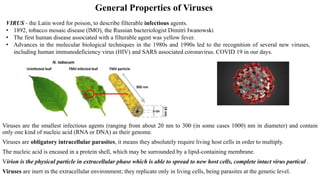
  • 1. General Properties of Viruses Viruses are the smallest infectious agents (ranging from about 20 nm to 300 (in some cases 1000) nm in diameter) and contain only one kind of nucleic acid (RNA or DNA) as their genome. Viruses are obligatory intracellular parasites, it means they absolutely require living host cells in order to multiply. The nucleic acid is encased in a protein shell, which may be surrounded by a lipid-containing membrane. Virion is the physical particle in extracellular phase which is able to spread to new host cells, complete intact virus partical . Viruses are inert in the extracellular environment; they replicate only in living cells, being parasites at the genetic level. VIRUS - the Latin word for poison, to describe filterable infectious agents. • 1892, tobacco mosaic disease (lMO), the Russian bacteriologist Dimitri Iwanowski • The first human disease associated with a filterable agent was yellow fever. • Advances in the molecular biological techniques in the 1980s and 1990s led to the recognition of several new viruses, including human immunodeficiency virus (HIV) and SARS associated coronavirus. COVID 19 in our days.
  • 2. The viral nucleic acid contains information necessary for programming the infected host cell to synthesize virus-specific macromolecules required for the production of viral progeny. During the replicative cycle, numerous copies of viral nucleic acid and coat proteins are produced. The coat proteins assemble together to form the capsid, which encases and stabilizes the viral nucleic acid against the extracellular environment and facilitates the attachment and penetration by the virus upon contact with new susceptible cells. Viruses have few or no enzymes of their own for metabolism; for example, they lack enzymes for protein synthesis and ATP generation. To multiply, viruses must take over the metabolic machinery of the host cell. The virus infection may have little or no effect on the host cell or may result in cell damage or death. The universe of viruses is rich in diversity. varying in size, shape, and genetic content; only some types possess a lipid envelope. Viruses vary greatly in structure, genome organization and expression, and strategies of replication and transmission. The host range for a given virus may be broad or extremely limited. Viruses are known to infect unicellular organisms such as mycoplasmas, bacteria, and algae and all higher plants and animals.
  • 3. TERMS AND DEFINITIONS IN VIROLOGY Capsid: The protein shell, or coat, that encloses the nucleic acid genome. Capsomeres: Morphologic units seen in the electron microscope on the surface of icosahedral virus particles. Capsomeres represent clusters of polypeptides, but the morphologic units do not necessarily correspond to the chemically defined structural units. Defective virus: A virus particle that is functionally deficient in some aspect of replication. Envelope: A lipid-containing membrane that surrounds some virus particles. It is acquired during viral maturation by a budding process through a cellular membrane. Virus encoded glycoproteins are exposed on the surface of the envelope. These projections are called peplomers. Nucleocapsid: The protein–nucleic acid complex representing the packaged form of the viral genome. The term is commonly used in cases in which the nucleocapsid is a substructure of a more complex virus particle. Structural units: The basic protein building blocks of the coat. They are usually a collection of more than one nonidentical protein subunit. The structural unit is often referred to as a protomer. Subunit: A single folded viral polypeptide chain. Enveloped virus with icosahedral symmetry. Not all icosahedral viruses have envelopes. Virus with helical symmetry
  • 4. Host Range The host range of a virus is the spectrum of host cells the virus can infect. There are viruses that infect invertebrates, vertebrates, plants, protists, fungi, and bacteria. However, most viruses are able to infect specific types of cells of only one host species. In rare cases, viruses cross the host-range barrier, thus expanding their host range. Viruses that infect bacteria are called bacteriophages, or phages. The particular host range of a virus is determined by the virus's requirements for its specific attachment to the host cell and the availability within the potential host of cellular factors required for viral multiplication. For the virus to infect the host cell, the outer surface of the virus must chemically interact with specific receptor sites on the surface of the cell. For some bacteriophages, the receptor site is part of the cell wall of the host; in other cases, it is part of the fimbriae or flagella. For animal viruses, the receptor sites are on the plasma membranes of the host cells.
  • 5. CLASSIFICATION OF ANIMAL VIRUSES The following properties have been used as a basis for the classification of viruses. Virion morphology, including • size, • shape, • type of symmetry, • presence or absence of peplomers, and • presence or absence of membranes. Virus genome properties, including • type of nucleic acid (DNA or RNA), • size of genome in kilobases (kb) or kilobase pairs (kbp), • strandedness (single or double), whether linear or circular, • sense (positive, negative), • segments (number, size), • nucleotide sequence, G + C content, and • presence of special features (repetitive elements, isomerization) Genome organization and replication, including • gene order, • number and position of open reading frames, • strategy of replication (patterns of transcription, translation), • cellular sites (accumulation of proteins, virion assembly, virion release). Virus protein properties, including • number, • size, • functional activities of structural and nonstructural proteins, • amino acid sequence, • modifications (glycosylation, phosphorylation, myristylation), • special functional activities (transcriptase, reverse transcriptase, neuraminidase, fusion activities). Physicochemical properties of the virion, including • molecular mass, • pH stability, • thermal stability, and • susceptibility to physical and chemical agents Biologic properties, including • natural host range, • mode of transmission, • vector relationships, • pathogenicity, • tissue tropisms, • pathology. Antigenic properties
  • 6. Viral Sizes and shapes Direct observation in the electron microscope is the most widely used method for estimating particle size. Another method that can be used is sedimentation in the ultracentrifuge. The relationship between the size and shape of a particle and its rate of sedimentation permits determination of particle size. Different viruses vary considerably in size. Although most are quite a bit smaller than bacteria, some of the larger viruses (such as the vaccinia virus) are about the same size as some very small bacteria (such as the mycoplasmas, rickettsias, and chlamydias). Viruses range from 20 to 1000 nm in length.
  • 7. Universal System of Virus Taxonomy Viruses are separated into major groupings—called families—on the basis of virion morphology, genome structure, and strategies of replication. Virus family names have the suffix -viridae. Within each family, subdivisions called genera are usually based on biological, genomic, physicochemical, or serologic differences. Criteria used to define genera vary from family to family. Genus names carry the suffix -virus. By 2000, the International Committee on Taxonomy of Viruses had organized more than 4000 animal and plant viruses into 56 families and 233 genera, with hundreds of viruses still unassigned. Of these, 24 families contained viruses that infect humans and animals.
  • 8. Families of Animal Viruses that Contain Members Able to Infect Humans
  • 9. Capsid and Envelope The nucleic acid of a virus is protected by a protein coat called the capsid. The structure of the capsid is ultimately determined by the viral nucleic acid and accounts for most of the mass of a virus, especially of small ones. Each capsid is composed of protein subunits called capsomeres. In some viruses, the proteins composing the capsomeres are of a single type; in other viruses, several types of protein may be present. The arrangement of capsomeres is characteristic of a particular type of virus. In some viruses, the capsid is covered by an envelope which usually consists of some combination of lipids, proteins, and carbohydrates. Some animal viruses are released from the host cell by an extrusion process that coats the virus with a layer of the host cell 's plasma membrane; that layer becomes the viral envelope. In many cases, the envelope contains proteins determined by the viral nucleic acid and materials derived from normal host cell components. Depending on the virus, envelopes may or may not be covered by spikes, which are carbohydrate-protein complexes that project from the surface of the envelope. Some viruses attach to host cells by means of spikes. Spikes are such a reliable characteristic of some viruses that they can be used as a means of identification. Viruses whose capsids are not covered by an envelope are known as nonenveloped viruses. The capsid of a nonenveloped virus protects the nucleic acid from nuclease enzymes in biological fluids and promotes the virus's attachment to susceptible host cells.
  • 10. PRINCIPLES OF VIRUS STRUCTURE Electron microscopy, cryoelectron microscopy, and x-ray diffraction techniques have made it possible to resolve fine differences in the basic morphology of viruses. Cubic Symmetry (Polyhedral viruses) Many animal, plant, and bacterial viruses are polyhedral, or manysided, viruses. All cubic symmetry observed with animal viruses is of the icosahedral pattern, a regular polyhedron with 20 triangular faces and 12 corners (vertices) and fivefold, threefold, and twofold axes of rotational symmetry. The vertex units have five neighbors (pentavalent), and all others have six (hexavalent). There are exactly 60 identical subunits on the surface of an icosahedron. To build a particle size adequate to encapsidate viral genomes, viral shells are composed of multiples of 60 structural units. An example of a polyhedral virus in the shape of an icosahedron are adenoviruses and polioviruses. Viral architecture can be grouped into three types based on the arrangement of morphologic subunits: (1) cubic symmetry (eg, adenoviruses), (2) helical symmetry (eg, orthomyxoviruses), and (3) complex structures (eg, poxviruses).
  • 11. Helical Symmetry In cases of helical symmetry, protein subunits are bound in a periodic way to the viral nucleic acid, winding it into a helix. The filamentous viral nucleic acid–protein complex (nucleocapsid) is then coiled inside a lipid-containing envelope. Thus, there is a regular, periodic interaction between capsid protein and nucleic acid in viruses with helical symmetry. All known examples of animal viruses with helical symmetry contain RNA genomes and, with the exception of rhabdoviruses, have flexible nucleocapsids that are wound into a ball inside envelopes. Helical viruses resemble long rods that may be rigid or flexible. The viruses that cause rabies and Ebola hemorrhagic fever are helical viruses. Enveloped viruses are roughly sphericaL When helical or polyhedral viruses are enclosed by envelopes, they are called enveloped helical or enveloped polyhedral viruses. An example of an enveloped helical virus is the influenza virus. An example of an enveloped polyhedral (icosahedral) virus is the herpes simplex virus.
  • 12. Complex Structures Some virus particles do not exhibit simple cubic or helical symmetry but are more complicated in structure. Some viruses, particularly bacterial viruses, have complicated structures and are called complex viruses. One example of a complex virus is a bacteriophage. Some bacteriophages have capsids to which additional structures are attached (Figure 13.5a). The capsid (head) is polyhedral and that the tail sheath is helicaL The head contains the nucleic acid. Another example of complex viruses are poxviruses, which do not contain clearly identifiable capsids but have several coats around the nucleic acid.
  • 13. CHEMICAL COMPOSITION OF VIRUSES Viral Protein The structural proteins of viruses have several important functions. Their major purpose is to facilitate transfer of the viral nucleic acid from one host cell to another. They serve to protect the viral genome against inactivation by nucleases, participate in the attachment of the virus particle to a susceptible cell. They provide the structural symmetry of the virus particle. The proteins determine the antigenic characteristics of the virus. Some surface proteins may also exhibit specific activities (eg, influenza virus hemagglutinin agglutinates red blood cells). Some viruses carry enzymes (which are proteins) inside the virions. The enzymes are present in very small amounts and are probably not important in the structure of the virus particles; however, they are essential for the initiation of the viral replicative cycle when the virion enters a host cell. Examples include an RNA polymerase carried by viruses with negative-sense RNA genomes (eg, orthomyxoviruses, rhabdoviruses) that is needed to copy the first mRNAs, and reverse transcriptase, an enzyme in retroviruses that makes a DNA copy of the viral RNA, an essential step in replication and transformation. At the extreme in this respect are the poxviruses, the cores of which contain a transcriptional system; many different enzymes are packaged in poxvirus particles. Lyzoceam - bacteriaphage
  • 14. Viral Nucleic Acid Viruses contain a single kind of nucleic acid—either DNA or RNA—that encodes the genetic information necessary for replication of the virus. The genome may be single or double stranded, circular or linear, and segmented or nonsegmented. The type of nucleic acid, its strandedness, and its size are major characteristics used for classifying viruses into families. The size of the viral DNA genome ranges from 3.2 kbp (hepadnaviruses) to 375 kbp (poxviruses). The size of the viral RNA genome ranges from about 4 kb (picobirnaviruses) to 32 kb (coronaviruses). All major DNA viral groups have genomes that are single molecules of DNA and have a linear or circular configuration. Viral RNAs exist in several forms. The RNA may be a single linear molecule (eg, picornaviruses). For other viruses (eg, orthomyxoviruses), the genome consists of several segments of RNA that may be loosely associated within the virion. The isolated RNA of viruses with positive-sense genomes (ie, picornaviruses, togaviruses) is infectious, and the molecule functions as an mRNA within the infected cell. The isolated RNA of the negative-sense RNA viruses, such as rhabdoviruses and orthomyxoviruses, is not infectious. For these viral families, the virions carry an RNA polymerase that in the cell transcribes the genomic RNA molecules into several complementary RNA molecules, each of which may serve as an mRNA. Viral nucleic acid may be characterized by its G + C content.
  • 15. Viral Lipid Envelopes A number of different viruses contain lipid envelopes as part of their structure. The lipid is acquired when the viral nucleocapsid buds through a cellular membrane in the course of maturation. Budding occurs only at sites where virus-specific proteins have been inserted into the host cell membrane. The budding process varies markedly depending on the replication strategy of the virus and the structure of the nucleocapsid. The specific phospholipid composition of a virion envelope is determined by the specific type of cell membrane involved in the budding process. For example, herpesviruses bud through the nuclear membrane of the host cell, and the phospholipid composition of the purified virus reflects the lipids of the nuclear membrane. Lipid-containing viruses are sensitive to treatment with desinfectants indicating that disruption or loss of lipid results in loss of infectivity. Nonlipid-containing viruses are generally resistant to ether.
  • 16. Viral Glycoproteins Viral envelopes contain glycoproteins. In contrast to the lipids in viral membranes, which are derived from the host cell, the envelope glycoproteins are virus encoded. However, the sugars added to viral glycoproteins often reflect the host cell in which the virus is grown. The surface glycoproteins of an enveloped virus attach the virus particle to a target cell by interacting with a cellular receptor. They are also often involved in the membrane fusion step of infection. The glycoproteins are also important viral antigens. Influenza virus membrane glycoproteins are hemagglutinin, neuraminidase.
  • 17. REACTION TO PHYSICALAND CHEMICALAGENTS Heat and Cold There is great variability in the heat stability of different viruses. Icosahedral viruses are heat stable, but enveloped viruses are much more heat labile. Viral infectivity is generally destroyed by heating at 50–60°C for 30 minutes, although there are some notable exceptions (eg, hepatitis B virus, polyomaviruses). Viruses can be preserved by storage at subfreezing temperatures, and some may withstand lyophilization and can thus be preserved in the dry state at 4°C or even at room temperature. Viruses that withstand lyophilization are more heat resistant when heated in the dry state. Enveloped viruses tend to lose infectivity after prolonged storage even at –90°C and are particularly sensitive to repeated freezing and thawing. Stabilization of Viruses by Salts Many viruses can be stabilized by salts in concentrations of 1 mol/L. The mechanism by which the salts stabilize viral preparations is not known. Viruses are preferentially stabilized by certain salts. MgCl2, 1 mol/L, stabilizes picornaviruses and reoviruses; MgSO4, 1 mol/L, stabilizes orthomyxoviruses and paramyxoviruses; and Na2SO4, 1 mol/L, stabilizes herpesviruses. The stability of viruses is important in the preparation of vaccines. The ordinary nonstabilized oral polio vaccine must be stored at freezing temperatures to preserve its potency. However, with the addition of salts for stabilization of the virus, potency can be maintained for weeks at ambient temperatures even in the high temperatures of the tropics.
  • 18. pH Viruses are usually stable between pH values of 5.0 and 9.0. Some viruses (eg, enteroviruses) are resistant to acidic conditions. All viruses are destroyed by alkaline conditions. Radiation Ultraviolet, x-ray, and high-energy particles inactivate viruses. The dose varies for different viruses. Infectivity is the most radiosensitive property because replication requires expression of the entire genetic contents. Irradiated particles that are unable to replicate may still be able to express some specific functions in host cells. Ether Susceptibility Ether susceptibility can be used to distinguish viruses that possess an envelope from those that do not. Detergents Nonionic detergents (eg, Nonidet P40 and Triton X-100) solubilize lipid constituents of viral membranes. The viral proteins in the envelope are released (undenatured). Anionic detergents (eg, sodium dodecyl sulfate) also solubilize viral envelopes; in addition, they disrupt capsids into separated polypeptides. Formaldehyde Formaldehyde destroys viral infectivity by reacting with nucleic acid. Viruses with single-stranded genomes are inactivated much more readily than those with double-stranded genomes. Formaldehyde has minimal adverse effects on the antigenicity of proteins and therefore has been used frequently in the production of inactivated viral vaccines.
  • 19. Photodynamic Inactivation Viruses are penetrable to a varying degree by vital dyes such as toluidine blue, neutral red, and proflavine. These dyes bind to the viral nucleic acid, and the virus then becomes susceptible to inactivation by visible light. Antibiotics and Other Antibacterial Agents Antibacterial antibiotics and sulfonamides, organic iodine compounds, quaternary ammonium compounds are not effective against viruses. However some antiviral drugs are available. Larger concentrations of chlorine are required to destroy viruses than to kill bacteria, especially in the presence of extraneous proteins. Alcohols, such as isopropanol and ethanol, are relatively ineffective against certain viruses, especially picornaviruses. Common Methods of Inactivating Viruses for Various Purposes Viruses may be inactivated for various reasons, such as to sterilize laboratory supplies and equipment, disinfect surfaces or skin, make drinking water safe, and produce inactivated virus vaccines. Different methods and chemicals are used for these purposes. Sterilization may be accomplished by steam under pressure, dry heat, ethylene oxide, and γ-irradiation. Surface disinfectants include sodium hypochlorite, glutaraldehyde, formaldehyde, and peracetic acid. Skin disinfectants include chlorhexidine, 70% ethanol, and iodophores. Vaccine production may involve the use of formaldehyde, β-propiolactone, psoralen + ultraviolet irradiation, or detergents (subunit vaccines) to inactivate the vaccine virus.
  • 20. Multiplication of Bacteriophages Bacteriophages The lytic cycle And lysogenic cycle Homework for practical course Growing Bacteriophages in the Laboratory
  • 21. Growing Animal Viruses in the laboratory In the laboratory, three methods are commonly used for culturing animal viruses. These methods involve using • living animals, • embryonated eggs, or • cell cultures. Viral Identification • Electron microscope. • Serological methods, such as Western blotting, are the most commonly used means of identification. In these tests, the virus is detected and identified by its reaction with antibodies. • Observation of cytopathic effects is also useful for the identification of a virus. • Restriction fragment length polymorphisms (RFLPs) • Polymerase chain reaction (PCR) was used to amplify viral RNA (West Nile virus in 1999 in the United Statesand the SARS-associated corona virus in China in 2002 and COVID19 these days
  • 22. Cells grown in vitro are central to the cultivation and characterization of viruses. A. Detection of Virus-Infected Cells Multiplication of a virus can be monitored in a variety of ways: 1. Development of cytopathic effects (ie, morphologic changes in the cells). Types of virus-induced cytopathic effects include cell lysis or necrosis, inclusion formation, giant cell formation, and cytoplasmic vacuolization 2. Appearance of a virus-encoded protein, such as the hemagglutinin of influenza virus. Specific antisera can be used to detect the synthesis of viral proteins in infected cells. 3. Detection of virus-specific nucleic acid. Molecular-based assays such as polymerase chain reaction provide rapid, sensitive, and specific methods of detection. 4. Adsorption of erythrocytes to infected cells, called hemadsorption, caused by the presence of virus-encoded hemagglutinin (parainfluenza, influenza) in cellular membranes. This reaction becomes positive before cytopathic changes are visible and in some cases occurs in the absence of cytopathic effects 5. Viral growth in an embryonated chick egg may result in death of the embryo (eg, encephalitis viruses), production of pocks or plaques on the chorioallantoic membrane (eg, herpes, smallpox, vaccinia), or development of hemagglutinins in the embryonic fluids or tissues (eg, influenza).
  • 23. B. Inclusion Body Formation In the course of viral multiplication within cells, virusspecific structures called inclusion bodies may be produced. They become far larger than the individual virus particle and often have an affinity for acid dyes (eg, eosin). They may be situated in the nucleus (herpesvirus), in the cytoplasm (poxvirus), or in both (measles virus). The presence of inclusion bodies may be of considerable diagnostic aid. The intracytoplasmic inclusion in nerve cells—the Negri body—is pathognomonic for rabies.
  • 24. Quantitation of Viruses A. Physical Methods • Quantitative polymerase chain reaction • Hemagglutination assays - are an easy and rapid method of quantitating these types of viruses. Certain viruses contain a protein (hemagglutinin) that has the ability to agglutinate red blood cells of humans or some animal. • Radioimmunoassays and enzyme-linked immunosorbent assays. These tests do not distinguish infectious from noninfectious particles and sometimes detect viral proteins not assembled into particles. • electron microscope (infectious virus particles cannot be distinguished from noninfectious ones). B. Biologic Methods Plaque assay - A widely used assay for infectious virus growing well in tissue culture. Monolayers of host cells are inoculated with suitable dilutions of virus and after adsorption are overlaid with medium containing agar or carboxymethylcellulose to prevent virus spreading throughout the culture. After several days, the cells initially infected have produced virus that spreads only to surrounding cells. Multiple cycles of replication and cell killing produce a small area of infection, or plaque. Under controlled conditions, a single plaque can arise from a single infectious virus particle, termed a plaqueforming unit. The cytopathic effect of infected cells within the plaque can be distinguished from uninfected cells of the monolayer with or without suitable staining, and plaques can usually be counted macroscopically. A more rapid method of assay is based on determination of the number of infected cells producing a viral antigen, such as by immunofluorescence. Certain viruses (eg, herpes and vaccinia) form pocks when inoculated onto the chorioallantoic membrane of an embryonated egg. Such viruses can be quantitated by relating the number of pocks counted to the viral dilution inoculated.
  • 25. REPLICATION OF VIRUSES Viruses multiply only in living cells. The host cell must provide the energy and synthetic machinery and the lowmolecular- weight precursors for the synthesis of viral proteins and nucleic acids. The viral nucleic acid carries the genetic specificity to code for all of the virus-specific macromolecules in a highly organized fashion. The unique feature of viral multiplication is that soon after interaction with a host cell the infecting virion is disrupted and its measurable infectivity is lost. This phase of the growth cycle is called the eclipse period. After the synthesis of viral nucleic acid and viral proteins, the components assemble to form new infectious virions. The yield of infectious virus per cell ranges widely, from modest numbers to more than 100,000 particles. The duration of the virus replication cycle also varies widely, from 6 to 8 hours (picornaviruses) to more than 40 hours (some herpesviruses). Not all infections lead to new progeny virus. • Productive infections occur in permissive cells and result in the production of infectious virus. • Abortive infections fail to produce infectious progeny, either because the cell may be nonpermissive and unable to support the expression of all viral genes or because the infecting virus may be defective, lacking some functional viral gene. • A latent infection may ensue, with the persistence of viral genomes, the expression of no or a few viral genes, and the survival of the infected cell.
  • 26. General Steps in Viral Replication Cycles Steps in viral replication include • attachment to a cell via binding to specific receptors on the cell surface, • entry into the cell, • uncoating of the viral genome, • regulated expression of viral transcripts, • synthesis of viral proteins, • replication of viral genomic nucleic acid, • assembly of new progeny viruses, and • release of new virions from the cell. The duration of replication cycles varies widely among different virus types. The infected cells may be killed or may survive with little damage. Not all infections lead to new progeny virus.
  • 27. Attachment, Penetration, and Uncoating The first step in viral infection is attachment, interaction of a virion with a specific receptor site on the surface of a cell. Receptor molecules differ for different viruses but are generally glycoproteins. Successful infection of a cell by a virus may involve interaction with more than one type of receptor. The presence or absence of receptors plays an important determining role in cell tropism and viral pathogenesis. The attachment step may initiate irreversible structural changes in the virion. After binding, the virus particle is taken up inside the cell. This step is referred to as penetration or engulfment. Uncoating occurs shortly after penetration. It is the physical separation of the viral nucleic acid from the outer structural components of the virion so that it can function. The genome may be released as free nucleic acid (picornaviruses) or as a nucleocapsid (reoviruses). The nucleocapsids usually contain polymerases. Uncoating may require acidic pH in the endosome.
  • 28. Expression of Viral Genomes and Synthesis of Viral Components The essential theme in viral replication is that specific mRNAs must be transcribed from the viral nucleic acid for successful expression and duplication of genetic information. After this is accomplished, viruses use cell components to translate the mRNA. Various classes of viruses use different pathways to synthesize the mRNAs depending on the structure of the viral nucleic acid. Negative-strand (negativesense) RNA viruses (eg, rhabdoviruses) carry RNA polymerases to synthesize mRNAs. Their single-strand RNA genome is complementary to mRNA, which is conventionally designated positive strand (positive sense). The negative-strand viruses must supply their own RNA polymerase because eukaryotic cells lack enzymes able to synthesize mRNA off an RNA template. Positive strand (positive sense) RNA viruses contain RNA which can act as mRNA. In the course of viral replication, all of the virus specified macromolecules are synthesized in a highly organized sequence. In some viral infections, notably those involving double-stranded DNA-containing viruses, early viral proteins are synthesized soon after infection and late proteins are made only late in infection after viral DNA synthesis begins. DNA viruses that replicate in the nucleus generally use host cell DNA and RNA polymerases and processing enzymes. The larger viruses (herpesviruses, poxviruses) are more independent of cellular functions than are the smaller viruses. This is one reason the larger viruses are more susceptible to antiviral chemotherapy because more virus-specific processes are available as targets for drug action. Viral DNA is usually replicated in the nucleus. Viral genomic RNA is generally duplicated in the cell cytoplasm, although there are exceptions.
  • 29.
  • 30. Morphogenesis and Release Newly synthesized viral genomes and capsid polypeptides assemble together to form progeny viruses. In general, nonenveloped viruses accumulate in infected cells, and the cells eventually lyse and release the virus particles. Enveloped viruses mature by a budding process. Virusspecific envelope glycoproteins are inserted into cellular membranes; viral nucleocapsids then bud through the membrane at these modified sites and in so doing acquire an envelope. Budding frequently occurs at the plasma membrane but may involve other membranes in the cell. Enveloped viruses are not infectious until they have acquired their envelopes. Viral components may accumulate and be involved in the formation of inclusion bodies in the cell. As a result of the profound deleterious effects of viral replication, cellular cytopathic effects eventually develop and the cell dies.
  • 31. The growth cycle of a nonenveloped, double- stranded DNA virus (1) After penetrating the host cell, viral DNA is uncoated and enters the nucleus. (2) Viral genes are transcribed. (3) The mRNAs are translated in the cytoplasm. Newly synthesized proteins enter the nucleus. (4) Viral DNA is replicated in the nucleus, sometimes with the help of newly synthesized viral replication proteins. (5) Viral DNA and viral structural proteins assemble in the nucleus to produce new progeny virions. (6) On rare occasions, viral DNA may be incorporated into cellular DNA as a side-effect of infection.
  • 32. The growth cycle of a positive-sense, single- stranded RNA virus (1) The virus enters the cell and the viral RNA genome is uncoated. (2) As a positive-sense, single-stranded genome, the RNA is directly translated, producing viral proteins. (3) A negative-sense RNA copy of the positive template is synthesized. (4) It is used to produce many positive-sense copies. (5) The newly synthesized positive-sense RNA molecules are assembled with viral structural proteins to produce new progeny virions.
  • 33.
  • 34. Interactions Among Viruses When two or more virus particles infect the same host cell, they may interact in a variety of ways. A. Recombination Recombination results in the production of progeny virus (recombinant) that carries traits not found together in either parent. The classic mechanism is that the nucleic acid strands break, and part of the genome of one parent is joined to part of the genome of the second parent. The recombinant virus is genetically stable, yielding progeny similar to itself upon replication. Viruses vary widely in the frequency with which they undergo recombination. In the case of viruses with segmented genomes (eg, influenza virus) the formation of recombinants is caused by reassortment of individual genome fragments rather than by an actual crossover event. B. Complementation This refers to the interaction of viral gene products in cells infected with two viruses, one or both of which may be defective. It results in the replication of one or both under conditions in which replication would not ordinarily occur. The basis for complementation is that one virus provides a gene product in which the second is defective, allowing the second virus to grow. The genotypes of the two viruses remain unchanged. If both mutants are defective in the same gene product, they will not be able to complement each other’s growth.
  • 35. C. Phenotypic Mixing A special case of complementation is phenotypic mixing, or the association of a genotype with a heterologous phenotype. This occurs when the genome of one virus becomes randomly incorporated within capsid proteins specified by a different virus or a capsid consisting of components of both viruses. If the genome is encased in a completely heterologous protein coat, this extreme example of phenotypic mixing may be called “phenotypic masking” or “transcapsidation.” Such mixing is not a stable genetic change because, upon replication, the phenotypically mixed parent will yield progeny encased in capsids homologous to the genotype. Phenotypic mixing usually occurs between different members of the same virus family; the intermixed capsid proteins must be able to interact correctly to form a structurally intact capsid. However, phenotypic mixing also can occur between enveloped viruses, and in this case, the viruses do not have to be closely related. D. Interference Infection of either cell cultures or whole animals with two viruses often leads to an inhibition of multiplication of one of the viruses, an effect called interference. Interference in animals is distinct from specific immunity. Furthermore, interference does not occur with all viral combinations; two viruses may infect and multiply within the same cell as efficiently as in single infections. Several mechanisms have been elucidated as causes of interference: (1) One virus may inhibit the ability of the second to adsorb to the cell, either by blocking its receptors (retroviruses, enteroviruses) or by destroying its receptors (orthomyxoviruses). (2) One virus may compete with the second for components of the replication apparatus (eg, polymerase, translation initiation factor). (3) The first virus may cause the infected cell to produce an inhibitor that prevents replication of the second virus.
  • 36. MODES OF TRANSMISSION OF VIRUSES Viruses may be transmitted in the following ways: 1. Direct transmission from person to person by contact. The major means of transmission include • droplet or aerosol infection (eg, influenza, measles, smallpox); • by sexual contact (eg, papillomavirus, hepatitis B, herpes simplex type 2, human immunodeficiency virus); • by hand– mouth, hand–eye, or mouth–mouth contact (eg, herpes simplex, rhinovirus, Epstein-Barr virus); • by exchange of contaminated blood (eg, hepatitis B, human immunodeficiency virus). 2. Indirect transmission • by the fecal–oral route (eg, enteroviruses, rotaviruses, infectious hepatitis A) • by fomites (eg, Norwalk virus, rhinovirus). 3. Transmission from animal to animal, with humans an accidental host. Spread may be • by bite (rabies) or by droplet or aerosol infection from rodent-contaminated quarters (eg, arenaviruses, hantaviruses). 4. Transmission by means of an arthropod vector (eg, arboviruses, now classified primarily as togaviruses, flaviviruses, and bunyaviruses).
  • 37. Emerging Viral Diseases Owing to wide-reaching changes in social attitudes, technology, and the environment—plus the decreased effectiveness of previous approaches to disease control—the spectrum of infectious diseases is expanding today. New agents appear, and diseases once thought to be under control are increasing in incidence as pathogens evolve and spread. The term “emerging infectious diseases” denotes these phenomena. Viral diseases emerge following one of three general patterns: • recognition of a new agent, • abrupt increase in illnesses caused by an endemic agent, and • invasion of a new host population. Combinations of factors contribute to disease emergence. Factors include (1) environmental changes (deforestation, damming or other changes in water ecosystems, flood or drought, famine), (2) human behavior (sexual behavior, drug use, outdoor recreation), (3) socioeconomic and demographic phenomena (war, poverty, population growth and migration, urban decay), (4) travel and commerce (highways, international air travel), (5) food production (globalization of food supplies, changes in methods of food processing and packaging), (6) health care (new medical devices, blood transfusions, organ and tissue transplantation, drugs causing immunosuppression, widespread use of antibiotics), (7) microbial adaptation (changes in virulence, development of drug resistance, cofactors in chronic diseases), and (8) public health measures (inadequate sanitation and vector control measures, curtailment of prevention programs, lack of trained personnel in sufficient numbers).
  • 38. Examples of emerging viral infections in different regions of the world include • Ebola virus, • Nipah virus, • hantavirus pulmonary disease, • human immunodeficiency virus infection, • dengue hemorrhagic fever, • West Nile virus, • Rift Valley fever, • COVID19 • bovine spongiform encephalopathy (the latter a prion disease). Of potential concern also is the possible use of animal organs as xenografts in humans (xenotransplantation). Concerns exist about the potential accidental introduction of new viral pathogens from the donor species into humans. Bioterrorism Agents Bioterrorism agents are microorganisms (or toxins) that could be used to produce death and disease in humans, animals, or plants for terrorist purposes. Such microorganisms could be genetically modified to increase their virulence, make them resistant to drugs or vaccines, or enhance their ability to be disseminated in the environment. Potential bioterrorism agents are classified into risk categories based on the ease of dissemination or transmission from person to person, mortality rates, ability to cause public panic, and requirement for public health preparedness. Viral agents in the highest risk category are smallpox and the viral hemorrhagic fevers; Highest risk bacteria include the agents of anthrax, botulism, plague, and tularemia.
  • 39. Viroids Viroids are small infectious agents that cause diseases of plants. Viroids are agents that do not fit the definition of classic viruses. They are nucleic acid molecules without a protein coat. Plant viroids are single-stranded, covalently closed circular RNA molecules consisting of about 360 nucleotides and with a highly base-paired rod like structure. Viroids replicate by an entirely novel mechanism. Viroid RNA does not encode any protein products; the devastating plant diseases induced by viroids occur by an unknown mechanism. Plant viruses cause many diseases of economically important crops, including beans (bean mosaic virus), corn and sugarcane (wound tumor virus), and potatoes (potato yellow dwarf virus). Viruses can cause color change, deformed growth, wilting, and stunted growth in their plant hosts. Some hosts, however, remain symptomless and only serve as reservoirs of infection. Plant cells are generally protected from disease by an impermeable cell wall. Viruses must enter through wounds or be assisted by other plant parasites, including nematodes, fungi, and, most often, insects that suck the plant's sap. Once one plant is infected, it can spread infection to other plants in its pollen and seeds. In laboratories, plant viruses are cultured in protoplasts (plant cells with the cell walls removed) and in insect cell cuitures.
  • 40. Prions A few infectious diseases are caused by prions. In 1982, American neurobiologist Stanley Prusiner proposed that infectious proteins caused a neurological disease in sheep called scrapie. The infectivity of scrapie-infected brain tissue is reduced by treatment with proteases but not by treatment with radiation, suggesting that the infectious agent is pure protein. Prusiner coined the name prion for proteinaceous infectious particle. They are highly resistant to inactivation by heat, formaldehyde, and ultraviolet light that inactivate viruses. Prion diseases, called “transmissible spongiform encephalopathies,” include scrapie in sheep, mad cow disease in cattle, and kuru, fatal familial insomnia and Creutzfeldt-Jakob disease in humans. These diseases are caused by the conversion of a normal host glycoprotein called Prpc (for cellular prion protein) into an infectious form called PrpSc (for scrapie protein). The gene for Prpc is located on chromosome 20 in humans. Recent evidence suggests that Prpc is involved in regulating cell death. The actual cause of cell damage is not known. Fragments of PrpSc molecules accumulate in the brain, forming plaques; these plaques are used for postmortem diagnosis, but they do not appear to be the cause of cell damage.
  • 41. General Characteristics of Viruses I. Depending on one's viewpoint, viruses may be regarded as exceptionally complex aggregations of nonliving chemicals or as exceptionally simple living microbes. 2. Viruses contain a single type of nucleic acid (DNA or RNA) and a protein coat, sometimes enclosed by an envelope composed of lipids, proteins, and carbohydrates. 3. Viruses are obligatory intracellular parasites. They multiply by using the host cell's synthesizing machinery to cause the synthesis of specialized elements that can transfer the viral nucleic acid to other cells. Host Range 1. Host rauge refers to the spectrum of host cells in which a virus can multiply. 2. Most viruses infect only specific types of cells in one host species. 3. Host range is determined by the specific attachment site on the host cell's surface and the availability of host cellular factors. Viral Size 1. Viral size is ascertained by electron microscopy. 2. Viruses range from 20 to 1000 nm in length. Viral Structure A virion is a complete, fully developed viral particle composed of nucleic acid surrounded by a coat.
  • 42. Nucleic Acid 1. Viruses contain either DNA or RNA, never both, and the nucleic acid may be single- or double-stranded, linear or circular, or divided into several separate molecules. 2. The proportion of nucleic acid in relation to protein in viruses ranges from about 1 % to about 50%. Capsid and Envelope 1. The protein coat surrounding the nucleic acid of a virus is called the capsid. 2. The capsid is composed of subunits, capsomeres, which can be a single type of protein or several types. 3. The capsid of some viruses is enclosed by an envelope consisting of lipids, proteins, and carbohydrates. 4. Some envelopes are covered with carbohydrate-protein complexes called spikes. General Morphology 1. Helical viruses (for example, Ebola virus) resemble long rods, and their capsids are hollow cylinders surrounding the nucleic acid. 2. Polyhedral viruses (for example, adenovirus) are many-sided. Usually the capsid is an icosahedron. 3. Enveloped viruses are covered by an envelope and are roughly spherical but highly pleomorphic. There are also enveloped helical viruses (for example, influenza virus) and enveloped polyhedral viruses (for example, Simpiexvirus). 4. Complex viruses have complex structures. For example, many bacteriophages have a polyhedral capsid with a helical tail attached
  • 43. Isolation, Cultivation, and Identification of Viruses 1. Viruses must be grown in living cells. 2. The easiest viruses to grow are bacteriophages. Growing Bacteriophages in the laboratory 1. The plaque method mixes bacteriophages with host bacteria and nutrient agar. 2. After several viral multiplication cycles, the bacteria in the area surrounding the original virus are destroyed; the area of lysis is called a plaque. 3. Each plaque originates with a single viral particle; the concentration of viruses is given as plaque-forming units. Growing Animal Viruses in the Laboratory 1. Cultivation of some animal viruses requires whole animals. 2. Some animal viruses can be cultivated in embryonated eggs. 3. Cell cultures are cells growing in culture media in the laboratory. 4. Primary cell lines and embryonic diploid cell lines grow for a short time in vitro. 5. Continuous cell lines can be maintained in vitro indefinitely. 6. Viral growth can cause cytopathic effects in the cell culture, Viral Identification 1. Serological tests are used most often to identify viruses. 2. Viruses may be identified by RFLPs and PCR.
  • 44. Viral Multiplication 1. Viruses do not contain enzymes for energy production or protein synthesis. 2. For a virus to multiply, it must invade a host cell and direct the host's metabolic machinery to produce viral enzymes and components. Multiplication of Bacteriophages 1. During the lytic cycle, a phage causes the lysis and death of a host cell. 2. Some viruses can either cause lysis or have their DNA incorporated as a prophage into the DNA of the host cell. The latter situation is called lysogeny. 3. During the attachment phase of the lytic cycle, sites on the phage's tail fibers attach to complementary receptor sites on the bacterial cell. 4. In penetration, phage lysozyme opens a portion of the bacterial cell wall, the tail sheath contracts to force the tail core through the cell wall, and phage DNA enters the bacterial cell. The capsid remains outside. 5. In biosynthesis, transcription of phage DNA produces mRNA coding for proteins necessary for phage multiplication. Phage DNA is replicated, and capsid proteins are produced. During the eclipse period, separate phage DNA and protein can be found. 6. During maturation, phage DNA and capsids are assembled into complete viruses. 7. During release, phage lysozyme breaks down the bacterial cell wall, and the new phages are released. 8. During the lysogenic cycle, prophage genes are regulated by a repressor coded for by the prophage. The prophage is replicated each time the cell divides. 9. Exposure to certain mutagens can lead to excision of the prophage and initiation of the lytic cycle. 10. Because of lysogeny, lysogenic cells become immune to reinfection with the same phage and may undergo phage conversion. 11. A lysogenic phage can transfer bacterial genes from one cell to another through transduction. Any genes can be transferred in generalized transduction, and specific genes can be transferred in specialized transduction.
  • 45. Multiplication of Animal Viruses 1. Animal viruses attach to the plasma membrane of the host cell. 2. Entry occurs by endocytosis or fusion. 3. Animal viruses are uncoated by viral or host cell enzymes. 4. The DNA of most DNA viruses is released into the nucleus of the host cell. Transcription of viral DNA and translation produce viral DNA and, later, capsid proteins. Capsid proteins are synthesized in the cytoplasm of the host cell. 5. DNA viruses include members of the families Adenoviridae, Poxviridae, Herpesviridae, Papovaviridae, and Hepadnaviridae. 6. Multiplication of RNA viruses occurs in the cytoplasm of the host cell. RNA-dependent RNA polymerase synthesizes a doublestranded RNA. 7. Picornaviridae + strand RNA acts as mRNA and directs the synthesis of RNA-dependent RNA polymerase. 8. Togaviridae + strand RNA acts as a template for RNA-dependent RNA polymerase, and mRNA is transcribed from a new - RNA strand. 9. Rhabdoviridae - strand RNA is a template for viral RNA-dependent RNA polymerase, which transcribes mRNA. 10. Reoviridae are digested in host cell cytoplasm to release mRNA for viral biosynthesis. 11. Retroviridae reverse transcriptase (RNA-dependent DNA polymerase) transcribes DNA from RNA. 12. After maturation, viruses are released, One method of release (and envelope formation) is budding. Nonenveloped viruses are released through ruptures in the host cell membrane.
  • 46. Viruses and Cancer I. The earliest relationship between cancer and viruses was demonstrated in the early 1905, when chicken leukemia and chicken sarcoma were transferred to healthy animals by cell -free filtrates. The Transformation of Normal Cells Into Tumor Cells 1. When activated, oncogenes transform normal cells into cancerous cells. 2. Viruses capable of producing tumors are called oncogenic viruses. 3. Several DNA viruses and retroviruses are oncogenic. 4. The genetic material of oncogenic viruses becomes integrated into the host cell 's DNA. 5. Transformed cells lose contact inhibition, contain virus-specific antigens (TSTA and T antigen), exhibit chromosome abnormalities, and can produce tumors when injected into susceptible animals. DNA Oncogenic Viruses 1. Oncogenic viruses are found among the Adenoviridae, Herpesviridae, Poxviridae, and Papovaviridae. 2. The EB virus, a herpesvirus, causes Burkin's lymphoma and nasopharyngeal carcinoma. Hepadllavirus causes liver cancer. RNA Oncogenic Viruses 1. Among the RNA viruses, only retroviruses seem to be oncogenic. 2. HTLV-I and HTLV-2 have been associated with human leukemia and lymphoma. 3. The virus's ability to produce tumors is related to the production o f reverse transcriptase. The DNA synthesized from the viral RNA becomes incorporated as a provirus into the host cell's DNA. 4. A provirus can remain latent, can produce viruses, or can transform the host cell
  • 47. latent Viral Infections I. A la tent viral infection is one in which the virus remains in the host cell for long periods without producing an infection. 2. Examples are cold sores and shingles. Persistent Viral Infections I. Persistent viral infections are disease processes that occur over a long period and are generally fatal. 2. Persistent viral infections are caused by conventional viruses; viruses accumulate over a long period . Prions I. Prions are infectious proteins first discovered in the 1980s. 2. Prion diseases, such as CJD and mad cow disease, all involve the degeneration of brain tissue. 3. Prion diseases are the result of an altered protein; the cause can be a mutation in the normal gene for Prpc or contact with an altered protein (PrpSc) . Plant Viruses and Viroids I. Plant viruses must enter plant hosts through wounds or with invasive parasites, such as insects. 2. Some plant viruses also multiply in insect (vector) cells. 3. Viroids are infectious pieces of RNA that cause some plant diseases, such as potato spindle tuber disease.