

Recommended
More Related Content
What's hot
What's hot (20)
Viewers also liked
Similar to Ischemic heart diseae lecture
Similar to Ischemic heart diseae lecture (20)
More from memoalawad
More from memoalawad (20)
Recently uploaded
Recently uploaded (20)
Ischemic heart diseae lecture
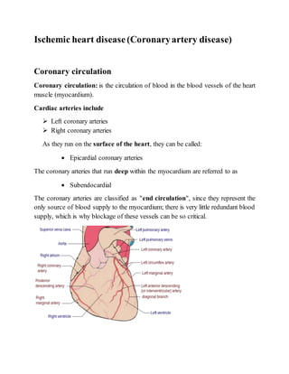
- 1. Ischemic heart disease(Coronaryartery disease) Coronary circulation Coronary circulation: is the circulation of blood in the blood vessels of the heart muscle (myocardium). Cardiac arteries include Left coronary arteries Right coronary arteries As they run on the surface of the heart, they can be called: Epicardial coronary arteries The coronary arteries that run deep within the myocardium are referred to as Subendocardial The coronary arteries are classified as "end circulation", since they represent the only source of blood supply to the myocardium; there is very little redundant blood supply, which is why blockage of these vessels can be so critical.
- 2. Myocardial ischemia: Occurs when there is an imbalance between the supply of oxygen and other nutrient and the myocardial demand for these substance Causes of myocardial ischemia 1. Reduction of blood flow to the heart that can be caused by stenosis, spasm, or acute occlusion (by an embolus) of the heart's arteries. 2. Resistance of the blood vessels. This can be caused by narrowing of the blood vessels; a decrease in radius. Blood flow is proportional to the radius of the artery to the fourth power. 3. Reduced oxygen-carrying capacity of the blood, due to several factors such as a decrease in oxygen tension and hemoglobin concentration. This decreases the ability to of hemoglobin to carry oxygen to myocardial tissue. Mechanicalobstructionof coronaryblood flow to myocardium causes by: Atheroma: is an accumulation of degenerative material in the tunica intima (inner layer) of artery walls. The material consists of (mostly) macrophage cells, or debris, containing lipids (cholesterol and fatty acids), calcium and a variable amount of fibrous connective tissue. The accumulated material forms a swelling in the artery wall, which may intrude into the channel of the artery, narrowing it and restricting blood flow. Atherosclerosis in a coronary artery
- 3. Veins do not develop atheroma , unless surgically moved to function as an artery, as inbypass surgery. Thrombosis Spasm Embolus Coronary ostial stenosis Coronary arteritis Decrease flow of the oxygenated blood to the myocardium Anemia Hypotension Coronary artery diseases(Ischemic heart diseases) Group of heart disease causes narrowing of the coronary arteries, leading to inadequate blood supply to the myocardium They includes: Stable angina Unstable angina Myocardial infarction Suddencoronary death Pathophysiology Limitation of blood flow to the heart causes ischemia (cell starvation secondary to a lack of oxygen) of the myocardial cells. Myocardial cells may die from lack of oxygen and this is called a myocardial infarction (commonly called a heart attack). It leads to heart muscle damage, heart muscle death and later myocardial scarring without heart muscle regrowth. Chronic high-grade stenosis of the coronary arteries can induce transient ischemia which leads to the induction of a ventricular arrhythmia, which may terminate into ventricular fibrillation leading to death.
- 4. Typically, coronary artery disease occurs when part of the smooth, elastic lining inside a coronary artery develops atherosclerosis with atherosclerosis, the artery's lining becomes hardened, stiffened, and swollen with calcium deposits, fatty deposits, and abnormal inflammatory cells to form a plaque Micrograph of a coronary artery with the most common form of coronary artery disease (atherosclerosis) and marked luminal narrowing. Masson's trichrome. Common symptom Chest pain or discomfort which may travel into the shoulder, arm, back, neck, or jaw. Occasionally it may feel like heartburn, Usually symptoms occur with exercise or emotional stress, last less than a few minutes, and gets better with rest. Shortness of breath may also occur and sometimes no symptoms are present. Heart attack., occasionally is the first sign. Risk factors include: Hypertension : Both systolic and diastolic hypertension are associated with CAD. So lowering blood pressure is recommended.
- 5. Diabetes mellitus: Diabetes increase the risk of CAD. High blood cholesterol: high cholesterol especially when associated with low level high density lipoprotein(HDL),family history of hypecholestrolaemia associated with high risk of CAD. Measuring fasting lipid profile (total cholesterol, low and high –density lipoprotein and triglyceride ) should be performed. Serum cholesterol can lowered by drugs(statin) , physical activity and dietary changes.HDL cholesterol is the friction that remove cholesterol from blood through the liver. Therefore low level of HDL associated with CAD. Age: CAD rates increase with age, atherosclerosis is rare in childhood, except in familial hyperlipidaemia. Gender: Men have higher incidence of CAD than premenopausal women. Post menopausal the incidence of atheroma in women approach that of men. The difference between gender probably relate to protective effect of oestrogen. Family history: CAD in several member of the same family because risk factors are familial. So it is better to refer first degree relative has dev elope ischaemic heart disease before age of 50 years. Smoking:In men the risk of developing CAD is directly related to the number of cigarettes smoked. It cause vasoconstriction, nicotine increase catecholamine and increase CO2 Diet and obesity: Diet high in fats are associated with ischaemic heart disease. as those with low intake of antioxidant (fruit and vegetable).Dietary changes which help in reducing rate of CAD include a reduction in fat particularly saturated fat intake, reduction in salt intake and increase in carbohydrate intake. Over weight (body mass index (BMI) over 30), patients have increased risk of CAD. Sedentary life style (Lack of exercise): It is recommended that adults should participate in a minimum of 30 minutes of at least moderate intensity activity(such as brisk walking, cycling or climbing the stairs) on 5 or more days of the week. Psychological well being: Four different types of psychological factors are discovered to associate with increase the risk of CAD: work stress,
- 6. lack of social support, depression (including anxiety) and personality (particularly hostility). Excessive alcohol:increase risk of CAD Genetic factors: Coagulation factors: Serum fibrinogen is strongly related to CAD risk. It affect on coagulation cascade, platelet aggregation, endothelial function and smooth muscle cell proliferation and migration. High level of coagulation factor VII also risk factor for CAD. Non steroidalanti – inflammatory drugs (NSAIDs): NSAIDs that specific inhibitors against cyclogenase-2(COX-2), have been shown to increase cardiovascular risk. Diagnoses Usual tests are used as in any patient with the suspicion of coronary artery disease are: Electrocardiography (ECG) Stress ECG testing: Taking ECG before during and after exercise treadmill or stationary bicycle
- 7. Exercise radioisotope test (nuclear stress test, myocardial scintigraphy) Echocardiography (including stress echocardiography): Coronaryangiogram By injecting contrast media into bloodstream and taking X ray for the coronary arteries to see blockage, malformation and stenosis. Coronary angiogram of a man Coronary angiogram of a woman Intravascular ultrasound Magnetic resonance imaging (MRI) Multifunction Cardiogram (MCG) X-ray of the chest and Blood tests may be performed.
- 8. Treatment involves 1. Prevention measures • Eating a healthy diet • Regular exercise • Maintaining a healthy weight • Don’t smoke • Check your blood sugar • Check your cholesterol • Control your blood pressure • Manage stress 2. Medications Aspirin, often used to prevent blood clots forming in the heart arteries in patients with coronary artery disease. Aspirin has been shown to improve survival after a heart attach. Use of Aspirin with unstable chest pain: , if you do not have a history of aspirin allergy or bleeding, emergency personnel may advise that you chew one full (325 mg) aspirin slowly. It's especially effective if taken within 30 minutes of the onset of symptoms. Beta blockers (reduce heart rate) Nitrogylcerin (vasodilator) may be recommended 3. Percutaneous coronary intervention (PCI) or coronary artery bypass surgery (CABG) may be used in severe disease. During PCI, a cardiologist feeds a deflated balloon or other device on a catheter from the inguinal femoral artery or radial artery up through blood vessels until they reach the site of blockage in the heart. X-ray imaging is used to guide the catheter threading. At the blockage, the balloon is inflated to open the artery, allowing blood to flow. A stent is often placed at the site of blockage to permanently open the artery.
- 9. 4. Coronary artery bypass grafting (CABG), commonly known as Heart Bypass, which bypasses stenotic arteries by grafting vessels usually Saphenous vein from the lower leg used as graft to bypass the occluded artery , is an alternative treatment. In those with stable CAD it is unclear if PCI or CABG in addition to the other treatments improve life expectancy or decreases heart attack risk.coronary stentsprovide a mechanical framework that holds the artery wall open, preventing stenosis, or narrowing, of coronary arteries
- 10. Complications include: 5. Heart failure or an irregular heart beat. Angina pectoris Definition Angina pectoris – commonly known as angina– is the sensation of chest pain, pressure, or squeezing, often due to ischemia of the heart muscle from obstruction or spasm of the coronary arteries.
- 11. The term derives from the Latin angere ("to strangle") and pectus("chest"), and can, therefore, be translated as "a strangling feeling in the chest" Cause Main cause is coronary artery disease An atherosclerotic process affecting the arteries feeding the heart Can derive from anemia, cardiac arrhythmias and heart failure Classification o Classical or Exertional or Stable angina o Unstable angina o Cardiac syndrome X Classical or Exertional or Stable angina Also known as effort angina, this refers to the classic type of angina related to myocardial ischemia. A typical presentation of stable angina is that of chest discomfort and associated symptoms precipitated by some activity (running, walking, etc.) with minimal or non-existent symptoms at rest or after administration of sublingual nitroglycerin. Symptoms typically abate several minutes after activity and recur when activity resumes. Other recognized precipitants of stable angina include cold windy weather, heavy meals, and emotional stress. The pathophysiology of stable angina The developing atheroma is protected with a fibrous cap. This cap may rupture in unstable angina, allowing blood clots to precipitate and further decrease the area of the coronary vessel's lumen. This explains why, in many cases, unstable angina develops independently of activity Unstable angina Unstable angina (UA) (also "crescendo angina"; this is a form of acute coronary syndrome) is defined as angina pectoris that changes or worsens.
- 12. It has at least one of these three features: 1. it occurs at rest, on lying down (or with minimal exertion), usually lasting 3– 5 minutes 2. it is severe and of new onset (i.e., within the prior 4–6 weeks) 3. it occurs with a crescendo pattern (i.e., distinctly more severe, prolonged, or frequent than before). UA may occur unpredictably at rest, which may be a serious indicator of an impending heart attack The pathophysiology of unstable angina Impaired left ventricular function as a result of coronary artery diseases The reduction of coronary flow due to transient platelet aggregation on apparently normal endothelium Coronary artery spasms Coronary thrombosis The process starts with Atherosclerosis, progresses through inflammation to yield an active unstable plaque, which undergoes thrombosis and results in acute myocardial ischemia, which, if not reversed, results in cell necrosis (infarction). Studies show that 64% of all unstable anginas occur between 10 PM and 8 AM when patients are at rest. Cardiac syndrome X (Microvascularangina) Characterized by angina-like chest pain, in the context of normal epicardial coronary arteries on angiography. The original definition of cardiac syndrome X also mandated that the patient display ischemic changes on exercise EKG (ST depressions with stress) despite normal coronary arteries. The primary cause of cardiac syndrome X is unknown, but factors which appear to be involved are endothelial dysfunction and reduced flow (perhaps due to spasm) in the tiny "resistance" blood vessels of the heart. Since microvascular angina is not characterized by major arterial blockages, it is harder to recognize and diagnose. It
- 13. is not completely clear why women are more likely than men to have it; however, hormones and other risk factors unique to women may play a role. Signs and symptoms Angina pectoris can be quite painful, but many patients with angina complain of chest discomfort rather than actual pain: the discomfort is usually described as a pressure, heaviness, tightness, squeezing, burning, or choking sensation. Anginal pains is central/retrosternal and may radiate. may also be experienced in the epigastrium (upper central abdomen), back, neck area, jaw, or shoulders. Typical locations for referred pain are arms (often inner left arm), shoulders, and neck into the jaw. Angina can range from mild ache to sever pain that provoke sweating and fear. Breathlessness may be present. Angina is typically precipitated by: Exertion or emotional stress. Exacerbated by having a full stomach Cold temperatures Treatment
- 14. Vasodilator (Nitroglycerin,Aspirin and other required according to the causative agents. Myocardial infarction Myocardial infarction occurwhen cardiac myocytes die due to prolonged myocardial ischaemia The symptoms of heart attack are: anxiety cough dizziness Tachycardia or bradycardia heaviness in or across the chest pain in the chest lasting more than 20 minutes, not respond to sublingual GTN. The pain may radiate to the back, jaw, and other areas of the upper body Dyspnoea (shortness of breath) Fatigue Syncope Pale and sweating Vomiting Diagnosing a Heart Attack Check blood pressure Check heart beats for irregularities and sounds and how fast it beats. Bloodtest , Cardiac troponin test – to check for heart damage Electrocardiogram– to measure the heart’s electrical activity should be repeated every 15 mint, stress test – to check how your heart reacts to certain situations, such as exercise
- 15. Treating Acute Myocardial Infarction Most treatments begin in the emergency room since heart attacks require immediate treatment. Angioplasty may be used to unblock the arteries upon arriving in the emergency room. Fibrinolyric therapy : Blood thinners may be given to dissolve or break up the blood clots in the arteries.