1. Pathology Inflammation Lecture 6 and 7
1
Tissue Repair: Regeneration & Healing
Repair : Host response to replace dead tissue by Healthy tissue
• It occurs by Two processes
• 1- Regeneration: Replacement of dead cells (tissue ) by proliferation of
parenchymal cells of same type (Return to Normal state )
• 2- Healing : Replacement by connective (fibrous) tissue resulting in permanent
Scar Formation
Important background facts
cellular proliferation
growth factors
the extracellular matrix
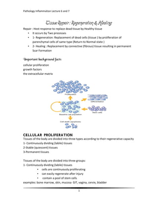
Tissues of the body are divided into three types according to their regenerative capacity
1- Continuously dividing (labile) tissues
2-Stable (quiescent) tissues
3-Permanent tissues
Tissues of the body are divided into three groups:
1- Continuously dividing (labile) tissues
• cells are continuously proliferating
• can easily regenerate after injury
• contain a pool of stem cells
examples: bone marrow, skin, mucosa GIT, vagina, cervix, bladder
2. Pathology Inflammation Lecture 6 and 7
2
2.Stable cells: a cell which stop multiplication when growth is complete after birth but
retain ability to multiply when there is a need for that e.g. liver cells ,pancreas ,kidney
fibroblast ,endothelial cell&smooth muscle
3.Permanent cells: Stop multiplication early in neonatal life e.g. neuron ,nerve cardiac
muscle &skeletal muscle
• Very important in tissue repair.
• Actions:
• stimulate cell division and proliferation
• promote cell survival
• Huge list! Usually have “GF” in name:
• EGF
• TGF
• PDGF FGF
• ECM is the network that surrounds cells
• Two forms: interstitial matrix and basement membrane
3. Pathology Inflammation Lecture 6 and 7
3
• Does lots of things!
• Sequesters water and minerals
• Gives cells a scaffold to adhere to
• Stores growth factors
• Bottom line: ECM regulates proliferation, movement, and differentiation of the
cells living in it.
• If you screw up your ECM, you can’t regenerate! You’ll form a scar instead.
REGENERATION
• Occurs all the time in labile tissues
• Cells are constantly being lost and replaced
• If demand increases, supply increases easily
• Occurs in limited form in stable tissues
• Remove one kidney: the other one undergoes hypertrophy and
hyperplasia
• Remove half of the liver: it will grow back
• Only occurs if residual tissue is intact!
SCARRING
• If injury is severe, regeneration can’t happen
• So, fibrosis (a scar) replaces the injured tissue
• Four components to this process:
• new vessel formation (angiogenesis)
• fibroblast proliferation
• synthesis of collagen (scar formation)
4. Pathology Inflammation Lecture 6 and 7
4
• remodeling of scar
Healing of skin wounds
1.Healing by primary union (Primary or first intention)
2.Healing by secondary union (Second intention).
Healing by First Intention
• Occurs in small clean wounds that close easily
• Epithelial regeneration predominates over fibrosis
• Healing is fast, with minimal scarring/infection
• Examples: Well-approximated surgical incisions
Healing by First Intention: Timeline
• By 24 hours
• By 3-7 days
• Weeks later
By 24 hours
• clot forms
• neutrophils come in
• epithelium begins to regenerate
By 3-7 days
• macrophages come in
• granulation tissue is formed
• new blood vessels
• fibroblasts
• collagen begins to bridge incision
• epithelium increases in thickness
Weeks later
• granulation tissue gone
• collagen is remodeled
• epidermis full, mature (but without dermal appendages)
• eventually, scar forms
By the end of first month : scar consists of C.T. without inflammatory cells and covered
by normal Epidermis . Dermal appendage are lost in the line of incision and No rete
ridges.
5. Pathology Inflammation Lecture 6 and 7
5
Healing by primary union
• Injury
• Acute inflammation
• Removal of the tinny blood clot
• Formation of scab at the surface
• growth of epithelial cells
• granulation tissue is formed between the edges of the wound
• mature in to fibrous tissue
• Tissue of Healing
• Is formed by day 3—5
• Soft , pink , granular tissue
• Consists of proliferating fibroblast , newly – formed B.Vs (capillaries ) in loose
ECM and inflammatory cells (macrophage)
• progressively G.T. change and mature into dense Fibrous Tissue "(fibrosis) and
scar formation
Wound which heal by secondary union
• Infected,
• contaminated wound,
• large blood clot,
• edges are widely separated
Healing by Second Intention
• Occurs in larger infected contaminated wounds that have gaps between wound
margins (widely separated edges)
• Fibrosis predominates over epithelial regeneration
• Healing is slower, with more inflammation and more granulation tissue
formation, and more scarring
6. Pathology Inflammation Lecture 6 and 7
6
• Examples:
• Infarction
• Large burns ,ulcers &abscess
Secondary union differs from primary union by followings
1- large tissue loss with large defect → large blood clot or scab is formed , require more
time to close.
2- Inflammation is more intense.
3- larger amounts of granulation tissue is formed to fill the gap or defect → large
Hypertrophic scar .(Keloid)
4- Wound contraction or contracture reduction in size of wound surface by 1/3 to 1/4
from its original size by contraction of myofibroblasts → limitation of joint movement .
Contracture → deformity in the extremities with limitation of joint movement which
cannot fully extend .
Second intention healing has:
• More inflammation
• More granulation tissue
• Wound contraction
7. Pathology Inflammation Lecture 6 and 7
7
Skin graft in wound healing
A large wound in which epithelialisation is delayed or impossible can be covered by skin
graft
Wound strength
• Sutured wound has 70% of normal skin strength because of the suture
• Suture is removed by the end of the first week wound strength is about 10%
of normal skin.
• By end of 3rd
month → 70- 80 % of normal skin and persist for life .
8. Pathology Inflammation Lecture 6 and 7
8
Complications of wound healing
• 1.Infections & Ulceration
• 2.Wound dehiscence
• 3.Implantation Epidermal cyst
• 4.Keloid formation.
• 5.Painfull scar.
• 6.Pigmented scar.
• 7.Weak scar.
• 8.Cicatrisation
• 9.Neoplastic changes
• 10.Exuberant granulation tissue
Keloid: Raised ugly hypertrophied scar tissue
Factors affecting wound Healing
1- Infection – is the single most important cause of delay in Healing .
2- Poor Blood supply: local venous obstruction, therosclerosis, DM will delay healing
pretibial skin wound heals much slower than face
3- Mechanical Factors : increased local pressure,Torsion, excessive movement →
wound to pull apart (wound Dehiscence)
4- Radiation therapy : inhibit cell proliferation ( radiotherapy for malignant tumour
is delayed until the surgical scar has been healed)
5- Foreign body in the wound
6- Adhesion to bony surface
7- Neoplastic changes (Tumour)
1- Protein – calorie malnutrition
2- Vit-c & Zink deficiency → inhibit collagen synthesis occurs in severe Burns &
intestinal fistula
3- Diabetes mellitus (DM)
4- Corticosteroid Treatment → inhibit fibrosis .
9. Pathology Inflammation Lecture 6 and 7
9
5- Renal Failure
6- Hematological diseases – leukemia
Neutropenia
7- Tumour cachexia.
Mechanisms of wound healing
1-Inflammation
2- Regeneration
3- Formation of new B.Vs (Angiogenesis).
4- Migration and proliferation of fibroblasts.
5- Deposition of ECM and collagen synthesis.
6- Maturation and scar remodeling by collagenase and gelatinase → collagen
degradation.
All these complicated process are controlled by variety of mechanism which involve
1-Chemical mediators
2-Growth factors:
e.g. macrophage G.F
Platelet G.F
Epidermal G.F. etc
3-Growth inhibiters: Tumour suppresser gene (Retinoblastoma gene)
4-Extracellular matrix (ECM)
• Not all injuries result in permanent damage; some are resolved almost
completely
• More often, there is some degree of scarring
• Scar is usually good (provides a resilient patch) but occasionally bad (can cause
permanent dysfunction)
With my best wishes