1. GRAM-POSITIVE TOXIC SHOCK SYNDROMES
Emma Lappin FCARCSI
Andrew Ferguson FRCA
Accepted Author!s Manuscript
NOTICE: this is the author's version of a work that was accepted for publication in The
Lancet Infectious Diseases. Changes resulting from the publishing process, such as peer
review, editing, corrections, structural formatting, and other quality control mechanisms
may not be reflected in this document. Changes may have been made to this work since it
was submitted for publication. A definitive version was subsequently published in The
Lancet Infectious Diseases, Volume 9, Issue 5, Pages 281 - 290, May 2009 doi:10.1016/
S1473-3099(09)70066-0.
2. 1
Gram-Positive Toxic Shock Syndromes: A Pathophysiological Review
Dr. Emma Lappin
Specialist Registrar, Department of Anaesthetics and Intensive Care Medicine
Craigavon Area Hospital, 68 Lurgan Road
Portadown BT63 5QQ
United Kingdom
Dr. Andrew J. Ferguson
Assistant Professor of Medicine (Critical Care) and Anesthesia
Dalhousie University
Room 377 Bethune Building, 1278 Tower Road
Halifax, Nova Scotia B3H 2Y9, Canada
Tel: 1 902 473 3606
Fax: 1 902 473 3610
Email: andrewferguson@dal.ca
Correspondence to Dr. Andrew Ferguson
Running title: Gram-positive toxic shock syndromes
Number of Tables: 1 Number of Panels: 2 Number of Figures: 1
Manuscript
3. 2
Summary:
Toxic shock syndrome is an acute, multi-system, toxin-mediated illness, often
resulting in multi-organ failure. It represents the most fulminant expression of a
spectrum of diseases caused by toxin-producing strains of Staphylococcus aureus and
Streptococcus pyogenes (Group A streptococcus, GAS). The importance of gram-
positive organisms as pathogens is increasing, and it is likely that toxic shock
syndrome is under diagnosed in patients with staphylococcal or GAS infection who
present with shock. TSS results from the ability of bacterial toxins to act as
superantigens, stimulating immune cell expansion and rampant cytokine expression in
a manner that bypasses normal MHC restricted antigen processing. A repetitive cycle
of cell stimulation and cytokine release results in a cytokine avalanche that causes
tissue damage, DIC, and organ dysfunction. Specific therapy centres on early
identification of the illness, source control, and administration on antimicrobial agents
including those drugs capable of suppressing toxin production (e.g. clindamycin,
linezolid). Intravenous immunoglobulin has the potential to neutralize superantigen
and mitigate against the subsequent tissue damage.
Keywords: Toxic shock syndrome, superantigen, cytokine, septic shock, gram-
positive shock, NF-!B, Toll-like receptor
Word count (body including conclusion): 4927
Word count (summary): 163
Acknowledgements: The authors wish to acknowledge the assistance of Dr. Richard
Hall who kindly reviewed the early drafts of this manuscript
4. 3
Search strategy and selection criteria
Data for this review were identified by a Medline search restricted to English
language articles. The search terms used were “toxic shock”, “staphylococcal sepsis”,
“streptococcal sepsis”, “superantigen”, “nuclear factor kappa B”, “toll-like receptor”,
“immunity”, “enterotoxin”, “exotoxin”, “t-cell receptor”, “septic shock” and
“immunoglobulin”. Further articles were identified through review of the references
in selected papers. No limit was set on publication dates or types.
5. 4
Introduction
Gram-positive infections are responsible for approximately 50% of sepsis cases in the
United States.1
In addition to “classical” sepsis syndromes, several gram-positive
species are also capable of producing disease through toxin production. Toxic shock
syndrome is an acute, multi-system, toxin-mediated illness, typically resulting in
shock and multi-organ failure early in its clinical course. It represents the most
fulminant expression of a spectrum of diseases caused by toxin-producing strains of
Staphylococcus aureus and Streptococcus pyogenes (Group A streptococcus, GAS).
Despite a mortality rate higher than meningococcal septicaemia, toxic shock
syndrome has not achieved the same level of awareness amongst healthcare
professionals, who will generally encounter very few recognised cases during their
career. TSS may present anywhere within the healthcare system, from occupational
health departments to specialist hospital units and may progress with a rapidity that,
once seen, is never forgotten. It is therefore essential that all healthcare practitioners
have a sound appreciation of the epidemiology, pathophysiology, clinical features and
management.
Epidemiology
Staphylococcal Toxic Shock Syndrome
Staphylococcal Toxic Shock Syndrome (TSS) was reported in 1978 and came to
prominence in the early 1980’s in the United States in association with the use of
“highly absorbent” tampons amongst young healthy women, with high percentages of
vaginal cultures yielding S. aureus.2
During this period, the peak incidence was
reported to be between 6·2 and 12·3 cases per 100,000 inhabitants per year in active
6. 5
surveillance programmes.3
With changes in tampon manufacture and usage advise the
incidence fell to around 1 case per 100,000 inhabitants per year in the US.4
Data from
a surveillance programme in Minneapolis-St. Paul for 2000 to 2003 suggest local
increases, with a rise from 0·9 to 3·4 cases per 100,000 inhabitants per year over the
4-year period.5
Currently, between 1 and 5% of healthy women have vaginal
colonization with a toxin producing strain of S. aureus. This is unchanged from 1980-
81 although overall staphylococcal colonization had increased.6
A French surveillance
study of 55 TSS cases over a 30-month period has suggested that non-menstrual
staphylococcal TSS is more prevalent than menstrual TSS, accounting for 62% of the
cases. There were no deaths in the menstrual TSS group compared with a mortality
rate of 22% for non-menstrual cases.7
Non-menstrual TSS may result from any primary staphylococcal infection, or indeed
from colonization with a toxin producing strain of S. aureus (including methicillin-
resistant S. aureus, MRSA). It can arise following disruption of the skin or mucous
membranes, in association with abscesses or burns, and after surgical procedures,
although commonly no source of infection is confirmed.8
In light of this, TSS should
be considered in patients with shock and infection with S. aureus.
Streptococcal Toxic Shock Syndrome
A second toxic shock-like syndrome attributed to S. pyogenes was reported in 1987.9
Streptococcal TSS (STSS) secondary to invasive GAS soft tissue infections had a
mortality of approximately 30% in some early series.10
Studies from Australia,
Denmark and the USA cite the incidence of invasive GAS infection at between 1·5
and 5·2 cases per 100,000 inhabitants per year, higher rates being found at the
extremes of age and amongst ethnic minorities.11-13
Between 5 and 14·4% of cases
7. 6
developed streptococcal TSS with an attendant case fatality rate ranging from 23 to
44%. Higher incidence was also observed in those with underlying chronic illness,
following varicella infection, and with non-steroidal anti-inflammatory use. Recently
published data from 11 European countries (Strep-EURO) gave an incidence of STSS
of 13% in streptococcal infection from any source. This increased dramatically to
50% in patients with necrotising fasciitis. The 7-day mortality rate from STSS was
44%.14
Pathophysiology
Superantigens trigger a cytokine avalanche
Bacterial toxins are pivotal to the pathogenesis of TSS and STSS. They act as
“superantigens”, which are protein toxins that share the ability to trigger excessive
and non-conventional T-cell activation with consequent downstream activation of
other cell types, and cytokine/chemokine release.15
In addition to gram-positive
organisms, some gram-negative bacteria, Mycoplasma, and certain viruses are
known to produce these proteins, and so-called “endogenous superantigens” are
found coded within the human genome (generally within endogenous retroviral
sequences). The staphylococcal and streptococcal superantigens identified to date
are single chain proteins expressed as precursor molecules, which are then cleaved
to release the functional extracellular toxin.16
The structure and function of S.
aureus and S. pyogenes superantigens are the best characterized.17-18
Superantigens bypass conventional mechanisms of major histocompatibility
complex (MHC) limited antigen processing, where antigens are processed into
peptide fragments within antigen presenting cells such as monocytes. These
fragments are then presented to the T cell via a specific peptide-binding groove of
the MHC class II molecule. T cells will only respond if they recognize the class II
8. 7
molecule and specific antigen fragment being presented. In contrast, superantigens
bind simultaneously as unprocessed intact proteins directly to the MHC class II
molecule and to the T cell receptor.18-19
They bind at sites distant to the
conventional peptide binding area, primarily to the variable region on the T cell
!"#"$%&!'()*+,'#-.."/'%0"'12'!"34&56'-.%0&730'-'89-..'579:"!'&;'87$erantigens
bind to the TCR ! chain.20-21
The interaction of superantigen with specific TCR
12'!"34&58'45/7#"8'#.&5-.'"<$!"884&5'&;')'#"..8'$&88"88453'%0&8"'8$"#4;4#'12')*+'
$-%%"!58=')048'-..&>8';&!'4/"5%4;4#-%4&5'&;'-'#0-!-#%"!48%4#'12'?8435-%7!"@';&!'%0"'
superantigen concerned and may be diagnostically useful.22-24
Binding activates up to 20-30% of host T cells whereas conventional antigen
presentation activates only around 0·01% of the host T cell population.18, 25-26
Interestingly, endogenous superantigen gene sequences appear to down-regulate
%0"'"<$!"884&5'&;')'#"..8'>4%0'%0"'12')*+'-$propriate to that superantigen. This
may prevent subsequent expansion of that T cell population in response to
exogenous superantigen challenge, offering a degree of protection to the host by
limiting the inflammatory consequences of the exposure.27
When superantigen binds to TCR and MHC Class II there is a rapid increase in
cytokine expression by T-cells (primarily TNF-", IL-2, and IFN-#) and by antigen
presenting cells such as monocytes (primarily TNF-!, IL-1", IL-6), likely linked
to activation of the transcription factor known as Nuclear Factor Kappa B (NF-
$B).28
NF-$B plays a central role in the generation and expansion of the
inflammatory response, activation of coagulation, and the development of organ
dysfunction (Figure 1). The degree of NF-$B activation also correlates with
mortality risk.29-30
Recently, antioxidant agents such as N-acetyl cysteine have
been shown to reduce T cell proliferation and cytokine expression through
9. 8
inhibition of NF-!B in a superantigen stimulated cell line model, and other
inhibitory approaches are under active investigation.31-32
T cell activation leads to recruitment of further T and B cells to the site of infection.
Clonal T cell expansion continues, as does activation of antigen presenting cells,
“winding up” the release of pro-inflammatory mediators and contributing to increased
procoagulant activity.33
There is a complex interplay between the cytokines released
during this pro-inflammatory avalanche, with IFN-" rapidly inducing TNF-# and IL-6
expression.
Superantigen structure-activity relationships
Superantigens have been grouped into five distinct populations (I-V) based on their
phylogenetic relationships.26
Superantigens take part in two key interactions, firstly
with MHC II and secondly with the TCR, using mechanisms that are thought to differ
across the five superantigen groups.34
Superantigens interact with the MHC-peptide antigen complex (pMHC) in four main
ways.35
1) Binding to the MHC #-subunit at a site that extends over the peptide surface
and contacts the $-subunit. This peptide-dependent interaction is exemplified
by TSST-1.
2) Binding to MHC #-subunit without any interaction with the peptide. This
peptide-independent interaction is seen with Group II superantigens such as
SEB and SEC3.
3) Binding to the MHC $-subunit in a zinc-dependent manner and involving
multiple sites of interaction with the peptide. This occurs at areas common to
10. 9
multiple peptides and is seen with Group IV and V superantigens such as
SpeC and SEK respectively.
4) Binding by a combination of methods 1 and 2 e.g. SEA.
The structural conformation of superantigen interaction with TCR V! has also been
studied.34-36
Although all superantigens appear to bind to the second complement
determining region (CDR2), the V! region contains multiple hypervariable elements
and superantigens vary in their binding specificity and cross-reactivity to these.
Superantigens with low specificity such as SEB and SEC3 require only a few of these
elements to complete binding e.g. CDR2 and HV4. As specificity increases (e.g.
SpeA) more and more of these hypervariable components are required, and hydrogen
bonds form between the superantigen and TCR. With even greater specificity (e.g.
SpeC) the complete TCR hypervariable element series including CDR1-3 and HV4 is
required. TSST-1 demonstrates the greatest degree of specificity, targeting a loop in
the third framework region (FR3) rather than relying on interaction with multiple
hypervariable elements. TSST-1 also requires the presence of a particular residue in a
particular location within the FR3 loop (Lys62) in order to activate T cells. The Group
V superantigen SEK possesses an extended "3-!8 loop with a specific residue that
binds to V! 5.1, FR4 and FR3 regions and is critical for T cell activation.
T cell activation may vary between Groups based on the overall affinity and
conformation of the MHC-superantigen-TCR complex.
1) TSST-1 (Group I) acts as a bridge between TCR and MHC molecules, with no
direct MHC-TCR contact. The affinity of the TSST-1-TCR and TSST-1MHC
interactions is comparable to that of conventional MHC-TCR interactions and
is an effective T cell activator.
11. 10
2) Group II superantigens such as SEB act as a wedge between MHC and TCR,
preventing contact between TCR and peptide antigen. However, there is direct
MHC-TCR contact. The SEB-MHC and SEB-TCR interactions are not
sufficient to achieve effective T cell activation. However, the additional MHC-
TCR interaction brings the total affinity to the point where T cell activation
occurs.
3) With Group IV superantigens such as SpeC there is a bridging of MHC and
TCR and again no direct MHC-TCR contact, as with TSST-1. However, the
resulting conformational planes are different. The combined affinities of the
zinc-dependent TCR interaction and the V! contact are sufficient for T cell
activation
Specific superantigen-disease associations
In menstrual TSS, there is a clear picture of the superantigen-disease relationship
with staphylococcal TSST-1 responsible for the vast majority (95%) of menstrual-
related TSS cases.37-38
This has traditionally been attributed to the ability of TSST-
1 to cross mucosal barriers, although SEB is also able to cross nasal, conjunctival
and vaginal mucosa39
It should be noted that TSST-1 is also detectable in around
50% of non-menstrual related TSS (NMTSS), the remaining cases being due
primarily to SEB and less often to other members of the family such as SEC, SEG
and SEI.40
Reports of TSST-1 in association with MRSA are becoming more
frequent. Highly virulent clones of MRSA harboring the TSST-1 gene (tst) have
been associated with TSS, a critical point to remember in managing patients with
MRSA and shock.41
12. 11
There are multiple associations between streptococcal superantigens and invasive
diseases. One of the most intriguing is soluble streptococcal M protein type M1. It
is well known that M1 streptococcal isolates are more virulent, and recent work
suggests that soluble M1 proteins may be superantigenic, preferentially activating
T cells with V!2 and V!4 TCR. M proteins also activate T-cells via Toll-like
receptor-2.42-43
The status of M protein as a superantigen remains contentious.
The expression of superantigen genes is also important. Four alleles of the
streptococcal pyrogenic exotoxin A (spe A) gene, designated spe A1-A4, have
been found in isolates from patients with severe invasive GAS disease.44
There is
a marked geographic distribution of genetic strains, with organisms expressing
SPE A2 and SPE A3 being responsible for the majority (60-90%) of streptococcal
TSS episodes in Europe and North America and Australia.45
In the Danish data
which contributed to the Strep-EURO study, either SpeA or SpeC was present in
all cases of STSS.12
Superantigen acts synergistically with endotoxin
Critically ill patients may be exposed to both endotoxin from gram-negative
organisms and superantigen from toxin producing gram-positive organisms, even
if the organism is simply colonizing the patient. In animal models, co-
administration of endotoxin and superantigen reduced the LD50 by a factor of up
to 50,000 compared with either toxin given alone.46
Immune effector cells
recognise so-called “pathogen associated molecular patterns” (PAMP) such as
lipopolysaccharide (LPS) from gram-negative and lipoteichoic acid (LTA) from
13. 12
gram-positive organisms.47
This recognition is intimately involved in the genesis
of the endotoxin-superantigen “double-hit”. Although there is a degree of overlap,
the detection system for LPS mainly involves activation of a Toll-like receptor
(TLR-4) and the co-receptor MD2, and that for gram-positive organisms mainly
involves lipoteichoic acid or peptidoglycan activation of TLR-2.48-49
The detailed
biology of these receptors has been well reviewed elsewhere.50-52
Activation of
each of these recognition systems results in pro-inflammatory mediator release
and further inflammatory stimulation via NF-!B. Superantigen-MHC binding up-
regulates the TLR-4/MD2 receptor system, priming monocytes for endotoxin
exposure, amplifying the expression of TNF-", IL-6 and IL-1#, and inducing
vasodilatation through type I interferon over-stimulation of inducible nitric oxide
synthetase (iNOS).53
In addition, streptococcal superantigens appear to up-regulate
TLR-2, which may become diagnostically useful in identifying streptococcal
toxin-mediated disease in a manner analogous to V# expansion.54
Superantigen genes are mobile within and across streptococcal strains
The genetic “plasticity” of the streptococcal genome results from the presence of
bacteriophages within the genome (so-called prophages) and may contribute to the
observed variability in virulence. 55
Prophage genetic material may account for up to
10% of the streptococcal genome.56
The majority of GAS superantigen genes are
found within these prophage sequences (also called pathogenicity islands or genomic
islands), and these phages are capable of transferring superantigen genes between
GAS strains, or indeed from GAS strains to group C and theoretically to group G
14. 13
streptococci.57
In so doing, they can convert a non-virulent or less virulent strain into
a highly virulent one. Invasive GCS and GGS incidence also appears to be increasing
along with the presence of superantigen genes within these organisms.58
An
Australian study has recently identified superantigen genes in GAS isolates and
correlated the superantigen with emm gene type (the gene encoding M protein).59
Twenty-six different superantigen profiles were present in 107 isolates, distributed
amongst 22 different emm types. These results were similar to previous reports and
support the hypothesis that conserved superantigen profiles result from surface M
proteins influencing the entry of bacteriophages in a selective manner.
Host-pathogen Interaction
Not all patients colonized or infected with a toxin producing strain of S. aureus or S.
pyogenes go on to develop TSS or STSS, and secondary infection rates are low. The
interaction between the host immune system and the pathogen may play a major role
in response to the bacterial and toxic challenge.
Deficient antibody titres predispose to TSS
The absence of antibodies to superantigens appears to be a major risk factor for the
development of TSS.25,60
More than 85% of women between 13 and 40 years of age
have TSST-1 antibody titres considered protective.38
Low or negative titres have been
demonstrated in 90·5% of patients with menstrual TSS and less than 50% of these
patients failed to sero-convert within 2 months of their illness.61
This may predispose
to repeated episodes of STSS and has been linked to the ability of TSST-1 to suppress
the action of immunoglobulin secreting cells.25
The superantigen-mediated cytokine
15. 14
response is associated with minimal T-helper type 2 cell (Th2) response, resulting in
failure to support B-cell proliferation and differentiation. In addition, high
concentrations of TSST-1 induce B-cell apoptosis. Levels of antibody to streptococcal
superantigens are lower in those with invasive disease than in healthy controls.
Immunogenetics - HLA haplotype variation modulates severity
The magnitude of the inflammatory response is closely linked to disease severity
and may be governed by host genetic factors such as MHC class II haplotype.62
The sites at which superantigens bind to HLA class II are polymorphic, and
differences in binding are reflected in a varying T cell and cytokine response. As
an example, the DRB1*15/DQB1*06 haplotype is associated with strong
protection from streptococcal TSS and reduced cytokine levels during GAS
infection, whereas the DRB1*14/DQB1*05 haplotype is associated with
predisposition to TSS.63-64
Gender alters response to sepsis and superantigen shock differently
There is a complex relationship between gender and susceptibility to sepsis with
17! oestradiol having variable effects on immune function (low concentrations
augmenting and high concentrations inhibiting IL-6 and TNF-" release), and
disagreement over the applicability of animal studies to the human setting.65
There
is broad agreement that male sex increases the risk of post-injury bacterial sepsis,
bacteraemia, referral to ICU, risk of septic shock, and mortality in conventional
sepsis. Females have been shown to have a more pronounced and prolonged
immune reaction to sepsis whereas males appear more prone to develop variable
degrees of immunoparesis after the initial immune response.66
However, there is a
16. 15
female preponderance in superantigen-mediated shock that extends to NMTSS.3
It
appears that something different is going on in superantigen-mediated shock that
alters the gender influence away from that found in septic shock. The exact nature
of this difference is unclear, but seems in part related to oestrogen. In a transgenic
mouse model, females were (a) more susceptible to S. pyogenes sepsis, (b) had a
significantly more pronounced TNF-!"#$%&'(%$")'"%*&$#+(),-$("./012")3+("4+5$%6"
(c) had lower levels of soluble TNF receptors (sTNF-R) I and II both at baseline
and on superantigenic challenge, suggesting deficient TNF-!"#$4'7+5"+(8".82"3+d
a greater degree of TNF-!-induced hepatic apoptosis and hence liver damage than
males.67
In addition, the authors were able to demonstrate that pre-treatment with
the oestrogen receptor modulator tamoxifen decreased both the early and late rise
in TNF-!6 reduced the level of hepatic apoptosis, and increased the levels of
sTNF-R. This is an area that requires cautious interpretation and further study.
Clinical Features and diagnosis
Toxic shock syndrome is characterised by an acute, progressive illness associated
with fever, rapid onset hypotension and accelerated multi-system failure. Multi-
system involvement is frequently established by the time of presentation. Clinical case
definitions for both syndromes have been proposed (Panels 1 and 2).68-69
Staphylococcal Toxic Shock Syndrome
Staphylococcal TSS presents abruptly with a flu-like prodromal illness consisting of
fever, gastrointestinal upset and severe myalgia followed commonly by confusion,
lethargy and agitation. Symptoms of hypovolaemia are frequent at presentation. If
present, a focus of infection is more likely to be superficial, may complicate burns or
17. 16
a surgical wound, or may result from a foreign body. Desquamation is a characteristic
late feature of staphylococcal TSS, occurring 10-21 days following disease onset. It is
important to note that blood cultures are positive in less than 5% of cases of
staphylococcal TSS.8
The clinical features of menstrual and non-menstrual TSS are identical in the
majority of cases. Up to 95% of patients diagnosed with menstrual TSS have an
onset of illness during menstruation.70
Patients with NMTSS are more likely to
have acquired the condition nosocomially and to have had prior antibiotic
treatment. Fever and rash are more prevalent in early illness and NMTSS is more
frequently associated with CNS manifestations and renal complications.8
Non-
SEA and non-TSST-1 superantigens appear to have greater neurotoxic potential.7
Post-operative NMTSS usually occurs within 48 hours of surgery and in many
cases evidence of clinically significant surgical site infection is lacking at the time
of presentation. Following the onset of symptoms, progression is rapid and multi-
organ failure can be present in as little as 8 to 12 hours. Recurrence of menstrual
TSS has been well documented but is rare in NMTSS. NMTSS must be
considered in the aetiology of shock states in patients with definite or suspected
staphylococcal infection.
Streptococcal Toxic Shock Syndrome
Streptococcal TSS more commonly arises from deep-seated invasive soft tissue
infections such as necrotising fasciitis, cellulitis and myositis. Pain may be severe
and relentless and is a common reason for seeking medical attention. A flu-like
18. 17
illness is also common in the early stages with fever, sore throat, swollen lymph
nodes and gastrointestinal upset. Those patients with a defined entry site may have
early and visible signs of inflammation. In the absence of a defined portal of entry
clinical evidence of a deep infection becomes more obvious as the illness
progresses. The initiating injury may be blunt trauma, muscle strain, and
haematoma or joint effusion and may appear trivial, so careful history taking is
essential. Examination may reveal bruising, haemorrhagic bullae, skin sloughing
and oedema. Hypotension and organ dysfunction are rapidly progressive.
The majority (60%) of patients with STSS have positive blood cultures.71
Presence or
absence of bacteraemia does not affect mortality. The diagnosis of STSS is confirmed
when GAS are cultured from normally sterile body fluids in patients with shock and
multi-organ failure. The mortality rate associated with streptococcal TSS is much
higher than with staphylococcal TSS, and has been quoted at up to 80% in association
with myositis.26
A murine model of the disease suggests that an early initial infection
may be followed up to 3 weeks later by bacteraemia, at which point symptoms and
signs of the disease appear and that trivial injury such as bruising amplified the
severity of the bacteraemia.72
Therapeutic Strategies
Supportive management and source control
Immediate intervention and resuscitation are required. In the early stages of illness,
the causative organism will be unknown and the same basic therapeutic strategy
should be applied as to any case of septic shock with active fluid resuscitation, early
use of vasopressors and/or inotropes, and intubation and mechanical ventilation if
19. 18
required. An appropriate antimicrobial regimen should begin immediately following
culture samples being taken.
A thorough search for infective focus is essential. The presence of necrotizing fasciitis
or myositis mandates immediate aggressive surgical debridement and is a true
surgical emergency. The underlying tissue infection may be much more extensive
than initially appreciated and the rate of spread may exceed the rate of debridement if
a conservative approach is taken. Surgical wounds should be considered potential
sources of infection, even in the absence of overt signs. Any infected wound should
be reopened and widely debrided, and packs or infected devices removed. In females,
a vaginal examination should be carried out and any tampon or foreign body removed.
Antimicrobial therapy to reduce toxin production as well as organism load
Inadequate initial antibiotic therapy increases mortality in intensive care patients with
severe sepsis and septic shock.73-75
Clinical trial data comparing antibiotic regimens in
TSS is lacking. Recommendations are based on in vitro studies and theoretical
principles and include the use of a !-lactam agent and a lincosamide pending culture
results.76
Therapy is focused on reducing both exotoxin production and organism
load. In cases where the causative organism is unknown, the antibiotic regimen should
cover both S. aureus (including MRSA if indicated) and S. pyogenes. There is an
increasing range of antimicrobial agents active against gram-positive organisms, and
definitive therapy decisions require knowledge of local drug availability, clinical
preferences, and sensitivity pattern. Potential therapeutic agents for various causative
organisms and strains are shown in Table I.
20. 19
Therapeutic principles
Group A streptococci remain exquisitely sensitive to ! lactam agents including
penicillin G, an agent often considered as part of first line therapy. It is usually given
with clindamycin, which has inhibitory actions on protein synthesis including
superantigen production. Although Penicillin G is bactericidal, it has been shown to
be less effective in the face of a higher organism load. This is perhaps due to the
reduced expression of penicillin binding proteins by bacteria in the stationary phase of
growth, which is reached more rapidly with large organism loads.77
Streptococcal
resistance to macrolides and fluoroquinolones appears to be increasing, especially in
Europe and Asia. In addition, macrolide resistance is linked to lincosamide
(clindamycin) resistance in so-called Macrolide-Lincosamide-Streptogramin B
resistant S. pyogenes (MLS).78-79
Therapy for MRSA has commonly included
vancomycin, however S. aureus strains with intermediate sensitivity (GISA) or
resistance (GRSA) to glycopeptides are increasing.80
The newer agents such as
linezolid, daptomycin and tigecycline are active against S. pyogenes, MRSA, GISA
and GRSA and represent effective (if expensive) agents to fall back on.
The rationale for clindamycin in initial therapy regimens
Clindamycin is a bacteriostatic lincosamide with efficacy unaffected by bacterial
growth phase or inoculum size. In a mouse model of S. pyogenes-induced
myositis, penicillin was ineffective if treatment was delayed by greater than 2
hours following onset of infection, whereas mice receiving clindamycin had
improved survival rates even if treatment was delayed.81
Clindamycin has been
shown to inhibit toxin production by both S. aureus and S. pyogenes. In vitro
21. 20
models comparing the effects of clindamycin, linezolid and penicillin on SpeA
release have shown a significant decrease in SpeA production in regimes
containing clindamycin and linezolid as opposed to penicillin G alone, despite the
theoretical ability of clindamycin to suppress synthesis of penicillin binding
proteins.82
This antagonistic effect does not seem to be clinically relevant with
adequate drug dosages. Linezolid and clindamycin have both been shown to
reduce TSST-1 production, and clindamycin significantly reduces SpeA
expression by of S. pyogenes compared to ampicillin.83
Linezolid has been used
successfully to treat staphylococcal TSS and has demonstrated reduced TSST-1
production.84
Effects and Mechanism of Intravenous Immunoglobulin
Patients with a deficient antibody response against TSST-1 are at increased risk of
primary or recurrent TSS, and patients with invasive GAS infections have
significantly lower levels of superantigen-neutralising antibodies.60
Case reports
published in the mid-1990’s suggested improved outcomes for patients with STSS
treated with intravenous immunoglobulin (IVIG). 85-87
Administration of IVIG can
block in vitro T cell activation by staphylococcal and streptococcal superantigens.
Factors beyond the presence of neutralising antibodies may contribute to the
efficacy of IVIG, at least in vitro, as the suppressive effect of whole IVIG on SEB-
induced T cell proliferation and cytokine production remains significant even after
removal of specific anti-SEB antibody from the IVIG.88-89
In a Canadian comparative observational study, the 21 patients receiving IVIG had
22. 21
a 30-day survival rate of 67% compared to 34% in the 32 control cases.90
Patients
treated with IVIG were more likely to have had surgery and to have received
clindamycin, and inclusion of historical controls may have introduced bias.
Analysis of plasma from 10 cases and 10 controls in this study demonstrated a
significant reduction in T cell triggered production of IL-!"#$%"&'()"#*+,-"#"./$01,"
dose of IVIG.
A subsequent multicentre, randomized, placebo-controlled trial studied the
efficacy of IVIG as adjunctive therapy in STSS.91
The trial was terminated due to
slow patient recruitment after 21 patients were enrolled, 10 receiving IVIG and 11
receiving placebo. The primary end-point was 28-day mortality but despite a 3·6
fold higher mortality rate being found in the placebo group (36% vs. 10% in IVIG
group) statistical significance was not reached. There was a greater improvement
in sepsis-related organ failure assessment (SOFA) score on days 2 and 3 of the
study in the IVIG group, and IVIG produced 87-100% inhibition of GAS strains
on in vitro testing.
S. aureus was isolated from blood culture in one patient in this trial and was found to
be inhibited to a lesser degree by IVIG. This prompted a comparison study to
investigate differential effects of IVIG on staphylococcal and streptococcal
superantigen production.92
Culture supernatants of S. pyogenes were consistently
inhibited to a greater degree than those of S. aureus. It was concluded that higher
doses of IVIG might be required to provide protective titres and clinical efficacy in
the treatment of staphylococcal TSS. In the original trial, the dose of IVIG used was
23. 22
1g/kg body weight on day 1 followed by subsequent doses of 0·5g/kg on days 2 and 3,
but a superior dose regimen for staphylococcal disease has not been confirmed.
Different IVIG preparations may vary in their neutralizing capacity, likely due to
differences (perhaps geographical) in organism exposure in the donor population.93
The mortality risk and rapidity of decline in TSS and STSS are such that delays in
effective therapy have significant potential to worsen outcome. On this basis we argue
that immunoglobulin therapy should not be unreasonably delayed in these cases.
There is no clear information from the literature on what constitutes a safe delay. The
United Kingdom Department of Health has issued guidance on the use of
immunoglobulin.94
For the management of invasive streptococcal disease (presumably
not just STSS) they advise that “IVIg may be added to adequate toxin-neutralising
antimicrobials, source control, and sepsis management when these approaches have
failed to elicit a response”. In the absence of a recommendation relating to time delay,
we advise that the same approach to timing be taken for STSS as is recommended for
staphylococcal TSS. In this setting the guidance states that “IVIg may be used for
TSS resulting from an infection refractory to several hours of aggressive therapy, in
the presence of an undrainable focus, or when there is persistent oliguria with
pulmonary oedema”. It is our approach to consider IVIg in cases where there has been
no clinical response within the first 6 hours of aggressive supportive therapy.
Conclusion
Toxic shock syndrome is a global disease entity caused by pathogens with the ability
to evolve in terms of superantigen generation and avoidance of our immune system.
24. 23
Despite intense research efforts, we do not yet have new clinically available therapies
capable of neutralizing superantigen-mediated T cell activation. Further research is
required addressing timing and components of therapy. In the real-world clinical
arena, a sound understanding of the pathophysiology, a high index of suspicion, early
diagnosis, and immediate intervention are the best ways to impact on the significant
mortality and morbidity rate of toxic shock syndrome. Given the supportive
background research and the severity of this syndrome, we recommend a therapeutic
approach in both TSS and STSS that incorporates prompt use of toxin-neutralising
antimicrobials such as clindamycin or linezolid, along with early IVIG where there is
failure to improve with aggressive support and source control.
Conflict of Interest Statement
The authors confirm that they have no interests or relationships, financial or
otherwise, that constitute a conflict of interest in the preparation or submission of this
manuscript.
25. 24
Panel 1: Staphylococcal Toxic Shock Syndrome Clinical Case Definition
1. Fever !"#$%&'("
2. Rash – diffuse macular erythroderma
3. Desquamation – 1-2 weeks after onset of illness, especially of palms and soles
4. Hypotension – SBP )&*++,-"./0"123456"
5. Multisystem involvement – 3 or more of the following
a. Gastrointestinal: vomiting or diarrhoea at the onset of illness
b. Muscular: severe myalgia or elevated creatine phosphokinase
c. Mucous membranes: vaginal, oropharyngeal, conjunctival hyperaemia
d. Renal: blood urea nitrogen or creatinine twice-upper limit of normal
e. Hepatic: total bilirubin twice-upper limit of normal
f. Haematological: platelets )7**8***9:"
g. CNS: disorientation or alterations in consciousness without focal
neurological signs
6. Negative results on the following tests
a. Blood, throat or CSF culture (blood culture may be positive for S.
aureus)
b. Rise in titre to Rocky Mountain spotted fever, leptospirosis or measles
Case Classification
Probable: case with 5 of the 6 clinical findings described above
Confirmed: case with all six of the clinical findings
26. 25
Panel 2: Streptococcal Toxic Shock Syndrome Clinical Case Definition
1. Isolation of Group A !-haemolytic streptococci
a. From a normally sterile site – blood, CSF, peritoneal fluid, tissue biopsy
b. From a non-sterile site – throat, vagina, sputum
2. Clinical signs of severity
a. Hypotension – SBP"#$%%&'()*(+,-./0(
b. 12(34(/56(43..37)*'(0)'*s
)8(96*+.()%:+);%6*/<(=;6+/)*)*6(>2%'?,@(A>BCCD%3.?@E(
ii. Coagulopathy: platelets "B$$F$$$D@(3;(GHI(
iii. Hepatic involvement: ALT, AST or total bilirubin twice the
upper limit of normal
iv. Adult respiratory distress syndrome
v. Generalised, erythematous, macular rash that may desquamate
vi. Soft tissue necrosis, including necrotising fasciitis, myositis or
gangrene
Case classification
Probable: Case fulfils 1b and 2 (a and b) if no other cause for the illness is found
Definite: Case fulfils 1a and 2 (a and b)
28. 27
References
1 Martin GS, Mannino DM, Eaton S, Moss M. The epidemiology of sepsis in
the United States from 1979 through 2000. N Engl J Med 2003; 348: 1546–
1554.
2 Todd J, Fishaut M, Kapral P, Welch T. Toxic-shock syndrome associated
with phage-group-I staphylococci. Lancet 1978; 2: 1116–1118.
3 Hajjeh RA, Reingold A, Weil A, Shutt K, Schuchat A, Perkins BA. Toxic
shock syndrome in the United States: surveillance update, 1979 1996.
Emerg Infect Dis 1999; 5: 807–810.
4 Gaventa S, Reingold AL, Hightower AW, Broome CV, Schwartz B, Hoppe
C, Harwell J, Lefkowitz LK, Makintubee S, Cundiff DR, Sitze S, and the
Toxic Shock Syndrome Study Group. Active surveillance for toxic shock
syndrome in the United States, 1986. Rev Infect Dis 1989; 11 (Suppl 1):
S28–S34.
5 Schlievert PM, Tripp TJ, Peterson ML. Reemergence of staphylococcal
toxic shock syndrome in Minneapolis-St. Paul, Minnesota, during the 2000-
2003 surveillance period. J Clin Microbiol 2004; 42: 2875–2876.
6 Schlievert PM, Case LC, Strandberg KL, Tripp TJ, Lin Y-C, Peterson ML.
Vaginal Staphylococcus aureus superantigen profile shift from 1980 and
1981 to 2003, 2004 and 2005. J Clin Microbiol 2007; 45: 2704–2707.
7 Descloux E, Perpoint T, Ferry T, Lina G, Bes M, Vandensch F,
Mhoammedi I, Etienne J. One in five mortality in non-menstrual toxic
shock syndrome versus no mortality in menstrual cases in a balanced
French series of 55 cases. Eur J Clin Microbiol Infect Dis 2008; 27: 37–43.
29. 28
8 !"##$%&'()&'*+,-./0/,.&$.1&2$.$-*2*.0&,3&40$56%7,+,++"8&$"#*"8&
0,9/.:2*1/$0*1&1/8*$8*)&!"#$%"&'$(&) ;<<=>&!" ?4"557&;#@&4A<BC4AAD)
9 E,.*&FGH&I,,1$#1&J'H&4+67/*K*#0&L!H&M,2,#%&N4)&E7/./+$7&$.1&
O$+0*#/,7,-/+&,O8*#K$0/,.8&,3&$&0,9/+&86,+P:7/P*&8%.1#,2*&1"*&0,&
40#*50,+,++"8&5%,-*.*8)&*&+",-&)&'$(&./01>&!$%@&AQBCAQD)
10 40*K*.8&JFH&M$..*#&!RH&I/.86/5&(H&4S$#08&'H&'/*8&T!H&4+67/*K*#0&L!H&
T$57$.&U)&4*K*#*&-#,"5&G&80#*50,+,++$7&/.3*+0/,.8&$88,+/$0*1&S/06&$&
0,9/+&86,+P:7/P*&8%.1#,2*&$.1&8+$#7*0&3*K*#&0,9/.&G)&*&+",-&)&'$( ADVD>&
!&$@&ACW)
11 O'Grady KA, Kelpie L, Andrews RM, Curtis N, Nolan TM, Selvaraj G et al.
The epidemiology of invasive group A streptococcal disease in Victoria,
Australia. Med J Aust 2007; 186: 565–569.
12 Luca-Harari B, Ekelund K, van der Linden M, Staum-Kaltoft M,
Hammerum AM, Jasir A. Clinical and epidemiological aspects of invasive
Streptococcus pyogenes infections in Denmark during 2003 and 2004. J
Clin Microbiol 2008; 46: 79–86.
13 O’Loughlin RE, Roberson A, Cieslak PR, Lynfield R, Gershman K, Craig
A, Albanese BA, Farley MM, Barrett NL, Spina NL, Beall B, Harrison LH,
Reingold A, Van Beneden CV. The epidemiology of invasive group A
streptococcal infection and potential vaccine implications: United States
2000-2004. Clin Infect Dis 2007; 45: 853–862.
14 Lamagni TL, Darenberg J, Luca-Harari B, Siljander T, Rfstratiou A,
Henriques-Normark B, Vuopio-Varkila J, Bouvet A, Creti R, Ekelund K,
Koliou M, Reinart RR, Stathi A, Strakova L, Ungureanu V, Schalen C,
Aftab J. Epidemiology of severe Streptococcus pyogenes disease in Europe.
30. 29
J Clin Microbiol 2008; 46: 2359-2367.
15 White J, Herman A, Pullen A, Kubo R, Kappler J, Marrack P. The V beta-
specific superantigen staphylococcal enterotoxin B: stimulation of mature
T-cells and clonal deletion in neonatal mice. Cell 1989; 56: 27–35.
16 Alouf JE, Muller-Alouf H. Staphylococcal and streptococcal superantigens:
molecular, biological and clinical aspects. Int J Med Microbiol 2003; 292:
429–440.
17 Igwe EI, Shewmaker PL, Facklam RR, Farley MM, van Beneden C, Beall
B. Identification of superantigen genes speM, ssa, and smeZ in invasive
strains of beta-hemolytic group C and G streptococci recovered from
humans. FEMS Microbiol Lett 2003; 229: 259–264.
18 Llewelyn M, Cohen J. Superantigens: microbial agents that corrupt
immunity. Lancet Infect Dis 2002; 2: 156–162.
19 Sriskandan S, Faulkner L, Hopkins P. Streptococcus pyogenes: insight into
the function of the streptococcal superantigens. Int J Biochem Cell Biol
2007; 39: 12–19.
20 Marrack P, Kappler J. The staphylococcal enterotoxins and their relatives.
Science 1990; 248: 705–711.
21 Pumphrey N, Vuidepot A, Jakobsen B, Forsberg G, Walse B, Lindkvist-
Petersson K. Cutting edge: Evidence of direct TCR alpha-chain interaction
with superantigen. J Immunol 2007; 179: 2700–2704.
22 Choi Y, Lafferty JA, Clements JR, Todd JK, Gelfand EW, Kappler J,
Marrack P, Kotzin BL. Selective expansion of T cells expressing V beta 2
in toxic shock syndrome. J Exp Med 1990; 172: 981–984.
31. 30
23 Wenisch C, Strunk D, Krause R, Smolle KH. Diagnostic value of V!2-
positive T-cell expansion in toxic shock syndrome. Int J Dermatology 2007;
46: 578–582.
24 Ferry T, Thomas D, Perpoint T, Lina G, Monneret G, Mohammedi I,
Chidiac C, Peyramond D, Vandenesch F, Etienne J. Analysis of
superantigenic toxin V!"T-cell signatures produced during cases of
staphylococcal toxic shock syndrome and septic shock. Clin Microbiol
Infect 2008; 14: 546–554.
25 Herman A, Kappler JW, Marrack P, Pullen AM. Superantigens:
mechanisms of T-cell stimulation and role in immune responses. Annu Rev
Immunol 1991; 9: 745–772.
26 McCormick JK, Yarwood JM, Schlievert PM. Toxic shock syndrome
and bacterial superantigens: an update. Annu Rev Microbiol 2001; 55:
77–104.
27 Rajagopalan G, Singh M, Sen MM, Murali NS, Nath KA, David CS.
Endogenous superantigens shape response to exogenous superantigens.
Clin Diag Lab Immunol 2005; 12: 1119–1122.
28 Trede NS, Castigli E, Geha RS, Chatila T. Microbial superantigens induce
NF-kappa B in the human monocytic cell line THP-1. J Immunol 1993; 150:
5604–5613.
29 Liu SF, Malik AB. NF-#B activation as a pathological mechanism of septic
shock and inflammation. Am J Physiol Lung Cell Mol Physiol 2006; 290:
L622–L645.
30 Zingarelli B. Nuclear factor-#B. Crit Care Med 2005; 33 (12 Suppl):
S414–416.
32. 31
31 Krakauer T, Buckley M. The potency of anti-oxidants in attenuating
superantigen-induced proinflammatory cytokines correlates with
inactivation of NF-kappaB. Immunopharmacol Immunotoxicol 2008; 30:
163–79.
32 Uwe S. Anti-inflammatory interventions of NF-!B signaling: potential
applications and risks. Biochem Pharmacol 2008; 75: 1567–1579.
33 Mattsson E, Herwald H, Egesten A. Superantigens from Staphylococcus
aureus induce procoagulant activity and monocytes tissue factor expression
in whole blood and mononuclear cells via IL-1". J Thromb Haemostasis
2003; 1: 2569–2576.
34 Sundgerg EJ, Deng L, Mariuzza RA. TCR recognition of peptide/MHC
class II complexes and superantigens. Semin Immunol 2007; 19: 262-271.
35 Fraser JD, Proft T. The bacterial superantigen and superantigen-like
proteins. Immunol Rev 2008: 225: 226-243.
36 Moza B, Varma AK, Buonpane RA, Zhu P, Herfst CA, Nicholson MJ,
Wilbuer A-K, Seth NP, Wucherpfennig KW, McCormick JK, Kranz DM,
Sundberg EJ. Structural basis of T-cell specificity and activation by the
bacterial superantigen TSST-1. EMBO J 2007; 26: 1187-1197.
37 Kass EH, Parsonnet J. On the pathogenesis of toxic shock syndrome. Rev
Infect Dis 1987; 9: S482–489.
38 Parsonnet J, Hansmann MA, Delaney ML, Modern PA, DuBois AM,
Wieland-Alter W, Wissemann KW, Wild JE, Jones MB, Seymour JL,
Onderdonk AB. Prevalence of toxic shock syndrome toxin 1-producing
Staphylococcus aureus and the presence of antibodies to this
superantigen in menstruating women. J Clin Microbiol 2005; 43: 4628–
33. 32
4634.
39 Rajagopalan G, Smart MK, Murali N, Patel R, David CS. Acute
systemic immune activation following vaginal exposure to
staphylococcal enterotoxin B--implications for menstrual shock. J
Reprod Immunol 2007; 73: 51–59.
40 Bohach GA, Fast DJ, Nelson RD, Schlievert PM. Staphylococcal and
streptococcal pyrogenic toxins involved in toxic shock syndrome and
related illnesses. Crit Rev Microbiol 1990; 17: 251–272.
41 Durand G, Bes M, Meugnier H, Enright MC, Forey F, Liassine N, Wenger
A, Kikuchi K, Lina G, Vandenesch F, Etienne J. Detection of new
methicillin-resistant Staphylococcus aureus clones containing the toxic
shock syndrome toxin 1 gene responsible for hospital- and community-
acquired infections in France. J Clin Microbiol 2006; 44: 847-853.
42 Påhlman LI, Mörgelin M, Eckert J, Johansson L, Russell W, Riesbeck K,
Soehnlein O, Lindbom L, Norrby-Teglund A, Schumann RR, Björck L,
Herwald H. Streptococcal M protein: a multipotent and powerful inducer of
inflammation. J Immunol 2006; 177: 1221–1228.
43 Påhlman LI, Olin AI, Darenberg J, Mörgelin M, Kotb M, Herwald H,
Norrby-Teglund A. Soluble M1 protein of Streptococcus pyogenes triggers
potent T cell activation. Cell Microbiol 2008; 10: 404–414.
44 Nelson K, Schlievert PM, Selander RK, Musser JM. Characterization
and clonal distribution of four alleles of the speA gene encoding
pyrogenic exotoxin A (scarlet fever toxin) in Streptococcus pyogenes. J
Exp Med 1991; 174: 1271–1274.
45 Proft T, Fraser JD. Streptococcal superantigens. Chem Immunol Allergy
34. 33
2007; 93: 1–23.
46 Schlievert PM. Enhancement of host susceptibility to lethal endotoxin
shock by staphylococcal pyrogenic exotoxin type C. Infect Immun 1982; 36:
123–128.
47 Sriskandan S, Altmann DM. The immunology of sepsis. J Pathol 2008;
214: 211–223.
48 van der Poll T, Opal SM. Host-pathogen interactions in sepsis. Lancet Infect
Dis 2008; 8: 32–43.
49 Chaplin DD. Overview of the human immune response. J Allergy Clin
Immunol 2006; 117: S430–435.
50 Hornef MW, Henriques-Normark B, Normark S. The function and
biological role of toll-like receptors in infectious diseases: an update. Curr
Opin Infect Dis 2008; 21: 304–12.
51 Akira S. Toll–like receptor signaling. J Biol Chem 2003; 278: 38105-38108.
52 Takeda K, Akira S. Toll–like receptors in innate immunity. Int Immunol
2005; 17: 1-14.
53 Hopkins PA, Fraser JD, Pridmore AC, Russell HH, Read RC, Sriskandan S.
Superantigen recognition by HLA class II on monocytes up-regulates toll-
like receptor 4 and enhances proinflammatory responses to endotoxin.
Blood 2005; 105: 3655–3662.
54 Hopkins PA, Pridmore AC, Ellmerich S, Fraser JD, Russell HH, Read RC,
Sriskandan S. Increased surface toll-like receptor 2 expression in
superantigen shock. Crit Care Med 2008; 36: 1267–76.
55 Banks DJ, Porcella SF, Barbian KD, Beres SB, Philips LE, Voyich JM,
DeLeo FR, Martin JM, Somerville GA, Musser JM. Progress toward
35. 34
characterization of the group A Streptococcus metagenome: complete
genome sequence of a macrolide-resistant serotype M6 strain. J Infect Dis
2004; 190: 727–738.
56 Banks DJ, Beres SB, Musser JM. The fundamental contribution of phages
to GAS evolution, genome diversification and strain emergence. Trends
Microbiol 2002; 10: 515–521.
57 Vojtek I, Pirzada ZA, Henriques-Normark B, Mastny M, Janapatla RP,
Charpentier E. Lysogenic transfer of group A Streptococcus superantigen
gene among Streptococci. J Infect Dis 2008; 197: 225–234.
58 Sachse S, Seidel P, Gerlach D, Günther E, Rödel J, Straube E, Schmidt KH.
Superantigen-like gene(s) in human pathogenic Streptococcus dysgalactiae,
subsp equisimilis: genomic localisation of the gene encoding streptococcal
pyrogenic exotoxin G (speG(dys)). FEMS Immunol Med Microbiol 2002;
34: 159–167.
59 Commons R, Rogers S, Gooding T, Danchin M, Carapetis J, Robins-
Browne R, Curtis N. Superantigen genes in group A streptococcal isolates
and their relationship with emm types. J Med Microbiol 2008; 57: 1238-
1246.
60 Basma H, Norrby-Teglund A, Guedez Y, McGeer A, Low DE, El-Ahmedy
O, Schwartz B, Kotb M. Risk factors in the pathogenesis of invasive group
A streptococcal infections: role of protective humoral immunity. Infect
Immun 1999; 67: 1871–1877.
61 Stolz SJ, Davis JP, Vergeront JM, Crass BA, Chesney PJ, Wand PJ,
Bergdoll MS. Development of serum antibody to toxic shock toxin among
36. 35
individuals with toxic shock syndrome in Wisconsin. J Infect Dis 1985;
151: 883–889.
62 Kotb M, Norrby-Teglund A, McGeer A, Green K, Low DE. Association of
human leukocyte antigen with outcomes of infectious diseases: the
streptococcal experience. Scand J Infect Dis 2003; 35: 665–669.
63 Nooh MM, El-Gengehi N, Kansal R, David CS, Kotb M. HLA transgenic
mice provide evidence for a direct and dominant role of HLA class II
variation in modulating the severity of streptococcal sepsis. J Immunol
2007; 178: 3076–3083.
64 Kotb M, Norrby-Teglund A, McGeer A, El Sherbini H, Dorak MT, Kurshid
K, et al. An immunogenetic and molecular basis for differences in outcomes
of invasive group A streptococcal infections. Nature Med 2002; 8: 1398–
1404.
65 Marriott I, Huet-Hudson YM. Sexual dimorphism in Innate Immune
Responses to Infectious Organisms. Immunol Res 2006; 34: 177–192.
66 van Eijk LT, Dorresteijn MJ, Smits P, van der Hoeven JG, Netea MG,
Pickkers P. Gender differences in the innate immune response and vascular
reactivity following the administration of endotoxin to human volunteers.
Crit Care Med 2007; 35: 1464–1469.
67 Faulkner L, Altmann DM, Ellmerich S, Huhtaniemi I, Stamp G, Sriskandan
S. Sexual dimorphism in superantigen shock involves elevated TNF-alpha
and TNF-alpha induced hepatic apoptosis. Am J Respir Crit Care Med
2007; 176: 473–482.
68 Wharton M, Chorba TL, Vogt RL, Morse DL, Buehler JW. Case definitions
for public health surveillance. MMWR Recomm Rep 1990; 39: 1–43.
37. 36
69 Defining the group A streptococcal toxic shock syndrome. Rationale and
consensus definition. The Working Group on Severe Streptococcal
Infections. JAMA 1993; 269: 390–391.
70 Davis JP, Osterholm MT, Helms CM, Vergeront JM, Wintermeyer LA,
Forfang JC, Judy LA, Rondeau J, Schell WL. Tri-state toxic-shock
syndrome study. II. Clinical and laboratory findings. J Infect Dis 1982; 145:
441–448.
71 Stevens DL. Streptococcal toxic shock syndrome. Clin Microbiol Infect
2002; 8: 133–136.
72 Seki M, Saito M, Iida K, Taniai H, Soejima T, Nakayama H, Yoshida S.
Onset of streptococcal toxic shock syndrome is accelerated by bruising in a
mouse model. Microb Pathog 2008; 44: 339–343.
73 MacArthur RD, Miller M, Albertson T, Panacek E, Johnson D, Teoh L,
Barchuk W. Adequacy of early empiric antibiotic treatment and survival in
severe sepsis: experience from the MONARCS trial. Clin Infect Dis 2004;
38: 284–288.
74 Garnacho-Montero J, Ortiz-Leyba C, Herrera-Melero I, Aldabó-Pallás T,
Cayuela-Dominguez A, Marquez-Vacaro JA, Carbajal-Guerrero J, Garcia-
Garmendia JL. Mortality and morbidity attributable to inadequate empirical
antimicrobial therapy in patients admitted to the ICU with sepsis: a matched
cohort study. J Antimicrob Chemother 2008; 61: 436–441.
75 Garnacho-Montero J, Garcia-Garmendia JL, Barrero-Almodovar A,
Jimenez-Jimenez FJ, Perez-Paredes C, Ortiz-Leyba C. Impact of adequate
empirical antibiotic therapy on the outcome of patients admitted to the
intensive care unit with sepsis. Crit Care Med 2003; 31: 274–-2751.
38. 37
76 Annane D, Clair B, Salomon J. Managing toxic shock syndrome with
antibiotics. Expert Opin Pharmacother 2004; 5: 1701–1710.
77 Stevens DL, Yan S, Bryant AE. Penicillin-binding protein expression at
different growth stages determines penicillin efficacy in vitro and in vivo:
an explanation for the inoculum effect. J Infect Dis 1993; 167: 1401–1405.
78 Mazzariol A, Koncan R, Bahar G, Cornaglia G. Susceptibilities of
Streptococcus pyogenes and Streptococcus pneumoniae to macrolides and
telithromycin: data from an Italian multicenter study. J Chemother 2007;
19: 500–507.
79 Richter SS, Heilmann KP, Dohrn CL, Beekmann SE, Riahi F, Garcia-de-
Lomas J, Ferech M, Goossens H, Doern GV. Increasing telithromycin
resistance among Streptococcus pyogenes in Europe. J Antimicrob
Chemother 2008; 61: 603–611.
80 Bulchandani D, Nachnani J, Fitzsimmons C, Jost P, Brewer J. Beyond
MRSA: the growing menace of hVISA and VISA. South Med J 2008; 101:
663-664.
81 Stevens DL, Gibbons AE, Bergstrom R, Winn V. The Eagle effect revisited:
efficacy of clindamycin, erythromycin, and penicillin in the treatment of
streptococcal myositis. J Infect Dis 1988; 158: 23–28.
82 Coyle EA, Cha R, Rybak MJ. Influences of linezolid, penicillin, and
clindamycin, alone and in combination, on streptococcal pyrogenic
exotoxin a release. Antimicrob Agents Chemother 2003; 47: 1752–1755.
83 Sriskandan S, McKee A, Hall L, Cohen J. Comparative effects of
clindamycin and ampicillin on superantigenic activity of Streptococcus
pyogenes. J Antimicrob Chemother 1997; 40: 275–277.
39. 38
84 Stevens DL, Wallace RJ, Hamilton SM, Bryant AE. Successful treatment of
staphylococcal toxic shock syndrome with linezolid: a case report and in
vitro evaluation of the production of toxic shock syndrome toxin type 1 in
the presence of antibiotics. Clin Infect Dis 2006; 42: 729–730.
85 Barry W, Hudgins L, Donta ST, Pesanti EL. Intravenous immunoglobulin
therapy for toxic shock syndrome. JAMA 1992; 267: 3315–3316.
86 Lamothe F, D'Amico P, Ghosn P, Tremblay C, Braidy J, Patenaude JV.
Clinical usefulness of intravenous human immunoglobulins in invasive
group A Streptococcal infections: case report and review. Clin Infect Dis
1995; 21: 1469–1470.
87 Perez CM, Kubak BM, Cryer HG, Salehmugodam S, Vespa P, Farmer D.
Adjunctive treatment of streptococcal toxic shock syndrome using
intravenous immunoglobulin: case report and review. Am J Med 1997; 102:
111–113.
88 Norrby-Teglund A, Kaul R, Low DE, McGeer A, Newton DW, Andersson
J, Andersson U, Kotb M. Plasma from patients with severe invasive group
A streptococcal infections treated with normal polyspecific IgG inhibits
streptococcal superantigen-induced T cell proliferation and cytokine
production. J Immunol 1996; 156: 3057–3064.
89 Kato K, Sakamoto T, Ito K. Gamma-globulin inhibits superantigen-induced
lymphocyte proliferation and cytokine production. Allergol Int 2007; 56:
439–444.
90 Kaul R, McGeer A, Norrby-Teglund A, Kotb M, Schwartz B, O'Rourke K,
Talbot J, Low DE. Intravenous immunoglobulin therapy for streptococcal
40. 39
toxic shock syndrome--a comparative observational study. The Canadian
Streptococcal Study Group. Clin Infect Dis 1999; 28: 800–807.
91 Darenberg J, Ihendyane N, Sjölin J, Aufwerber E, Haidl S, Follin P,
Andersson J, Norrby-Teglund A, StreptIg Study Group. Intravenous
immunoglobulin G therapy in streptococcal toxic shock syndrome: a
European randomized, double-blind, placebo-controlled trial. Clin Infect
Dis 2003; 37: 333–340.
92 Darenberg J, Söderquist B, Normark BH, Norrby-Teglund A. Differences in
potency of intravenous polyspecific immunoglobulin G against
streptococcal and staphylococcal superantigens: implications for therapy of
toxic shock syndrome. Clin Infect Dis 2004; 38: 836–842.
93 !"#$%&'()*(+,%-(.*(/%-&(01*(2$%3'$(4+*(1$567(89(+:66'$'-7(;$';%$%7:5-3(56(
:-7$%<'-5,3(:==,-5&>5?,>:-(<%$@(:-(7#':$('66:"%"@(75(-',7$%>:A'(
37$';75"5""%>(3,;'$%-7:&'-3B(:=;>:"%7:5-3(65$(7$'%7='-7(56(
37$';75"5""%>(75C:"(3#5"D(3@-E$5='9(!"#$%&$'()*%+#, FGGHI !"B JKLMJKH9
94 N>:-:"%>(.,:E'>:-'3(65$(O==,-5&>5?,>:-(P3'9(!'"5-E(QE:7:5-9(05-E5-*(
+(-./*0($*%1'%2(."*3*(R%@(FGGS9
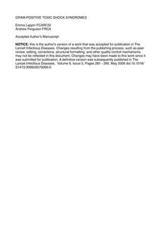
41. 40
Figure 1: NF-!B plays a central role in the generation and propagation of the
inflammatory response. Activation of toll-like receptor (TLR)-2 pathways by gram-
positive components, TLR-4 pathways by gram-negative products, and superantigenic
stimulation, all bring about a sequence of events that allow free NF-!B to pass into
the nucleus and bind to DNA. This leads to 1) expression of inflammatory mediators
and “wind-up” of the inflammatory cascade; 2) neutrophil adhesion and activation; 3)
activation of tissue factor and plasminogen activator inhibitor 1 to reduce fibrinolysis
and enhance coagulability; 4) inducible nitric oxide synthetase acceleration with
consequent vasodilatation and hypotension; and 5) induction of cyclo-oxygenase 2
and 5-lipoxygenase systems elaborating pro-inflammatory prostanoids, leukotrienes
and thromboxane A2. This figure has been adapted from 29.