2. We reasoned that the explanation for the limited immune re-
sponse to vaccines such as anthrax and plague could lie in this
initial interaction between the vaccine and immature DC (iDC). In
the present study, therefore, we used an in vitro strategy for the
examination of vaccine-DC encounters and subsequent DC matu-
rity, secretory capacity, and induction of T cell polarization.
Through this approach, we demonstrate that inadequate stimula-
tion of DCs by the anthrax vaccine may be a factor underlying its
limited immunogenicity in vivo. In contrast, at high concentra-
tions, the plague vaccine is capable of full DC maturation and
induction of effector responses. Adapting our in vitro strategy to
examine the potential for beneficial or negative effects of vaccine
combinations and adjuvants, we were able to show that combina-
tions that include powerful adjuvant agents such as whole-cell per-
tussis (WCP) can enhance the limited stimulatory effects of the
anthrax vaccine, a concept supported by analysis of recall re-
sponses in anthrax/WCP vaccinees.
Materials and Methods
We generated iDCs from five healthy human donors and exposed them to
vaccines to examine the major checkpoints in the events leading to T cell
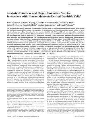
priming in the lymph node. Our overall strategy is shown in Fig. 1. This
study was approved by the local research ethics committee.
Generation of immature monocyte-derived DCs
PBMCs from healthy laboratory donors were isolated by density gradient
centrifugation on Lymphoprep (Nycomed). PBMCs were then layered onto
a Percoll (Amersham Biosciences) gradient, consisting of three density
layers (1.076, 1.059, and 1.045 g/ml), and centrifuged at 1750 ϫ g for 45
min. The light density fraction, containing predominantly monocytes, was
seeded into 24-well culture plates at a density of 0.5 ϫ 106
cells/ml in
IMDM (Invitrogen Life Technologies) containing 86 g/L gentamicin
(Sigma-Aldrich), 2 mM L-glutamine, 100 IU/ml penicillin, and 100 g/ml
streptomycin (Invitrogen Life Technologies) supplemented with 1% FCS
(PAA Laboratories). Cytofluorimetric analysis showed that the purification
procedure yielded Ͼ90% pure CD14ϩ
cells. After 1 h at 37°C, nonadherent
cells were removed, and adherent cells were cultured in IMDM/FCS sup-
plemented with IL-4 (250 IU/ml) and GM-CSF (500 IU/ml; both cytokines
from Strathmann Biotec) to obtain immature monocyte-derived DCs. On
day 3, the supplemented media was refreshed, and after 6 days, the iDCs
were ready for stimulation. iDCs were washed and analyzed by cytofluo-
rimetric analysis for CD1a, CD14, and CD3 expression. The mean per-
centage (ϮSD) of CD1aϩ
, CD14ϩ
, and CD3ϩ
cells was 92% (2.6), 7.5%
(2.4), and 2.8% (1.6), respectively, from five experiments.
Vaccine preparations
The United Kingdom human anthrax vaccine consists of a protein precip-
itate from the supernatant fluid of cultures of the Sterne strain of B. an-
thracis. The major immunogen is protective Ag (PA), the nontoxic, cell-
binding component of the anthrax toxin complex. The vaccine also
contains lethal factors (LFs) and edema factors (EFs). The concentration of
PA in the vaccine preparation is 1.3–2.2 g/ml (whole molecule and frag-
ments), and the concentration of LF is 0.4–0.7 g/ml (G. Griffiths and M.
Hudson, personal communication; provisional data to be updated and val-
idated using GLP functional assays/immunoassays). An EF is present in
very low levels below the detection limit of the assay method. This alum-
precipitated human anthrax vaccine (product license no. PL1511/0037) was
produced by The Centre for Applied Microbiology and Research (Porton
Down, Salisbury, Wiltshire, U.K.) for the United Kingdom Department of
Health. In our study, the anthrax vaccine was used over a range of quan-
tities, which achieved a PA concentration of 0.13–43 pg/ml (i.e., vaccine
diluted between 1/10,000 and 1/30).
The plague vaccine (CSL Limited) consists of a suspension of agar-
grown, heat-killed organisms of Y. pestis in saline at 3 ϫ 109
organisms/ml
and was used in our studies in the range 0.3–100 ϫ 106
/ml (i.e., vaccine
diluted between 1/10,000 and 1/30). The strains used in this preparation
were obtained from the Haffkine Institute (Mumbai/Bombay, India), and
their virulence was confirmed by demonstration of lethal effect in rats.
The WCP vaccine consists of killed whole-cell preparation of Bordetella
pertussis W28, prepared at The Centre for Applied Microbiology and Re-
search to the original Burroughs-Wellcome procedure for the preparation
of a single WCP vaccine and provided at a strength of 4 ϫ 1010
organ-
isms/ml (product license no. 208/10/99). In our studies, the pertussis vac-
cine was used in the concentration range 0.4–4 ϫ 107
organisms/ml (i.e.,
vaccine diluted between 1/10,000 and 1/30).
In preliminary studies to examine DC maturation and effector function
by analysis of accessory molecule expression, cytokine production, and
cellular toxicity, vaccines were used across the range of concentrations
stated above. Single concentrations were then selected as those at which
optimal DC activation was observed at 48 h. These selected concentrations
were used in time-course studies (6, 16, 24, 48, and 72 h) and in the
examination of DC interaction with CD40L, MLR, and polarization of
effector T cell responses.
Examining cytokine potential of iDCs and degree of maturation
Immature DCs were stimulated at a density of 8 ϫ 104
cells/200 l in
96-well plates in IMDM and 1% FCS with either vaccines or various con-
trol preparations representing maturation-inducing factors (MFs) IL-1 (10
ng/ml), TNF-␣ (50 ng/ml) (both cytokines from Strathmann Biotec AG),
and LPS (100 ng/ml) (Sigma-Aldrich); the Th1-type stimulant IFN-␥ (1000
U/ml; R&D Systems), which drives the development of DCs that promote
Th1 responses; and PGE2 (10Ϫ6
M; Sigma-Aldrich), which promotes Th2
responses. Supernatants were harvested and stored at Ϫ80°C for subse-
quent analysis of cytokine secretion using the cytokine bead array (CBA)
inflammatory kit assay (BD Biosciences) and a FACSCalibur flow cytom-
eter (BD Biosciences) according to the manufacturer’s instructions. In
brief, we used four bead populations with distinct fluorescence intensities,
coated with capture Ab specific for IL-12p70, IL-6, IL-10, and TNF-␣
proteins. Supernatant samples were incubated with human cytokine capture
beads and stained with PE detection reagent. After incubation for 3 h at
room temperature, samples were washed and acquired using the FACS-
Calibur. Data were analyzed using CellQuest and CBA Analysis Software
1.1 (both from BD Biosciences). The lower detection limit for IL-12p70
was 1.9 pg/ml, for IL-6 was 2.5 pg/ml, for IL-10 was 3.3 pg/ml, and for
TNF-␣ was 3.7 pg/ml.
iDCs were also cultured under identical conditions to examine the ef-
fects of vaccines on maturation. DCs were then harvested, washed exten-
sively to remove all supplements, and stained for expression of cell-surface
molecules representing maturation and activation by flow cytometry.
Mouse anti-human mAbs against the following molecules were used:
CD1a-FITC, CD83-FITC, and CD86-PE (all from Serotec); and HLA-DR-
PerCP, CD3-FITC, CD14-FITC, CD19-FITC, and CD56-FITC (all from
BD Biosciences). In addition, cellular toxicity was evaluated using 7-ami-
no-actinomycin D (7-AAD; final concentration, 1 g/ml; Calbiochem). At
least 5000 events gated on forward and side scatter were analyzed using the
CellQuest program (BD Biosciences), with dead (7-AADϩ
) cells excluded.
Corresponding isotype Abs were used to establish the quadrants and mark-
ers for analysis.
Effects of vaccines on the T cell stimulatory potential of DCs
Vaccine effects on the T cell stimulatory potential of DCs were examined
using the MLR. Immature DCs were exposed to vaccines for 48 h at the
following concentrations, which had elicited optimal effects on DCs in the
preliminary studies: anthrax at 0.43 pg/ml of PA; plague at 10 ϫ 106
organisms/ml; pertussis at 1 ϫ 106
organisms/ml. DCs were harvested,
FIGURE 1. Scheme used to examine checkpoints in DC maturation and
generation of Th effector cells. mDCs, Mature DCs.
7236 BIOWARFARE VACCINES AND DC INTERACTIONS
3. washed extensively, and cultured at a range of cells/well (100–10,000) in
96-well flat-bottom plates in the presence of 50,000/well allo-CD4ϩ
T
cells, purified from PBMCs using the Isolation kit II (Miltenyi Biotec).
After 5 days, [3
H]thymidine (0.5 Ci/well) was added, cells were har-
vested, and proliferation was measured by liquid scintillation spectroscopy.
Effects of vaccines on cytokine production by mature DCs
To examine vaccine effects on cytokine production by mature DCs, iDCs
were first matured for 48 h in the presence of vaccines as described above,
with the addition of the conventional MFs (IL-1 at 10 ng/ml, TNF-␣ at 50
ng/ml, and LPS at 100 ng/ml final concentrations). In these experiments,
vaccines were used at the following concentrations, which had elicited
optimal effects on DCs in the preliminary studies: anthrax at 0.43 pg/ml of
PA; plague at 10 ϫ 106
organisms/ml; pertussis at 1 ϫ 106
organisms/ml.
Mature DCs were then harvested, washed extensively, and cultured (4 ϫ
104
cells/well) in 96-well flat-bottom plates in the presence of an equiva-
lent number of human CD40L-expressing mouse plasmacytoid cells (J558
cells; a gift from Dr. P. Lane, Medical Research Council Center for Im-
mune Regulation, University of Birmingham, Birmingham, U.K.) in
IMDM containing 10% FCS in a final volume of 200 l. Supernatants were
harvested after 24 h and stored at Ϫ80°C for analysis of cytokine secretion
measured using the CBA inflammatory kit as described above.
Effects of vaccines on naive Th cell polarization by mature DCs
To examine vaccine effects on naive Th cell polarization by mature DCs,
iDCs were first matured for 48 h in the presence of vaccines (at the same
concentrations as above) supplemented with conventional MFs (IL-1 (10
ng/ml), TNF-␣ (50 ng/ml), and LPS (100 ng/ml)).
Naive (CD4ϩ
CD45RAϩ
CD45ROϪ
) cells were purified from PBMCs
by negative selection using a CD4ϩ
/CD45ROϪ
kit (R&D Systems) and
were Ͼ98% pure as assessed by flow cytometry. Naive CD4ϩ
T cells (2 ϫ
104
cells/200 l of IMDM with 10% FCS) were cocultured with 5 ϫ 103
DCs matured in the presence of vaccines plus MF and staphylococcal en-
terotoxin B (final concentration, 100 pg/ml; Sigma-Aldrich), in 96-well
flat-bottom plates. On day 5 of these cultures, recombinant human IL-2
(final concentration, 10 U/ml; Cetus Corporation) was added, and the cul-
tures were expanded for the next 9 days. On day 14, the quiescent Th cells
were restimulated with PMA (10 ng/ml; Sigma-Aldrich) and ionomycin (1
g/ml; Sigma-Aldrich) for 6 h, and during the last 5 h, brefeldin A (10
g/ml; Sigma-Aldrich) was present, to detect the intracellular production
of IL-4 and IFN-␥ using specific mAbs (both from BD Pharmingen) by
flow cytometry. Briefly, the cells were fixed in paraformaldehyde (2%;
Sigma-Aldrich), permeabilized with saponin (0.1%; Sigma-Aldrich), and
labeled with FITC-conjugated anti-IFN-␥ mAb (BD Biosciences) and PE-
conjugated anti-IL-4 (BD Biosciences). The cells were evaluated by FAC-
SCalibur (BD Biosciences). At least 10,000 events gated on forward and
side scatter were analyzed using the CellQuest program (BD Biosciences).
Corresponding isotype Abs were used to establish the quadrants for
analysis.
Modulation of vaccine effects on DCs by adjuvant
To investigate the potential for effects of anthrax vaccine to be countered
by adjuvant, all of the studies were also conducted in the presence of WCP
extract.
ELISPOT analysis of recall T cell responses to the anthrax
vaccine
Blood samples were obtained from volunteers attending the Gulf War Ill-
ness Research Unit at King’s College London as part of a cross-sectional
stage II immunological analysis of veterans of the 1990–1991 Persian Gulf
War, the details of which have been reported previously (15). The study
had ethical committee approval, and informed consent was obtained from
each subject. Acquisition for the current study was between 2002 and 2005.
In all cases, exposure to the anthrax vaccine with WCP as an adjuvant was
recorded in personal medical records, and vaccination was conducted
as described previously (16) (͗www.mod.uk/issues/gulfwar/info/medical/
mcm.htm͘). Fresh heparinized blood was obtained from 25 veterans and 8
healthy control subjects without history of anthrax/anthrax vaccine expo-
sure. Cytokine ELISPOT analysis was performed as described previously
(17, 18) using U-Cytech kits for IFN-␥, IL-2, IL-4, and IL-13 and the
following stimuli: medium alone, anthrax vaccine (diluted 1/3000), and
tetanus toxoid vaccine (1/1000). Plates were dried, and spots of Ͼ80 m
were counted in a BioReader 3000 (Biosys). Results are reported as spots
per 300,000 cells.
Statistical analysis
Comparisons of cytokine secretion by DCs under different experimental
conditions were made using one-way ANOVA and the Dunn multiple com-
parison test. Comparison of ELISPOT responses were made using the
Mann-Whitney U test. Calculations were made using Prism 4 software, and
p values Ͻ0.05 were considered significant.
Results
Phenotypic analysis of DC maturation and cytokine production
in the presence of anthrax and plague vaccines
We first studied the direct effects of the anthrax and plague vac-
cines on the maturation of monocyte-derived iDCs and their cy-
tokine production. DCs cultured in the presence of control prepa-
rations comprising TNF-␣, IL-1, and LPS (MF) showed
maturation as expected after 48 h. This was evident from the in-
duction of CD83 expression and the marked up-regulation of
HLA-DR and CD86, the key molecules for T cell stimulation (Fig.
2, B–D, filled bars), as well as the production of the Th1-stimula-
tory cytokine IL-12p70 and proinflammatory cytokines TNF-␣,
IL-6, and IL-10 (Fig. 3, A–D, filled bars).
Our initial experiments aimed to establish optimal vaccine con-
centrations for additional in vitro studies. After 48-h cultures with
vaccines, iDCs were assessed for viability, surface molecule ex-
pression, and cytokine production. As shown in Fig. 2A, cell via-
bility of iDCs was preserved in the presence of a range of con-
centrations of the plague vaccine. On the basis that it offered
optimal expression of accessory molecules (Fig. 2, B–D) and se-
cretion of cytokines (Fig. 3, A–D), a dilution of the plague vaccine
of 1/300 (equivalent to 107
organisms/ml) was selected for use in
subsequent experiments. At this dilution, the plague vaccine was
able to activate iDCs to a similar level, in terms of cytokine pro-
duction and expression of CD83, HLA-DR, and CD86, to that
achieved with MF. In contrast, for the anthrax vaccine, cell death
increased markedly at dilutions of Ͻ1/300 (Fig. 2A). At dilutions
between 1/300 and 1/10,000, cell death was equivalent to non-
treated iDCs, and across this range, surface molecule expression
and cytokine production did not vary greatly (Figs. 2, B–D, and 3,
A–D). In subsequent experiments, a dilution of 1/3000 was
selected.
These selected dilutions were used to examine the optimal time
course for iDC stimulation. iDCs showed optimal cell viability and
stimulation, as judged by the balance between surface marker ex-
pression, cytokine production, and cell death, at 48 h in the pres-
ence of either MF or the biowarfare vaccines (Figs. 2, E–G, and 3,
E and F).
Under the optimal conditions that we established, it was evident
that the two biowarfare vaccines behaved differently in terms of
iDC maturation. Whereas the plague vaccine was able to up-reg-
ulate surface molecules and induce cytokine secretion to levels
equivalent to those observed with MF, the anthrax vaccine, in di-
rect contrast, induced very low levels of maturation, such that the
surface phenotype and cytokine secretion of DCs cultured in their
presence resembled the immature state (Figs. 2 and 3). Although
stimulants such as MF can up-regulate HLA-DR expression by
three to five times, levels of HLA-DR expression on DCs after a
48-h stimulation with the anthrax vaccine were unchanged. Sim-
ilarly, although MF up-regulated costimulatory molecule (e.g.,
CD86) expression by three to eight times, anthrax vaccine-exposed
DCs had levels of CD86 expression similar to those observed in
the immature state. Likewise, CD83, a surface molecule charac-
teristically appearing on DCs along with the process of maturation,
as shown here, increased 4- to 5-fold after induction with MF but
was expressed at very low levels after coculture of iDCs with the
anthrax vaccine (Fig. 2D). The anthrax vaccine triggered very little
7237The Journal of Immunology
4. IL-12p70 secretion (Fig. 3A), achieving levels similar to or less
than unstimulated iDCs and significantly lower than the level
achieved with MF alone ( p Ͻ 0.01 compared with mean levels in
seven independent experiments on five different donors at an an-
thrax vaccine dilution of 1/3000). Likewise, TNF-␣, IL-6, and
IL-10 were undetectable or were produced in amounts similar to or
below the level observed for iDCs and significantly lower than in
the presence of MF ( p Ͻ 0.01 compared with means from seven
independent experiments on five different donors).
Overall, these data on the acquisition of T cell-stimulatory mol-
ecules and cytokine secretion after coculture with biowarfare
vaccines indicate that the anthrax vaccine does not induce DC
maturation or cytokine production when assessed using the con-
ventional markers shown here.
Effects of vaccines on the T cell-stimulatory potential of DCs
In light of these findings, we next sought to examine whether DCs
exposed to biowarfare vaccines acquired the capacity to stimulate
MLR. This was assessed using a fixed number of allogeneic re-
sponder CD4ϩ
T cells and varying numbers of DCs that had been
exposed to vaccines for 48 h. We found that plague and pertussis
vaccines have a high capacity to stimulate allogeneic CD4ϩ
T cells
that is directly comparable to DCs exposed to MF. In contrast, DCs
exposed to the anthrax vaccine stimulated CD4ϩ
T cells subopti-
mally, but at a level higher than iDCs (Fig. 4A).
CD40L-induced cytokine production by DCs matured in the
presence of vaccines
We next sought to establish the cytokine milieu generated by DCs
matured in the presence of the anthrax and plague vaccines on
encountering CD40L-expressing cells as a surrogate for interaction
with lymph node T cells.
Because the anthrax vaccine alone did not induce a mature
DC phenotype, and the state of maturity influences the capacity
of DCs to drive Th1 or Th2 responses, to perform these exper-
iments, DCs were matured in the presence of vaccines as well
as conventional MFs (IL-1, TNF-␣, and LPS). Under these
conditions, maturation in the presence of the anthrax vaccine
was equivalent to that achieved with MF alone (Table I and Fig.
5). IL-12p70, TNF-␣, IL-6, and IL-10 production reached levels
similar to or beyond those achieved in the presence of MF
alone. However, it is noteworthy that in each of five experi-
ments the presence of anthrax induced IL-12p70 production at
levels higher than those seen with MF alone, although this
difference did not reach statistical significance. Overall, these
FIGURE 2. Optimization of vaccine concentration and time course for detection of DC maturation. Bar charts show flow cytometric analysis of
immature monocyte-derived DCs cultured in the presence of medium alone and remaining iDCs, or in the presence of MFs (IL-1, TNF-␣, and LPS), or
in the presence of a range of concentrations of anti-biowarfare vaccines for anthrax (ATX) and plague (PL). DCs have been labeled with 7-AAD for
live/dead analysis (A), anti-HLA-DR (B), anti-CD86 (C), and anti-CD83 (D). Combining data for cytokine production under the same conditions (see Fig.
3, A–D) indicates that the optimal dilutions were 1/3000 for ATX and 1/300 for PL. These concentrations were used in time course studies (E–G) that
indicate an optimal culture period of 48 h for DC maturation. Bars represent mean levels (error bars are SDs) for triplicate measurements on the same
cultures and are representative of seven independent experiments on five different donors. MFI, Mean fluorescence intensity.
7238 BIOWARFARE VACCINES AND DC INTERACTIONS
5. observations indicate that the anthrax vaccine had no inhibitory
effect on mature DC interaction with T cells.
Nature of Th effector cell polarization by anthrax and plague
vaccines
Mature effector DCs obtained after a 48-h coculture in the presence
of vaccines supplemented with MF were used to stimulate naive
CD4 T cells in the presence of staphylococcal enterotoxin B to
generate polarized effector Th cells. In keeping with previous reports,
DCs matured with conventional MFs (IL-1, TNF-␣, and LPS) in-
duce the development of a mixture of IFN-␥-producing, Th1- and
IL-4-producing Th2 effector cells; the addition of IFN-␥ leads to a
strongly polarized Th1 effector cell response, and the addition of
PGE2 biases responses toward IL-4-producing Th2 cells (Fig. 6).
DCs matured in the presence of MF supplemented with the an-
thrax or plague vaccine induced a mixed effector Th response, in
that both Th1 and Th2 cells were generated (Fig. 6). For both
vaccines, the percentage of Th1 cell outgrowth was less than that
seen in the presence of IFN-␥ but more than or equivalent to that
seen in the presence of PGE2 or MF alone. For both vaccines,
generation of IL-4-producing Th2 cells was similar to that
achieved with the PGE2 control and greater than that seen for MF
alone. These data suggest that both vaccines are capable of stim-
ulating naive Th outgrowth. In the case of plague, this result might
be expected on the basis of cytokine production and influence on
DC maturation (Figs. 2 and 3). However, the result for anthrax is
surprising, given the failure of the anthrax vaccine to stimulate
cytokine production or DC maturation as measured by conven-
tional markers (Figs. 2 and 3).
Effect of biowarfare vaccines on DCs matured with LPS
Because the anthrax vaccine did not induce DC maturation as as-
sessed by conventional markers, we examined whether there was
evidence for an inhibitory effect of this agent on DCs. iDCs were
exposed to biowarfare vaccines for 24 h, washed extensively, and
stimulated with LPS (1, 10, or 100 ng/ml) for 16 h. There was no
evidence for inhibition of LPS-induced expression of maturation
markers or cytokine production by the anthrax vaccine (Fig. 7) at
any LPS concentration. In fact, in six experiments with two sep-
arate donors, LPS-induced IL-12p70 and TNF-␣ production was
always enhanced by the addition of the anthrax vaccine (Fig. 7).
These data indicate that the anthrax vaccine, although showing
limited stimulatory activity alone, is able to synergize with other
agents to influence DC function.
FIGURE 3. Bar charts showing multiplex cytokine analysis of immature monocyte-derived DCs cultured in the presence of medium alone and remaining
iDCs, or in the presence of MFs (IL-1, TNF-␣, and LPS), or in the presence of a range of concentrations of anti-biowarfare vaccines for anthrax (ATX)
and plague (PL). DC supernatants have been analyzed for secretion of IL-12p70 (A), TNF-␣ (B), IL-6 (C), and IL-10 (D). Combining data for surface marker
expression under the same conditions (see Fig. 2, A–D) indicates that the optimal dilutions were 1/3000 for ATX and 1/300 for PL. These concentrations
were used in time course studies (E and F) for IL-12p70 and TNF-␣ that indicate an optimal culture period of 48 h for DC maturation. Bars represent mean
levels (error bars are SDs) for triplicate measurements on the same cultures and are representative of seven independent experiments on five different donors.
7239The Journal of Immunology
6. Modulation of DC maturation status and function by adjuvant
Our results indicate that the anthrax vaccine fails to induce DC
maturation or cytokine production as assessed using conventional
markers. As a consequence, expansion of effector T cells in vivo
could, in theory, be limited or less sustained. These limitations
could theoretically be counteracted through the use of an appro-
priate adjuvant. To examine this, we analyzed the adjuvant effect
of whole-cell extract of B. pertussis on the different checkpoints of
DC function. Pertussis was selected because it has previously been
coadministered with the anthrax vaccine to enhance immunoge-
nicity and protection from potential biological warfare attack.
Using our in vitro approach, we assessed the effects of adding
WCP extract to DCs cocultured with the anthrax biowarfare vac-
cine. The WCP extract alone proved a powerful stimulator of DC
maturation. At dilutions Ͻ1/3000, WCP was toxic to DCs as as-
sessed by staining with 7-AAD. However, at a dilution of 1/3000,
WCP induced DC maturation (91% CD83ϩ
DCs; mean fluores-
cence intensity for HLA-DR and CD86 staining was 456 and 189
units, respectively). Likewise, WCP diluted 1/3000 induced a
mean 1955 pg/ml (SD 54) TNF-␣ and 1260 pg/ml (SD 192) IL-
12p70. These markers of DC maturation for WCP were similar to
those obtained with MF alone as shown in Figs. 2 and 3 (filled
bars). In addition, WCP appeared to have a strong overall Th1
priming effect (Fig. 6).
Using WCP in cocultures with the anthrax vaccine, there was no
apparent modulatory effect on DC maturation or cytokine produc-
tion by the anthrax vaccine. Thus, iDCs showed appropriate mat-
uration in the presence of WCP and the anthrax vaccine combined
(Fig. 8). Cytokine production by mature DCs in the combined
presence of WCP and the anthrax vaccine was similar to that for
WCP alone (Fig. 5), whereas the effect of adding WCP to biowarfare
vaccines was a polarization of naive CD4 T cells toward Th1 similar
to that observed for WCP alone (Fig. 6). These data indicate that the
anthrax vaccine, despite inducing only limited DC maturation when
judged by conventional markers, does not lead to any sustained in-
hibitory effects on DC maturation or effector function.
Detection of recall responses to the anthrax vaccine in a cohort
of vaccinees
Our in vitro data suggested that the limited DC stimulation
achieved by the anthrax vaccine could be overcome in the presence
of an adjuvant such as WCP. Indeed, this is a strategy used in vivo
to enhance the immunogenicity of the anthrax vaccine. To examine
the efficacy of this approach, we next investigated whether recall T
cell responses against the anthrax vaccine could be detected by
cytokine ELISPOT in control subjects (anthrax vaccine naive) and
vaccinated subjects (military personnel who had received the an-
thrax vaccine coadministered with WCP as an adjuvant). Anthrax-
specific recall responses were clearly detectable for both Th1 and
Th2 cytokines in the vaccinated group but not in the naive controls
(Fig. 9). There were significant differences in the number of an-
thrax-specific spots between vaccinated and naive individuals for
IFN-␥ ( p Ͻ 0.05), IL-2 ( p Ͻ 0.01), and IL-13 ( p Ͻ 0.0001).
Discussion
In the present study, we have used existing technologies for ex-
amining DC responses to pathogens to develop an in vitro strategy
for examining the interaction between vaccines and DCs. We have
applied this approach to increase our understanding of the immune
response engendered by two vaccine preparations, anthrax and
plague, that may be of critical importance in protection from ex-
posure to the respective biological warfare agents. Our data show
that the plague vaccine preparation triggers maturation and effector
function of DCs. In contrast, using the same measures of DC ac-
tivation, the anthrax vaccine invokes minimal maturation and very
limited effector function. In the case of the anthrax vaccine, this
may explain the requirement for repeated dosing to obtain protec-
tion in vivo.
The anthrax and plague vaccines are known to be inefficient
immunogens, requiring repeated and frequent administration. As a
consequence, the ability of public health systems to plan for pos-
sible bioterrorism is severely impaired. Not only is the degree of
protection unpredictable, but so is the length of time taken to
achieve it, and any possible confounding effects of multiple vac-
cinations given in the same short space of time. An in vitro model,
in which it is possible to play out interaction between vaccine and
iDCs, may therefore prove useful in evaluating vaccine effects.
Vaccines could fail, or engender only limited protection, for at
least two possible reasons: either as a result of not stimulating DC
maturation or through the presence of toxins and proteins with
inhibitory effects on DC function. The in vitro model can be used
to explore the relative contribution of these scenarios and also to
examine measures that could be used to redress the balance, such
as the use of adjuvants.
Table I. Phenotypic characteristics of DCs exposed to vaccines in the
presence of MFs
Cell Type
Maturation
Stimulus
Maturation Markers
HLA-DR CD86 CD83
iDC 90 36 6
Mature DC MF 243 98 66
Mature DC Anthrax ϩ MF 257 150 60
Mature DC Plague ϩ MF 318 222 62
Mature DC Pertussis ϩ MF 424 306 78
FIGURE 4. MLR of 50,000 allogeneic CD4ϩ
T cells to a range of DC
numbers after DC exposure to different vaccine conditions. A, Proliferation
of CD4ϩ
T cells after coculture with iDC (f), anthrax vaccine (), plague
vaccine (F), pertussis vaccine (ࡗ), and MFs (Œ). The plague and pertussis
vaccines give rise to DCs that evoke robust MLR response equivalent to
MF alone. MLR response to anthrax vaccine-treated DCs is higher than for
iDCs but falls short of that achieved with MF. B, Proliferation of CD4ϩ
T
cells after coculture with iDCs (f), MF alone (Œ), and anthrax vaccine plus
pertussis (ƒ), or plus MF (). These data indicate that the anthrax vaccine
has no sustained inhibitory effect on the ability of DCs to induce MLR.
Data represent means of triplicates from a single experiment, and the error
bars are SEMs. Data from a single donor are shown and are representative
of data from three independent experiments from three different donors.
7240 BIOWARFARE VACCINES AND DC INTERACTIONS
7. Our studies indicate that during its encounter with iDCs, the
anthrax vaccine induces limited maturation or production of key
proinflammatory cytokines. This finding resonates with the recent
report that the anthrax lethal toxin, present in our vaccine prepa-
ration, severely impairs DC function through disruption of immune
cell MAPK signaling networks (19). In that report, DCs exposed to
lethal toxin failed to up-regulate costimulatory molecules or make
proinflammatory cytokines and did not effectively stimulate Ag-
specific T cells in vivo. There are some similarities between these
findings and those made in the present study when DCs were ex-
posed to the vaccine, although we were unable to demonstrate
inhibition of LPS-induced responses. This may reflect differences
in the preparations used, or the fact that the final concentrations of
PA and LF achieved in our cultures are below those showing clear
inhibitory effects in the study by Agrawal et al. (19). An additional
possibility is that factors present in the anthrax vaccine prepara-
tion, but not LF or PA, are responsible for effects on DCs. The
combination of PA, a nontoxic, cell-binding component of the an-
thrax toxin complex, with EFs produces an edema toxin that in-
duces increased intracellular cAMP levels in susceptible cells (20).
This inhibits neutrophil phagocytosis (21) and differentially down-
regulates LPS-induced production of TNF-␣ and IL-6 by increas-
ing the intracellular cAMP levels in monocytes (22). Additionally,
factors that up-regulate cAMP, such as PGE2 (23), cholera toxin
(24), and histamine (25), are all known to induce Th2-type re-
sponses, and in the present study, anthrax-primed, mature DCs
generated a predominant Th2-type response.
Clearly, there are important implications both for the generation
of effective immunity during encounter with wild-type B. anthra-
cis and also for anthrax vaccine design. However, our strategy of
in vitro studies has the capacity to indicate manipulations that may
overcome any undesirable effects. First, we were able to show that,
for the most part, the limited DC maturation and cytokine production
induced by the anthrax vaccine was correctable. For example, DCs
matured in the presence of conventional MFs plus vaccine produced
appropriate amounts of some of the major proinflammatory cytokines
FIGURE 5. Bar charts showing cytokine production by DCs exposed at the mature stage for 24 h in the presence of CD40 ligation to various control
stimuli, vaccines, or combinations of vaccines, always in the presence of MFs. IFN-␥ and PGE2 (plus MF) are used to represent Th1 and Th2 polarizing
stimuli, respectively. Bars represent mean (SD) cytokine levels measured in triplicates from cultures obtained in a single representative experiment of five
replicated studies. ATX, Anthrax; PL, plague.
FIGURE 6. Dot plot flow cytomet-
ric analyses of IFN-␥ (x-axis) vs IL-4
(y-axis) staining of T cells expanded
and polarized by DCs matured for 48 h
under the conditions shown. The fig-
ures in quadrants represent the percent-
age of stained cells. Representative
data from one of five different donors
are shown.
7241The Journal of Immunology
8. (TNF-␣, IL-6) and were less liable to Th2 polarization when com-
pared with vaccine alone. This has important implications for the
possible use of adjuvants to overcome poor immunogenicity. It is
also consistent with the fact that repeated, short-term vaccination
achieves effective host protection, presumably as a result of induc-
ing escalating levels of local inflammation that in turn provides DC
maturation signals.
It is of interest that under conditions designed to examine Th
cell outgrowth, both the anthrax and plague vaccines promote the
generation of Th2 effector cells. It is possible that this response
occurs naturally as a result of exposure to wild-type B. anthracis
or Y. pestis organisms, but to date no study on the T cell response
under these conditions has been conducted. The current literature
is limited to observations on the type of Ab response generated as
the result of natural infection compared with vaccine administra-
tion. IgG1 and IgG3 class Abs are generally detected after natural
infection compared with all of the IgG subclasses after vaccine
administration, and it is noteworthy that IgG4 isotype Abs are
indicative of a Th2 immune response (26). A final possibility, in
the case of the anthrax vaccine at least, is the effect of alum, a
known Th2-polarizing adjuvant. However, it is known that alum
does not have direct effects on DC activation (27).
Despite the fact that the anthrax vaccine invoked limited DC
maturation, it was able to induce a predominant Th2 response.
This is not typical of iDCs and is more surprising given the high
production of IL-12p70 after CD40 ligation. It is possible that
Th2-type vaccine effector responses are due to OX40 ligand
interaction, which is known to promote Th2 cells (28, 29), or
other factors, which are as yet unknown. Equally possible is
that the anthrax vaccine, by not maturing DCs and not trigger-
ing the expression of costimulatory molecules on DCs, results
in an iDC-like phenotype that is known to produce more IL-
12p70 than the mature cells (30).
To counter the limited immunogenicity of the anthrax and
plague vaccines, WCP extract was added to the vaccination regi-
men as an adjuvant for United Kingdom troops deployed to the
first Persian Gulf War. Pertussis has a powerful effect on DC mat-
uration and activation, with marked proinflammatory cytokine pro-
duction and Th1 polarization (29). In our study, examination of the
effect of pertussis on DC-anthrax vaccine interactions showed that
the limited effects of this biowarfare vaccine could be overcome. A
mature DC phenotype was achieved along with good Th1 out-
growth. Indeed, our ex vivo cytokine ELISPOT data indicate that
Th1 and Th2 anthrax-specific responses are induced and long-lived
in individuals receiving combined anthrax and pertussis vaccines,
although we were unable to document the relative benefit of ad-
juvant because subjects that received the anthrax vaccine alone are
not available for study. In contrast with the limited effects of the
FIGURE 7. Effect of anthrax biowarfare vaccine on LPS-stimulated
DCs. The graph shows cytokine production by iDCs exposed initially to
anthrax and plague vaccines and subsequently stimulated with LPS for
24 h. Data are representative of six experiments on two donors. Bars rep-
resent the means of triplicates, and error bars represent the SD. ATX,
Anthrax; PL, plague vaccine.
FIGURE 8. Effects of WCP in combination with the
anthrax vaccine on DC maturation. Histograms of flow
cytometric analysis of acquisition of maturation markers
on DCs exposed at the immature stage to control stimuli,
single vaccine preparations, and combinations of vaccines
for 48 h are shown. The thin line represents isotype con-
trol Ab staining; the bold line represents the designated
mAb. The histograms represent mean fluorescent intensity
for HLA-DR and CD86 and the percentage of positive
cells set at the 99th percentile of the isotype control for
CD83. Representative data from one of five different do-
nors are shown.
7242 BIOWARFARE VACCINES AND DC INTERACTIONS
9. anthrax vaccine, the plague vaccine was efficient in induction of
DC maturation and effector function. It seems unlikely, therefore,
that the requirement for repeated multiple administrations of the
plague vaccine is explained by an inability to activate the immune
system via DCs.
In summary, anthrax and plague vaccines are poor immunogens
in vivo; in the case of anthrax, this may be a result, at least in part,
of its limited ability to trigger the maturation process of iDCs and
generate an appropriate proinflammatory cytokine milieu, which
are crucial for initiation of effector immune responses. Our study
also supports, through in vitro and in vivo data, the potential of
DC-activating adjuvants to overcome such effects.
Acknowledgments
We are grateful to colleagues at Defense Science and Technology and
CAMR (Porton Down, Salisbury, U.K.) for discussions and contributions
to this work.
Disclosures
The authors have no financial conflict of interest.
References
1. O’Toole, T., and T. V. Inglesby. 2000. Facing the biological weapons threat.
Lancet 356: 1128–1129.
2. Rotz, L. D., A. S. Khan, S. R. Lillibridge, S. M. Ostroff, and J. M. Hughes. 2002.
Public health assessment of potential biological terrorism agents. Emerg. Infect.
Dis. 8: 225–230.
3. Gruchalla, R. S., and J. Jones. 2003. Combating high-priority biological agents:
what to do with drug-allergic patients and those for whom vaccination is con-
traindicated? J. Allergy Clin. Immunol. 112: 675–682.
4. Goodman, L. 2004. Taking the sting out of the anthrax vaccine. J. Clin. Invest.
114: 868–869.
5. Inglesby, T. V., T. O’Toole, D. A. Henderson, J. G. Bartlett, M. S. Ascher,
E. Eitzen, A. M. Friedlander, J. Gerberding, J. Hauer, J. Hughes, et al. 2002.
Anthrax as a biological weapon, 2002: updated recommendations for manage-
ment. J. Am. Med. Assoc. 287: 2236–2252.
6. Marshall, J. D., Jr., P. J. Bartelloni, D. C. Cavanaugh, P. J. Kadull, and
K. F. Meyer. 1974. Plague immunization. II. Relation of adverse clinical reac-
tions to multiple immunizations with killed vaccine. J. Infect. Dis. 129(Suppl.):
S19–S25.
7. Inglesby, T. V., D. T. Dennis, D. A. Henderson, J. G. Bartlett, M. S. Ascher,
E. Eitzen, A. D. Fine, A. M. Friedlander, J. Hauer, J. F. Koerner, et al. 2000.
Plague as a biological weapon: medical and public health management. Working
Group on Civilian Biodefense. J. Am. Med. Assoc. 283: 2281–2290.
8. Banchereau, J., and R. M. Steinman. 1998. Dendritic cells and the control of
immunity. Nature 392: 245–252.
9. Steinman, R. M., and M. C. Nussenzweig. 2002. Avoiding horror autotoxicus: the
importance of dendritic cells in peripheral T cell tolerance. Proc. Natl. Acad. Sci.
USA 99: 351–358.
10. Hart, D. N. 1997. Dendritic cells: unique leukocyte populations which control the
primary immune response. Blood 90: 3245–3287.
11. Kalinski, P., C. M. Hilkens, E. A. Wierenga, and M. L. Kapsenberg. 1999. T-cell
priming by type-1 and type-2 polarized dendritic cells: the concept of a third
signal. Immunol. Today 20: 561–567.
12. Mellman, I., and R. M. Steinman. 2001. Dendritic cells: specialized and regulated
antigen processing machines. Cell 106: 255–258.
13. Banchereau, J., F. Briere, C. Caux, J. Davoust, S. Lebecque, Y. J. Liu,
B. Pulendran, and K. Palucka. 2000. Immunobiology of dendritic cells. Annu.
Rev. Immunol. 18: 767–811.
14. Liu, Y. J. 2001. Dendritic cell subsets and lineages, and their functions in innate
and adaptive immunity. Cell 106: 259–262.
15. Skowera, A., M. Hotopf, E. Sawicka, R. Varela-Calvino, C. Unwin, V. Nikolaou,
L. Hull, K. Ismail, A. S. David, S. C. Wessely, and M. Peakman. 2004. Cellular
immune activation in gulf war veterans. J. Clin. Immunol. 24: 66–73.
16. Hotopf, M., A. David, L. Hull, K. Ismail, C. Unwin, and S. Wessely. 2000. Role
of vaccinations as risk factors for ill health in veterans of the Gulf War: cross
sectional study. Br. Med. J. 320: 1363–1367.
17. Arif, S., T. I. Tree, T. P. Astill, J. M. Tremble, A. J. Bishop, C. M. Dayan,
B. O. Roep, and M. Peakman. 2004. Autoreactive T cell responses show proin-
flammatory polarization in diabetes but a regulatory phenotype in health. J. Clin.
Invest. 113: 451–463.
18. Schloot, N. C., G. Meierhoff, M. Karlsson Faresjo, P. Ott, A. Putnam,
P. Lehmann, P. Gottlieb, B. O. Roep, M. Peakman, and T. Tree. 2003. Compar-
ison of cytokine ELISpot assay formats for the detection of islet antigen autore-
active T cells: Report of the Third Immunology of Diabetes Society T-Cell Work-
shop. J. Autoimmun. 21: 365–376.
19. Agrawal, A., J. Lingappa, S. H. Leppla, S. Agrawal, A. Jabbar, C. Quinn, and
B. Pulendran. 2003. Impairment of dendritic cells and adaptive immunity by
anthrax lethal toxin. Nature 424: 329–334.
20. Leppla, S. H. 1982. Anthrax toxin edema factor: a bacterial adenylate cyclase that
increases cyclic AMP concentrations of eukaryotic cells. Proc. Natl. Acad. Sci.
USA 79: 3162–3166.
21. O’Brien, J., A. Friedlander, T. Dreier, J. Ezzell, and S. Leppla. 1985. Effects of
anthrax toxin components on human neutrophils. Infect. Immun. 47: 306–310.
22. Hoover, D. L., A. M. Friedlander, L. C. Rogers, I. K. Yoon, R. L. Warren, and
A. S. Cross. 1994. Anthrax edema toxin differentially regulates lipopolysaccha-
ride-induced monocyte production of tumor necrosis factor ␣ and interleukin-6
by increasing intracellular cyclic AMP. Infect. Immun. 62: 4432–4439.
23. Snijdewint, F. G., P. Kalinski, E. A. Wierenga, J. D. Bos, and M. L. Kapsenberg.
1993. Prostaglandin E2 differentially modulates cytokine secretion profiles of
human T helper lymphocytes. J. Immunol. 150: 5321–5329.
24. Xu-Amano, J., H. Kiyono, R. J. Jackson, H. F. Staats, K. Fujihashi,
P. D. Burrows, C. O. Elson, S. Pillai, and J. R. McGhee. 1993. Helper T cell
subsets for immunoglobulin A responses: oral immunization with tetanus toxoid
and cholera toxin as adjuvant selectively induces Th2 cells in mucosa associated
tissues. J. Exp. Med. 178: 1309–1320.
25. van der Pouw Kraan, T. C., A. Snijders, L. C. Boeije, E. R. de Groot,
A. E. Alewijnse, R. Leurs, and L. A. Aarden. 1998. Histamine inhibits the pro-
duction of interleukin-12 through interaction with H2 receptors. J. Clin. Invest.
102: 1866–1873.
26. Rodriguez, V., M. Centeno, and M. Ulrich. 1996. The IgG isotypes of specific
antibodies in patients with American cutaneous leishmaniasis; relationship to the
cell-mediated immune response. Parasite Immunol. 18: 341–345.
27. Sun, H., K. G. Pollock, and J. M. Brewer. 2003. Analysis of the role of vaccine
adjuvants in modulating dendritic cell activation and antigen presentation in vitro.
Vaccine 21: 849–855.
28. Flynn, S., K. M. Toellner, C. Raykundalia, M. Goodall, and P. Lane. 1998. CD4
T cell cytokine differentiation: the B cell activation molecule, OX40 ligand, in-
structs CD4 T cells to express interleukin 4 and upregulates expression of the
chemokine receptor, Blr-1. J. Exp. Med. 188: 297–304.
29. de Jong, E. C., P. L. Vieira, P. Kalinski, J. H. Schuitemaker, Y. Tanaka,
E. A. Wierenga, M. Yazdanbakhsh, and M. L. Kapsenberg. 2002. Microbial
compounds selectively induce Th1 cell-promoting or Th2 cell-promoting den-
dritic cells in vitro with diverse th cell-polarizing signals. J. Immunol. 168:
1704–1709.
30. Cella, M., D. Scheidegger, K. Palmer-Lehmann, P. Lane, A. Lanzavecchia, and
G. Alber. 1996. Ligation of CD40 on dendritic cells triggers production of high
levels of interleukin-12 and enhances T cell stimulatory capacity: T-T help via
APC activation. J. Exp. Med. 184: 747–752.
FIGURE 9. Recall T cell responses detected by cytokine ELISPOT
against tetanus toxoid (TT) and anthrax (ATX) vaccines in control subjects
(anthrax vaccine naive) and military personnel who had received anthrax
vaccine coadministered with WCP as an adjuvant. Bars represent the mean
number of spots counted for each cytokine in triplicate, and error bars are
SEM. Anthrax-specific recall responses are clearly detectable for both Th1
and Th2 cytokines in the vaccinated group but not in the naive controls.
There are significant differences in the number of anthrax-specific spots be-
tween vaccinated and naive individuals for IFN-␥ (,ء p Ͻ 0.05), IL-2 (,ءء p Ͻ
0.01), and IL-13 (,ءءء p Ͻ 0.0001). Mean background (medium alone) spots
ranged between 0 and 4 and were not different between the groups.
7243The Journal of Immunology