No Advance 931~602~0077 Goa ✂️ Call Girl , Indian Call Girl Goa For Full nig...
05
1. Acta Clin Croat, Vol. 47, No. 2, 2008 91
S. Peršiæ et al.: Oral lesions in patients with lichen planusActa Clin Croat 2008; 47:91-96 Professional Paper
ORAL LESIONS IN PATIENTS WITH LICHEN PLANUS
Sanja Peršiæ1
, Liborija Lugoviæ Mihiæ2
, Jozo Budimir1
, Mirna Šitum2
, Vedrana Bulat2
and Iva Krolo2
1
School of Dental Medicine, University of Zagreb; 2
University Department of Dermatology and Venereology, Sestre
milosrdnice University Hospital, Zagreb, Croatia
SUMMARY – Forty patients with lichen planus admitted to University Department of Dermatology and
Venereology, Sestre milosrdnice University Hospital in Zagreb during the 2004-2006 period were assigned
to this retrospective study. In these 40 patients (27 female and 13 male), lichen planus was diagnosed on
the basis of clinical presentation, laboratory findings and histopathologic analysis. The results obtained
indicated an increased prevalence of lichen planus in middle-aged patients (40% of patients were aged
40-60), with a significant female predominance (67.5% vs. 32.5%). The majority of patients with lichen
planus presented with both cutaneous and oral lesions (62.5%), one third of cases had only cutaneous
lesions (35%), and only one patient had isolated oral lesions (2.5%). The initial symptoms in patients
with lichen planus usually manifested on the skin (82.5%), in oral cavity (5%), or both simultaneously.
Oral lesions usually developed on buccal mucosa (88.5%) in the form of Wickham’s striae. All patients
were administered topical therapy (corticosteroids, keratolytics), while 55% of patients were given both
systemic and topical therapy (corticosteroids, retinoids). Phototherapy was used in 27.5% of patients.
The management of patients with oral lichen planus lesions requires multidisciplinary approach including
dermatologists and oral pathologists, general practitioners, as well as ENT specialists, internal medicine
specialists, and others.
Key words: Lichen Planus; Mouth Mucosa – pathology; Lichen Planus, Oral – diagnosis; Lichen Planus, Oral –
pathology; Lichen Planus, Oral – complications
Correspondence to: Liborija Lugoviæ Mihiæ, MD, PhD, University De-
partment of Dermatology and Venereology, Sestre milosrdnice
University Hospital, Vinogradska c. 29, HR-10000 Zagreb, Croatia
E-mail: liborija@yahoo.com
Received May 5, 2008, accepted June 17, 2008
Introduction
Lichen planus (lichen ruber planus, lichen), is a non-
infectious, pruritic, distinctive papular skin disease of
unknown etiology, commonly affecting mucous mem-
branes1
. This relatively common dermatosis is usually
seen in middle-aged patients, with mean age at onset
of 40 years, and predominantly affecting women1-3
. Li-
chen planus can manifest on the skin, mucous mem-
branes or both. The prevalence of solitary skin lesions
is 0.9%-1.2%, and of oral lesions 0.1%-2.2%. According
to literature data, oral lesions as the only clinical mani-
festation occur in 30%-70% of patients with lichen pla-
nus2
.
The etiopathogenesis of lichen planus is largely un-
known, with several potential etiologic factors2,4
. Lichen
planus has been associated with chronic liver disease,
primary biliary cirrhosis, hepatitis B and C, diabetes
mellitus, ulcerative colitis, Crohn’s disease, a wide vari-
ety of medications (thiazides, diuretics, β-blockers, pen-
icillamine, salicylic acid, lithium, ketoconazole, strep-
tomycin)1,2-7
. Some dental materials have also been re-
ported as potential etiologic factors in oral lichen (aller-
gic or toxic reaction to particular components of dental
reconstructive materials)2
.
The characteristic skin lesion is a smooth, flat, red-
dish-blue, polygonal papule on cutaneous tension lines.
The surface of lichen papules shows a network of white
lines (Wickham’s striae) due to histologic focal thick-
ening of the stratum granulosum1-3
. Papules may coa-
lesce resulting in lichen skin plaques.
According to clinical features and histopathologic
analysis, there are several variants including exanthe-
2. 92 Acta Clin Croat, Vol. 47, No. 2, 2008
S. Peršiæ et al.: Oral lesions in patients with lichen planus
matous, localized, linear, hypertrophic, bullous, erosive,
palmoplantar, nodular and many other as well as mucos-
al forms (oral, genital and perianal), which are often ther-
apy-resistant9-13
(Tables 1 and 2).
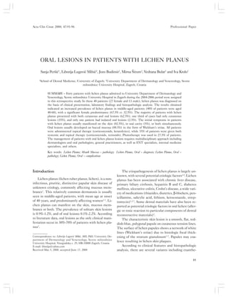
The estimated prevalence of oral lichen planus is
about 50% in patients with lichen planus. It commonly
presents as slightly raised whitish linear lines (Wick-
ham’s striae) on buccal mucosa, lips and occasionally
tongue. Squamous cell carcinoma has been reported in
patients with non-healing erosive oral lichen lesions.
Such lesions are rather therapy-resistant.
Oral lichen planus is usually recognized on time, and
can appear in several clinical forms including lichen ru-
ber planus (at the mucosal level), bullous type of oral
lichen planus (above the level of oral mucosa), and ero-
sive type of oral lichen planus (below the level of oral
mucosa)2
. Although with heterogeneous clinical pres-
entation, lichen planus has identical characteristic his-
topathologic features. Papular form of oral lichen planus
is characterized by slightly raised papules on buccal
mucosa, which may coalesce into Wickham’s striae. A
lace-like pattern on the buccal mucosa and the tongue
is referred to as the reticular form of lichen planus. The
plaque form of oral lichen planus is characterized by
plaque-like lesions, most frequently on the dorsum of
the tongue and gingiva, clinically resembling oral leu-
koplakia. Annular form arises from expansion of the mid-
dle portion of the reticular net, and is characterized by
erosive base with elevated hyperkeratotic edges2
.
Atrophic form is characterized by atrophic, inflam-
matory mucosa, most commonly on the dorsum of the
tongue, which eventually becomes smooth, without pa-
pillae (post-inflammatory absence of lingual papillae).
The bullous form is a very rare variant of oral lichen (2%
of patients), characterized by the formation of bullae of
oral mucosa, which rupture rapidly, thus forming residu-
al erosive and ulcerative lesions2
.
According to the World Health Organization criteria
from 1978, oral lichen planus is considered to be a pre-
malignant lesion which may evolve into squamous cell
Table 1. Clinical variants of lichen planus (according to Braun-Falco, 2000)3
Lichen planus exanthematicus multiple skin lesions, potential progression into erythroderma
Lichen planus localisatus solitary lesions on the neck, penis and lower trunk
Lichen planus linearis linear streaks of fused papules, potentially resembling nevus verrucosus
Lichen planus hypertrophicus localized on dorsa of the feet and lower extremities, severe itching
Lichen planus bullosus very rare, vesicles and bullae that rupture rapidly
Lichen planus erosivus erosions can evolve into painful ulcerations, slow healing
Lichen planus palmoplantaris localized on hands and feet, painful keratotic plaques, resistant to therapy
Lichen planus actinicus photoexposed areas, brown plaques
Lichen planus nodularis hyperkeratotic papules coalesce into larger nodules
Lichen planus annularis papules form hyperkeratotic rings with central clearing
Lichen planus atrophicans localized on lower extremities, well-defined lesions without hair or follicles
“Lichen planus – lupus variant of these two diseases
erythematosus” overlap
Lichen planus follicularis localized on flexural side of the extremities, hyperkeratotic, follicular papules
Lichen planopilaris skin lichen lesions on the scalp, more often in females, may evolve into cicatri-
Lichen planus unguium cial alopecia longitudinal ridges and thickening on the nails, pterygium
or “twenty nail dystrophy”
Table 2. Clinical forms of lichen planus according to mucosal lesions
Lichen planus mucosae oris Hyperkeratotic papules, striae, plaques, bullae and erosions on buccal mucosa, lips,
gingiva and tongue, precancerous lesions
Lichen planus genitalis Annular lesions on glans in male patients, hyperkeratotic lesions on labia minora in
female patients
Lichen planus perianalis Wickham’s striae, common cause of pruritus, biopsy needed
3. Acta Clin Croat, Vol. 47, No. 2, 2008 93
S. Peršiæ et al.: Oral lesions in patients with lichen planus
carcinoma1-3,14,15
. The prevalence of malignant transfor-
mation in longstanding, non-healing oral lichen planus
varies from 1.3% to 2.2%2
. Malignant transformation is
more common in atrophic, erosive and ulcerative forms
of oral lichen planus, in lesions situated on the ventral
side of the tongue or sublingual regions2,15
.
The characteristic histopathologic features of lichen
planus include hyperkeratosis, orthokeratosis with focal
thickening of the granular layer, acanthosis with inter-
cellular edema, epidermal sawtoothing with keratiniza-
tion of the basal layer, and liquefactive degeneration of
epidermal basal cells associated with a dense band of
lymphocytes in the papillary dermis1-3
.
The management of the disease involves topical and
systemic therapy16-19
. Vitamin A derivatives are largely
used, e.g., acitretin (25-50 mg/day), with efficient mor-
bistatic effect, or isotretinoin (0.3-0.5 mg/kg/day)3,18
.
Treatment can include systemic corticosteroids, e.g.,
prednisolone, 20-40 mg/day for several weeks (with grad-
ual dose reduction) or parenteral application of triamci-
nolone-acetonide1-3
. Phototherapy is applicable for wide-
spread skin lesions and PUVA bath for largely expanded
pruritic forms1,3
.
The majority of our patients were administered top-
ical corticosteroid therapy, frequently under occlusion.
Topical corticosteroid therapy is beneficial for oral li-
chen planus lesions; intralesional corticosteroid thera-
py can also be very effective (triamcinolone acetonide
diluted with topical anesthetic or saline solution at 1:5
ratio)1
. Oral lesions can also be treated with topical an-
tiseptics, antibiotics, antimycotics, vitamin A, retinoic
acid derivatives or keratolytics2
. Topical corticosteroid
lotions and solutions are used for scalp lesions.
Exanthematous lichen planus lesions may relapse in
two years, but typically resolve within the same period
of time3,19
. Some lichen planus variants are more thera-
py-resistant and tend to be more persistent, such as
hypertrophic lichen planus, oral lichen, lichen planopi-
laris and nail lichen planus1
.
The aim of the study was to obtain information re-
garding oral lesion prevalence in patients with lichen
planus, with comparison to other studies. The study in-
cluded data on age and sex, habits, lesion localization,
onset of symptoms and therapy. The results obtained in
this study were compared with the results reported by
other authors in order to achieve better treatment out-
come for patients with lichen planus in the future.
Material and Methods
This retrospective study included patients admit-
ted to University Department of Dermatology and Ve-
nereology, Sestre milosrdnice University Hospital in
Zagreb for lichen planus during the 2004-2006 period.
Medical data kept at Department were used for research
purposes. Preliminary evaluation was made for each pa-
tient, including medical history, clinical picture and bi-
opsy with histopathologic findings, in order to confirm
the diagnosis of lichen planus. Data on patient age and
sex, habits, lesion localization, onset of symptoms and
therapy were analyzed.
Results
Our study included 40 patients, 27 female and 13
male, diagnosed with lichen planus according to clini-
cal, laboratory and histopathologic findings. According
to the results obtained, lichen planus predominantly
manifested between the age of 40 and 60 (45%) (Table
3). Lichen planus was more prevalent in female (67.5%)
than in male patients (32.5%).
Table 3. Characteristic features in patients with lichen planus
Age (yrs) 20-40 7/40 (17.5%)
40-60 18/40 (45%)
60-80 15/40 (37.5%)
Sex M 13/40 (32.5%)
F 27/40 (67.5%)
Habits Smoking 11/40 (27.5%)
Alcohol 4/40 (10%)
Underlying Diabetes 5/40 (12.5%)
diseases mellitus
Hypertension 5/40 (12.5%)
Chronic liver 2/40 (5%)
disease
Primary localization Oral mucosa 2/40 (5%)
Skin 33/40 (82.5%)
Both 5/40 (12.5%)
We noticed that skin lesions preceded the onset of
oral lesions in 82.5% patients with lichen planus, while
oral lesions preceded cutaneous lesions in only 5% of
cases; a simultaneous onset of oral and skin lesions was
recorded in 12.5% of patients.
Skin lesions were associated with oral lesions in the
majority of patients with lichen planus (62.5%) (Table
4. 94 Acta Clin Croat, Vol. 47, No. 2, 2008
S. Peršiæ et al.: Oral lesions in patients with lichen planus
4). One third of our patients had isolated cutaneous le-
sions (35%), and only one patient had oral lesions alone
(2.5%). Oral lesions were most often localized on buccal
mucosa (88.5%) and most commonly presented as Wick-
ham’s striae (65.4%) (Table 5). All patients were treat-
ed by topical therapy (corticosteroids, keratolytics),
while approximately one half also received systemic ther-
apy (corticosteroids, retinoids) along with topical agents
(Table 6). Phototherapy was used in 27.5% of patients.
Discussion
Lichen planus is a noninfectious, pruritic, papular
skin disease, commonly affecting mucous membranes,
characterized by the appearance of characteristic smooth,
flat, reddish-blue polygonal papules, often with whitish
freckles (Wickham’s striae). The papules may coalesce,
resulting in lichen skin plaques with various clinical fea-
tures1-3
. We acquired important clinical experience from
the study through monitoring patients with lichen pla-
nus during the aforementioned period of time.
Lichen planus predominantly affects middle-aged
and elderly people; mean age at onset is 40 years, and
shows a female predominance, which is consistent with
literature data1,2
. Our study results indicated that lichen
planus primarily affected middle-aged, 40- to 60-year-
old individuals (45%). With respect to sex predilection,
lichen planus predominantly affected women (67.5% vs.
32.5%), which is also consistent with literature reports.
Table 6. Treatment of patients with lichen planus
Therapy Corticosteroids 34/40 (85%)
Topical Keratolytics 5/40 (12.5%)
Antimycotics 4/40 (10%)
Intralesional
1/40 (2.5%)
corticosteroids
Corticosteroids 3/40 (7.5%)
Systemic Antihistaminics 15/40 (37.5%)
Retinoids 5/40 (12.5%)
Other Phototherapy 11/40 (27.5%)
Lichen planus has been associated with chronic liv-
er disease, primary biliary cirrhosis, hepatitis B and C,
diabetes mellitus and other diseases1-3
. Results obtained
from our study revealed lichen planus association with
other diseases in several patients, most often in those
with diabetes (12.5%), hypertension (12.5%) and chronic
liver disease (5%).
There were patients with isolated cutaneous lesions,
sole oral lesions or both. According to the literature, the
incidence of lichen planus varies. There are data on the
incidence of lichen planus on the skin ranging from 0.9%
to 1.2%, and of oral lichen planus from 0.1% to 2.2%2
.
According to other sources, solitary oral lesions (with-
out skin manifestations) are common and appear in 30%
Table 4. Localization of skin and mucosal lesions in patients with lichen planus
Involvement Skin 14/40 (35%)
Oral mucosa 1/40 (2.5%)
Skin + oral mucosa 25/40 (62.5%)
Most common localization Trunk 19/39 (48.7%)
of skin lesions Limbs 32/39 (82%)
Scalp 3/39 (7.7%)
Time from diagnosis to therapy <1 month 5/40 (12.5%)
1 month – 6 months 22/40 (55%)
6 months – 1 year 5/40 (12.5%)
>1 year 3/40 (7.5%)
Table 5. Mucosal lesions in patients with lichen planus
Mucosal lesions Oral mucosa 26/40 (65%)
Genital mucosa 6/40 (15%)
Oral lesions Localization
gingiva 2/26 (7.7%)
buccal mucosa 23/26 (88.5%)
tongue 3/26 (7.7%)
Form
plaque 5/26 (19.2%)
papular 4/26 (15.4%)
reticular 17/26 (65.4%)
(Wickham’s striae)
erosive 3/26 (11.5%)
bullous 1/26 (3.8%)
5. Acta Clin Croat, Vol. 47, No. 2, 2008 95
S. Peršiæ et al.: Oral lesions in patients with lichen planus
to 70% of cases2
. According to dermatological practice
reports, solitary oral lesions are rare. Results obtained
in our study showed the majority of patients with lichen
planus to have cutaneous and oral lesions (62.5%), while
35% of patients presented with isolated skin lesions,
and only one patient had solitary oral lesion (2.5%).
While literature reports describe the occurrence of
oral manifestations without skin lesions in 30% to 70%
of patients with lichen planus, our results yielded a low-
er incidence of oral lichen planus (2.5% of patients). The
lower incidence of oral lesions in our dermatological prac-
tice could probably be attributed to the fact that the
majority of patients with oral lesions had been diagnosed
and treated exclusively by oral pathologists, whereas
those with skin lesions were managed by dermatologists.
With respect to the disease onset, we found oral le-
sions to have preceded the onset of skin lesions in 5% of
our patients, while simultaneous appearance of oral and
skin lesions occurred in 12.5% of our patients. Accord-
ing to literature reports, oral lesions appear in 50% of
patients with lichen planus, whereas our study showed
oral lesions in 65% of patients, predominantly localized
on buccal mucosa (88.5%), usually in the form of Wick-
ham’s striae.
In some patients, it took several months to up to
one year to reach the accurate diagnosis, pointing to the
necessity of timely recognition as an imperative for ap-
propriate treatment and prognosis. Several diagnostic
procedures such as thorough medical history, clinical
picture and histopathologic analysis may frequently be
needed to make an accurate diagnosis2
. On taking med-
ical history we pay due attention to personal habits, sys-
temic diseases or medications in order to identify the
etiology of the disease. Treatment of underlying disor-
ders improves the course and prognosis of lichen pla-
nus. It is important to specify clinical findings, such as
inflammation, hyperkeratosis, size of lesions, and type
of lesions (bullae, erosions) in order to establish an ac-
curate clinical diagnosis2
.
There are various therapeutic modalities available
for the treatment of lichen planus. All our patients were
treated with topical therapy, and about one half received
systemic therapy in adjunction to the respective topical
management. Phototherapy was used in 27.5% of our
patients. Topical corticosteroids and keratolytics under
occlusion were often applied. Systemic retinoids were
used in 12.5% and systemic corticosteroids in 7.5% of
our patients.
Oral lichen planus is a chronic disease characterized
by remissions and relapses. The prognosis of oral lichen
planus is unpredictable and depends upon the adequa-
cy of care provided to these patients1-3
. It is of vital im-
portance to treat underlying diseases through specialist
care. Various agents can be used to enhance keratiniza-
tion of the oral epithelium, in order to prolong the time
of remission2
. Malignant transformation of longstanding,
non-healing oral lichen planus is possible2
. Prevention
and timely recognition of premalignant oral lesions is
mandatory, with follow up, repeat oral lesion biopsies
(every 5-12 months) and retinoic acid derivative thera-
py2
.
In the management of patients with oral lichen pla-
nus lesions, multidisciplinary care including a derma-
tologist, oral pathologist, general practitioner, ENT spe-
cialist, internal medicine specialist and other special-
ized care, is of utmost importance and contributes greatly
to the improved prognosis of the disease.
References
1. DOBRIÆ I, et al. Dermatovenerologija. Zagreb: Grafoplast, 2005.
2. CEKIÆ-ARAMBAŠIN A, et al. Oralna medicina. Zagreb: Školska
knjiga, 2005.
3. BRAUN-FALCO O, PLEWIG G, WOLFF HH, BURGDORF
WHC, editors. Dermatology. 2nd
completely revised edition.
Berlin: Springer-Verlag, 2000.
4. BLACK MM. What is going on in lichen planus. Clin Exp
Dermatol 1977;2:303-10.
5. BELLMAN B, REDDY RK, FALANGA V. Lichen planus
associated with hepatitis C. Lancet 1995;346:1234.
6. WILSON E. On lichen planus. J Cutan Med Dis Skin 1869;3:
117-32.Fig. 1. Oral lesions in lichen planus (www.lindeberg.suite.dk).
6. 96 Acta Clin Croat, Vol. 47, No. 2, 2008
S. Peršiæ et al.: Oral lesions in patients with lichen planus
7. ELLGEHAUSEN P, ELSNER P, BURG G. Drug-induced
lichen planus. Clin Dermatol 1998;16:325-32.
8. POWELL FC, ROGERS RS, DICKSON ER, et al. An
association between HLA DR1 and lichen planus. Int J
Dermatol 1986;114:473-8.
9. FELLNER MJ. Lichen planus. Int J Dermatol 1980;19:71-5.
10. SHKLAR P. Erosive and bullous oral lesions of lichen planus.
Arch Dermatol 1968;97:411-6.
11. CRAM DL, KIERLAND RR, WINKELMANN RK. Ulcerative
lichen planus of the feet. Arch Dermatol 1966;93:692-701.
12. KATZENELLENBOGEN I. Lichen planus actinicus (lichen
planus in subtropical countries). Dermatologica 1962;124:10-
20.
13. PLOTNICK H, BURNHAM TK. Lichen planus and coexisting
lupus erythematosus versus lichen planus-like lupus
erythematosus. Clinical, histologic, and immunopathologic
considerations. J Am Acad Dermatol 1986;14:931-8.
Sažetak
PROMJENE NA SLUZNICI USNE ŠUPLJINE KOD BOLESNIKA S LIHEN PLANUSOM
S. Peršiæ, L. Lugoviæ Mihiæ, J. Budimir, M. Šitum, V. Bulat i I. Krolo
Ovo retrospektivno istraživanje obuhvatilo je bolesnike hospitalizirane zbog lihen planusa u Klinici za dermatovenerologiju
KB „Sestre milosrdnice“ u Zagrebu u razdoblju od sijeènja 2004. do kraja 2006. godine. Obuhvaæeno je 40 bolesnika (27 žena
i 13 muškaraca) koji su bolovali od lihen planusa, a dijagnoza je postavljena na temelju klinièko-laboratorijskih pretraga te
patohistološkog nalaza. Prema našim rezultatima lihen se najèešæe javljao u dobi od 40. do 60. godine (45%), èešæe kod žena
(67,5%) nego kod muškaraca (32,5%). Veæina bolesnika je istodobno imala promjene na koži i sluznici (62,5%), kod oko treæine
bolesnika promjene su bile iskljuèivo na koži (35%), dok je samo jedan bolesnik imao promjene iskljuèivo na sluznici usne
šupljine (2,5%). Bolest je najèešæe zapoèinjala na koži (82,5%), zatim na sluznici usne šupljine (5%), dok je istodobni poèetak
pojave promjena na sluznici usne šupljine i koži zabilježen u 12,5% bolesnika. Promjene usne šupljine najèešæe su bile
lokalizirane na bukalnoj sluznici (88,5%), uglavnom u obliku Wickhamovih strija (65,4%). Kod svih bolesnika se primijenila
lokalna terapija (kortikosteroidi, keratolitici), dok je 55% bolesnika uz lokalnu primilo i sistemsku terapiju (kortikosteroidi,
retinoidi). Kod 27,5% bolesnika je provedena fototerapija. S obzirom na to da se promjene kod lihen planusa èesto javljaju na
sluznici usne šupljine potreban je multidisciplinski pristup koji ukljuèuje suradnju specijalista dermatovenerologa, oralnog
patologa, lijeènika obiteljske medicine, ORL, internista i drugih.
Kljuène rijeèi: Lichen planus; Sluznica usne šupljine – patologija; Lichen planus oralni – dijagnostika; Lichen planus oralni – patologija;
Lichen planus oralni – komplikacije
14. CAMISA C, HAMATY FG, GAY JD. Squamous cell carcinoma
of the tongue arising in lichen planus: a case report and review
of the literature. Cutis 1998;62:175-8.
15. KRONENBERG K, FRETZING D, POTTER B. Malignant
degeneration of lichen planus. Arch Dermatol 1971;104:304-7.
16. CRIBIER B, FRANCES C, CHOSIDOW O. Treatment of
lichen planus. An evidence-based medicine analysis of efficacy.
Arch Dermatol 1998;134:1521-30.
17. OLIVER GF, WINKELMANN RK. Treatment of lichen planus.
Drugs 1993;45:56-65.
18. WOO TY. Systemic isotretinoin treatment of oral and cutaneous
lichen planus. Cutis 1985;35:385-93.
19. RAGAZ A, ACKERMAN B. Evolution, maturation and
regression of lesions of lichen planus. New observations and
correlation of clinical and histologic findings. Am J
Dermatopathol 1981;3:5-25.