Antifungal and Molecular aspect of Selenium Nanoparticles
1. Antifungal and Molecular aspect of Selenium Nanoparticles
Presented By: Anil Kumar
Centre for Nano-Sciences, CUG, Gandhinagar, Gujarat.
Email ID: kmr.nano@yahoo.com
Introduction: In recently more cases become in newspapers focused on fungus infections (Diseases) pose significant global health challenges, especially in view of the fact that the
emergence of resistant dermitophyte (keratins) fungus and the adverse side effects associated with prolonged use continue to slow down the application of effective antifungal
therapies. This makes imperative the need for the development of safe and potent alternatives to conventional antifungal drugs. In the present sequence of events and master plan on
Nano scale materials have emerged as novel antifungal agents for the possibilities offered by their unique chemical and physical properties. Selenium nano-particles have mainly been
studied for their antimicrobial potential may be active against several types of Fungus. Selenium (Se) is an essential trace element, but is toxic at high concentrations. Its toxicity is
related to the oxyanions selenate and selenite as they are water soluble and bioavailable, selenium against teratogenicity and cytotoxicity action. Sodium selenate inhibited the growth
of cultures of some keratin fungus. its known as reach selenocystein amino acid (21). The use of nonmetal nano-particles provides an interesting opportunity for novel antifungal
therapies. Since nonmetals may attack a broad range of targets in the fungus there is a lower possibility to develop resistance as compared to conventional antifungal. The present
study focuses on the development of methods for the production of selenium nanoparticles and on their use as antifungus therapeutics against pathogenic dermitophyte (keratins)
fungus.
Materials and Methods :Collection of soil samples From Athner village field of
Betul Districts located in vidarbh zone of M.P., India.
The Agrobacterium isolated were also grown on YEMA media The initial pH of the
medium was 6.5 in prasences of selenite (Na2SeO3) materials. Concentrations of
selenium were in approximate 20 µg mL−1. During growth in 2 4-48 hours, respectively
trasfer in both culture properly growth for Selenium Isolation.
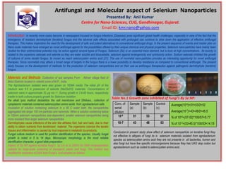
Table No.1 Growth zone inhibited of Fungi’s By Se NP:
the alkali lysis method destabilize the cell membrane and DNAses., collection of
cytoplasmic materials contained selenocystien amino acids from agrobacterium cells Conc. of Sample Sample Control Average(10-6)=51+53/2=52
incubation of solution containing selenium in a 90 C water bath, the nanoparticles serial (a) (b) (c)
aggregated into larger 100 nm particles and nanorods. When a solution containing below dilution Average(10-7)=43+48/2=45.5
to 100nm selenium nanoparticles size-dependent, smaller selenium nanoparticles being 10-6 51 53 57
more resistant than larger selenium nanoparticles
% of 10-6=(57-52)*100/57=5.77
Dermitophytes cause infections of the skin like athletes foot, hair and nails, due to their 10-7 43 48 53 % of 10-7=(53-45.5)*100/53=14.15
ability to obtain nutrients from keratinized material. The organisms colonize the keratin
tissues and inflammation is caused by host response to metabolic by-products.
Conclusion:in present study show effect of selenium nanoparticle on keratine fungi they
Fungal culture medium is used for positive identification of the species. Usually fungal
not effective to alltypes of fungi its is selenium materials isolated from agrobacterium
growth is noted in 5 to 14 days. Microscopic morphology is the most reliable
species as selenocystien amino acid they are not presente in all bacterilas, humen and
identification character, a good slide preparation
also fungi but have few specific microorganisms because they has UAG stop codon but
Aspect of Se NP against keratine fungi’s by Lin et al (2001) for DNA minipreparation
agrobacterium such as coded to selenocystien amino acid,
suitable for large-scale screening of transgenic plants and fungi. The method was
modified for extraction of DNA from Fungi’s and adopted as follows: