DIAGNOSIS OF A CERVICAL TUMEFACTION - F HERAN
•Download as PPTX, PDF•
1 like•437 views

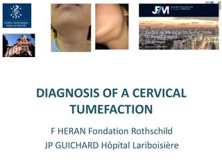
F HERAN Fondation Rothschild JP GUICHARD Hôpital Lariboisière

Recommended
More Related Content
What's hot
What's hot (20)
Similar to DIAGNOSIS OF A CERVICAL TUMEFACTION - F HERAN
Similar to DIAGNOSIS OF A CERVICAL TUMEFACTION - F HERAN (20)
More from JFIM - Journées Francophones d'Imagerie Médicale
More from JFIM - Journées Francophones d'Imagerie Médicale (20)
Recently uploaded
Recently uploaded (20)
DIAGNOSIS OF A CERVICAL TUMEFACTION - F HERAN
- 1. DIAGNOSIS OF A CERVICAL TUMEFACTION F HERAN Fondation Rothschild JP GUICHARD Hôpital Lariboisière
- 2. PATIENT Age : < 40 YO : inflammation, congenital > 40 YO : often malignant 80 % persistent cervical masses in adult = tumor Alcohol, smoker Known neoplasia LOCATION ONSET Sudden or progressive occurrence, other clinical signs (infection…) ELEMENTS THAT HELP US FOR THE DIAGNOSIS LOCATION : anatomy STRUCTURE (signal, density, echogenicity) CLINICAL DATA IMAGING DATA
- 3. Adenopathy Dermoïd cyst of the floor of the mouth Ectopic thyroïd gland Lipoma Schwannoma MIDDLINE LESIONS CYSTIC SOLID OR HETEROGENOUS Thyroglossal duct cyst
- 4. Frequent : 40 % of the cervical congenital malformations Failure to close the thyreoglossal canal (canal of Bochdalek) Developed along the migration path of the thyroïd gland Base of the tongue (foramen caecum) thyroïd gland Median lesion, centered on, above or under the hyoid bone body, Often lateralized if it is located under it. Cyst, with thin wall, sometimes compartimentalized Acute inflammatory episodes THYROGLOSSAL DUCT CYST (TDC)
- 5. Low CTD (under hyoïd bone)
- 7. DERMOID CYST
- 8. DERMOID: Development lesions found inside normal organs or tissues Defect in the fusion of the embryonic lateral mesenchymatic mass Inclusion of tissue Traumatism Floor of the mouth =1/5 of the dermoïd cyst of head and neck Upward displacement of the tongue, slurred speech, and difficulty in swallowing, If located between the mylohyoid muscle and the neck's cutaneous muscle: geniohyoid cyst (resembles a double chin) Surgery Baliga M, Shenoy N, Poojary D, Mohan R, Naik R Epidermoid cyst of the floor of the mouth . Natl J Maxillofac Surg. 2014 Jan;5(1):79-83. Patient du Pr Guy Princ
- 9. LATERAL LESIONS Plunging ranula Cystic hemolymphangioma Branchial cleft cyst Lymphocele Thymic cyst Laryngocèle CYSTIC
- 10. MUCOIDE CYST, SALIVAY CYST, RANULA Sublingual gland salivary cyst due to retention within a sublingual or accessory gland. Superficial swelling over the floor of mouth, bluish appearance (frog’s belly), May extend through or around the mylohyoid muscle complex = plunging ranula which presents as a neck lump Former trauma or inflammatory episods. US : Cyst CT scan : hypodense with thin walls. MRI : cystic, variation of the signal depending on the composition (protids…)
- 11. RANULA
- 12. PLUNGING RANULA
- 13. Plunging ranula (salivary cyst)
- 17. Complex vascular malformation, association of abnormal lymphatic and venous vessels. First clinical signs before 20 YO Multiloculated ETN infections hemorrhage, new cysts, acute increase of size Treatment Punction of the hematoma Embolization, sclerosis CYSTIC LYMPHANGIOMA et HEMOLYMPHANGIOMA IRM 3DT2 FATSAT : extension
- 18. MALFORMATIONS OF THE BRANCHIAL CLEFTS Abnormality in the obliteration of the clefts in between the branchial arches 4 types Cyst Fistula Both Complications: mostly infections.
- 19. Most frequent (95 %) Often young adult (20 - 30 YO), episode of infection, Fistula: very seldom Ovoid, well demarcated, thin wall (thickening if inflammation) Content: depends on its composition Infection, inflammation Between Submandibulary gland Sternocleidomastoïd muscle Carotid artery/ jugular vein SECOND BRANCHIAL CLEFT CYST (Amygdaloïd cyst) Harnsberger
- 22. LYMPHOMA
- 23. LATEROCERVICAL MASS EVEN IF IT SEEMS TO BE A TYPICAL CYST DIFFUSION INJECTION
- 24. FIRST BRANCHIAL CLEFT CYST Type I superficial periauricular cyst External Auditory Duct Kyste type II Kyste type I Rare (<10 %) Type II deep kyste (or fistula) Parotid area Harnsberger Harnsberger
- 25. . Fastenberg J, Nassar M First Branchial Cleft Cyst. N Engl J Med.2016 Oct 20;375(16):e33 Otoscopy keratin debris and purulence
- 26. Very seldom, located in the posterior cervical space Fistula with the pyriform sinus Deep infections Between Carotid artery/ jugular vein Sternocleidomastoïd muscle THIRD BRANCHIAL CLEFT CYST Harnsberger
- 27. Very seldom Lower anterior neck Between pyriform sinus and left thyroid lobe Recurrent abscesses or suppurative thyroiditis Harnsberger FORTH BRANCHIAL CLEFT CYST (pyriform sinus fistula)
- 28. Schmidt K et al Rapidly enlarging neck mass in a neonate causing airway compromise. Proc (Bayl Univ Med Cent).2016 Apr;29(2):183-4. neck mass in a neonate rhabdomyosarcoma teratoma venolymphatic malformations, fibromatosis colli, branchial cleft cyst
- 29. OTHERS LYMPHOCELE OF NECK Benign lymph-filled cysts Leaking disrupted lymphatic channels Along the jugular lymphatics Supraclavicular fossa THYMIC CYST Benign lymph-filled cysts Leaking disrupted lymphatic channels Along the jugular lymphatics Supraclavicular fossa
- 30. LATERAL LESIONS Adenopathy Salivary lesion (parotid or submandibular gland) lesion Thyroïd gland tumor Schwannoma Paraganglioma Lipoma Malignant tumor (lymphoma, sarcoma, extension of a pharyngolaryngeal tumor) Fibromatosis of the head and neck SOLID
- 31. SUBMANDIBULAR GLAND PEOMOPHIC ADENOMA dynamic contrast- enhanced T1-weighted MRI
- 33. LIPOMA
- 34. Vascular mass splaying ECA and ICA Rapid contrast uptake dynamic study Salt and pepper Between Carotid artery/ jugular vein Sternocleidomastoïd muscle CAROTID BODY PARAGANGLIOMA
- 37. FIBROMATOSIS (DESMOID TUMOR) Aggressive, ill-defined or sharp margins Slow growing, painless Strong enhancement Most cases sporadic May follow surgery, trauma In adults, associated with polyposis syndromes (Gardner, familial adenomatous polyposis) Complete resection
- 38. SCHWANNOMA
- 40. LATERAL LESIONS SOLID AND CYSTIC Infections Malignant tumor Necrotic adenopathy Cystic schwannoma Vascular lesions Malformations Carotid artery aneuvrysm
- 42. SARCOMA
Editor's Notes
- Classification de Bailey