Adult Diagnosis of Swyer James Mcleod Syndrome
•
1 like•374 views

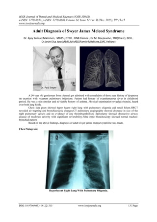
A 50-year-old man presented with a 3-year history of dyspnea on exertion and recurrent pulmonary infections. He had a history of exanthematous fever in childhood. Physical exam revealed rhonchi in both lung fields. Chest X-ray showed a hyperlucent right lung with pulmonary oligemia and a small hilum. HRCT revealed air trapping and bronchiectatic changes. CT pulmonary angiography showed decreased size of the right pulmonary vessels with no evidence of thromboembolism. Spirometry showed obstructive airway disease with reversibility. Bronchoscopy showed a normal tracheobronchial pattern. Based on these findings, the patient was diagnosed with adult Swyer James Mac

Recommended
Recommended
More Related Content
What's hot
What's hot (9)
Similar to Adult Diagnosis of Swyer James Mcleod Syndrome
Similar to Adult Diagnosis of Swyer James Mcleod Syndrome (20)
More from iosrjce
More from iosrjce (20)
Recently uploaded
Recently uploaded (20)
Adult Diagnosis of Swyer James Mcleod Syndrome
- 1. IOSR Journal of Dental and Medical Sciences (IOSR-JDMS) e-ISSN: 2279-0853, p-ISSN: 2279-0861.Volume 14, Issue 12 Ver. II (Dec. 2015), PP 13-15 www.iosrjournals.org DOI: 10.9790/0853-141221315 www.iosrjournals.org 13 | Page Adult Diagnosis of Swyer James Mcleod Syndrome Dr. Ajoy Samuel Mammen, MBBS , DTCD , DNB trainee , Dr.M. Deepaselvi , MD(Chest), DCH , Dr.Jesin Elsa Jose,MBBS,M.MED(Family Medicine,CMC Vellore) A 50 year old gentleman from chennai got admitted with complaints of three year history of dyspnoea on exertion with recurrent pulmonary infections. Patient had history of exanthematous fever in childhood period. He was a non smoker and no family history of asthma. Physical examination revealed rhonchi, heard over both lung fields. Chest skia gram showed hyper lucent right lung with pulmonary oligemia and small hilum.HRCT revealed air trapping and bronchiectactic changes.CT pulmonary angiography showed decrease in size of the right pulmonary vessels and no evidence of any thromboembolism. Spirometry showed obstructive airway disease of moderate severity with significant reversibility.Fibre optic bronchoscopy showed normal tracheo- bronchial pattern Based on the above findings, diagnosis of adult swyer james mcleod syndrome was made. Chest Skiagram: Hyperlucent Right Lung With Pulmonary Oligemia.
- 2. Adult Diagnosis of Swyer James Mcleod Syndrome DOI: 10.9790/0853-141221315 www.iosrjournals.org 14 | Page Hrct Chest Hrct Chest Relieved Air Trapping And Bronchiectic Changes. Contrast Ct Showing Narrowing Of Right Pulmonary Artery Ct Pulmonary Angiography Showed Decrease In The Size Of Right Pulmonary Vessels And No Evidence Of Thromboembolism.
- 3. Adult Diagnosis of Swyer James Mcleod Syndrome DOI: 10.9790/0853-141221315 www.iosrjournals.org 15 | Page