Optometer.pptx

Optometers are subjective if the patient judges the clarity of the retinal image or objective when the machine or examiner does it. Subjective optometers control the focus of the retinal image and are used to determine when a target is conjugate to the retina. Objective optometers measure the defocus or disconjugacy of the retinal image and the stimulus target. There are also measures of accommodation that measure characteristics of the crystalline lens such as front surface curvature via the third Purkinje image. SIMPLE OPTOMETER: The simple optometer is a plus lens placed in the anterior focal plane or spectacle plane of the eye. The virtual image of objects placed before the lens can be imaged from infinity to close to the spectacle plane, simply by moving the target from the anterior focal plane of the lens to the lens plane respectively. The virtual image distance is calculated from the Gaussian equation 1/u + F = 1/v where: u= object distance, v=image distance, F = focal power One problem with the simple optometer in the measurement of accommodation is that the image increases in size with proximity so that you have both size and blur cues to accommodation. BADAL OPTOMETER: Invented by Jules Badal in 1876, who is French scientist The Badal optometer utilizes a plus lens placed so that its posterior focal plane is coincident with the anterior focal plane of the eye. This instrument keeps image size constant while varying target distance and stimulus to accommodation. The optical system is telecentric in both the object and image space, that is, the rays are parallel. NAGEL OPTOMETER: The Nagel optometer is based on a similar concept. Here ,a plus lens whose posterior focal plane is coincident with the nodal point of the eye. It also keeps image size constant with changing object distance. SUBJECTIVE OPTOMETERS For Δz = 0, the light emerging from the lens is collimated (i.e. object at infinity) For Δz > 0, the light emerging from the lens is diverging. The object appears in front of eye, so will be in focus for myopes. For Δz < 0, the light emerging from the lens is converging. The virtual image is behind the eye, so will be in focus for hyperopes. STIGMATOSCOPY: Combine of Simple and Badal lens optometers with various visual stimuli to enhance the sensitivity of subjective measures by improving sensitivity to blur detection. The stigmascope enhances blur perception with a small point source as the target viewed through the optometer lens. When the image of the point source is seen clearly and sharply, it is optically conjugate to the fovea. At the same time, this image may be introduced so that the eye can be fixating some other target which acts as the stimulus to accommodation such as Snellen chart. Bracketing the measures of positive and negative blur of the stigma allows you to estimate the accommodative response. SCHEINER’S PUPIL: Scheiner developed a double pupil that causes images viewed through it to app

Recommended
More Related Content
What's hot
What's hot (20)
Similar to Optometer.pptx
Similar to Optometer.pptx (20)
More from Indrani Sirivella
Recently uploaded
Recently uploaded (20)
Optometer.pptx
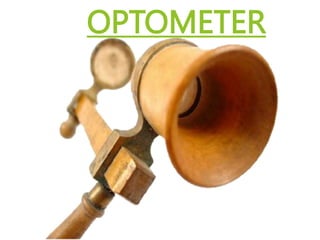
- 1. OPTOMETER
- 2. HISTORY William Porterfield invented the optometer, which was useful for measuring sight It measures the limits of distinct vision , and determining with great exactness the strength and weakness of sight. This is the root name for the profession of optometry.
- 4. OPTOMETER PRINCIPLE It involved a convex lens placed in front of the eye at its focal length from the eye (or the spectacle plane) and the movable target is viewed .t Vergence of light in the focal of lens is linearly related to the displacement of target. the lens
- 5. TYPES OF OPTOMETERS: Optometers are subjective if the patient judges the clarity of the retinal image or objective when the machine or examiner does it. Subjective optometers control the focus of the retinal image and are used to determine when a target is conjugate to the retina. Objective optometers measure the defocus or disconjugacy of the retinal image and the stimulus target. There are also measures of accommodation that measure characteristics of the crystalline lens such as front surface curvature via the third Purkinje image.
- 6. STIMULUS OPTOMETERS SIMPLE OPTOMETER: The simple optometer is a plus lens placed in the anterior focal plane or spectacle plane of the eye. The virtual image of objects placed before the lens can be imaged from infinity to close to the spectacle plane, simply by moving the target from the anterior focal plane of the lens to the lens plane respectively. The virtual image distance is calculated from the Gaussian equation 1/u + F = 1/v where: u= object distance, v=image distance, F = focal power One problem with the simple optometer in the measurement of accommodation is that the image increases in size with proximity so that you have both size and blur cues to accommodation.
- 7. BADAL OPTOMETER: Invented by Jules Badal in 1876, who is French scientist The Badal optometer utilizes a plus lens placed so that its posterior focal plane is coincident with the anterior focal plane of the eye. This instrument keeps image size constant while varying target distance and stimulus to accommodation. The optical system is telecentric in both the object and image space, that is, the rays are parallel.
- 8. NAGEL OPTOMETER: The Nagel optometer is based on a similar concept. Here ,a plus lens whose posterior focal plane is coincident with the nodal point of the eye. It also keeps image size constant with changing object distance.
- 9. SUBJECTIVE OPTOMETERS For Δz = 0, the light emerging from the lens is collimated (i.e. object at infinity) For Δz > 0, the light emerging from the lens is diverging. The object appears in front of eye, so will be in focus for myopes. For Δz < 0, the light emerging from the lens is converging. The virtual image is behind the eye, so will be in focus for hyperopes.
- 10. STIGMATOSCOPY: Combine of Simple and Badal lens optometers with various visual stimuli to enhance the sensitivity of subjective measures by improving sensitivity to blur detection. The stigmascope enhances blur perception with a small point source as the target viewed through the optometer lens. When the image of the point source is seen clearly and sharply, it is optically conjugate to the fovea. At the same time, this image may be introduced so that the eye can be fixating some other target which acts as the stimulus to accommodation such as Snellen chart. Bracketing the measures of positive and negative blur of the stigma allows you to estimate the accommodative response.
- 11. SCHEINER’S PUPIL: Scheiner developed a double pupil that causes images viewed through it to appear double unless the image is in focus. When the Scheiner pupil is combined with a Badal optometer it is called a coincidence optometer. With a coincidence optometer, the stigmascope is added to a two hole disc. When the point source is not in focus on the fovea, it will not only be blurred but will also be doubled. The greater the separation of the apertures in the Scheiner disc, the greater the separation of the images on the retina for a given nonfocal condition, thus increasing the precision.
- 12. It can be use dto differentiate myopia from hypermetropia.
- 13. OBJECTIVE OPTOMETERS: An objective optometer consists of two parts: an optical system to throw a bright image on the retina of the subject and an ophthalmoscope which, in being focused on the retinal image, discloses the state of refraction of the eye. Most autorefractors used clinically are objective optometers. Many use the Scheiner principal in which the two beams of the double Scheiner pupil are alternately interrupted (chopped) to facilitate the handling of the electronic signal
- 14. AUTOREFRACTOMETER: Autorefractors are devices that automatically and objectively measure refractive error in patients. They usually have very repeatable measurements, tend to be slightly off from a patient’s subject refraction. Therefore, they are good for clinical studies to track changes in refraction and as a starting point for a subjective refraction.
- 15. REFERENCE: [1] Benjamin, W., Benjamin, W. and Borish, I. (2006). Borish's Clinical Refraction. 2nd ed. Saintt Louis: Elsevier Health Sciences. [2] Wp.optics.arizona.edu. 2019 [cited 18 August 2019]. Available from: https://wp.optics.arizona.edu/visualopticslab/wp- content/uploads/sites/52/2016/08/Class08_08.pdf