Evolution of tmj and its development
•
5 likes•1,685 views

The Indian Dental Academy is the Leader in continuing dental education , training dentists in all aspects of dentistry and offering a wide range of dental certified courses in different formats.for more details please visit www.indiandentalacademy.com

Recommended
More Related Content
What's hot
What's hot (20)
Viewers also liked
Similar to Evolution of tmj and its development
Similar to Evolution of tmj and its development (20)
More from Indian dental academy
More from Indian dental academy (20)
Recently uploaded
Recently uploaded (20)
Evolution of tmj and its development
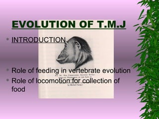
- 1. EVOLUTION OF T.M.J INTRODUCTION Role of feeding in vertebrate evolution Role of locomotion for collection of food
- 2. Early stages in the development of functional activity of the jaws D ism e m b e rin g (c a rn ivo u ro u s ca n in e s) C ro p p in g (h e rb ivo ro u s in cis o rs ) T e a rin g G n a w in g (ro d e n t in c iso rs) S licin g (ca rn ivo ro u s ch e e k te e th ) G rin d in g (ro d e n t a n d h e rb ivo ro u s ch e e k te e th ) C u ttin g C ru sh in g co m m in u tio n P ie rc in g P re h e n tio n
- 3. Nomenclature Jaw joint Primitive jaw joint or Reptilian jaw joint or Quadrate articular articulation. Mammalian type of jaw joint or Dentary- squamosal articulation.
- 4. Evolutionary history of dentary- squamosal joint Hinge development in the first viceral arch. Mobility of upper jaw and lower jaw. Dentary squamosal contact. Influence of muscle activity upon jaw development. Development of inter -articulating disk.
- 5. Skulls Of Earlier And Later Mammal-like Reptiles From South Africa
- 6. Progressive upgrowth of the dentary bone of the lower jaw to form a new joint with the skull.
- 7. Development of Inter- articular disk
- 8. Variations Of Functional activity Of T.M.J. PREHENSION Ability to grasp and prevent the escape of a slippery prey. Interlocking canines Development of hinged articulation Development of preglinoid process laterally and post-glinoid process mesially prevents dislocation.
- 10. TEARING Simplest way of comminution of food to make swallowing possible and enable digestion to occur. PULL developed by neck musculature. Greater risk of DISLOCATION of condyle ,leads to development of preglenoid process in carnivorous animals.
- 11. CRUSHING IN PRIMITIVE MAMMALS Ability to bring post canine teeth together. GIANT PANDA-A herbivorous carnivorous Molar teeth richly covered with cusp and tubercles. Transverse cylinder rotating with a hinge movement in a deep groove. Articular disk thin. Level of occlution is lower than the articulation which helps in antero- posterior GRINDING movement to the closing movement. This represent the adaptation for herbivorous diet within the carnivorous.
- 12. CUTTING IN THE CARNIVORE Condyle is cylindrical and at the same level as the occlusal plane. Coronoid process is very large and projects vertically.It provide large area of insertion for the temporalis muscle(prevent dislocation). Well developed masseter muscle with large depression on the lat. side of ramus. Prominent preglenoid and postglenoid processes. Interarticular disk present with upper and lower synovial cavities. Joint capsules and ligaments permits slight lat. Translatory movement.
- 13. CUTTING IN RODENTS Sliding postero-anterior movement of condyle with relatively little hinge like rotation present. Inter-articular disk well developed.It helps in gnawing activity of rodents. GRINDING IN RODENTS GNAWING: Lower jaw shifted forward to bring the incisors together. SHIFTING: Mandible shift posteriorly to bring molars in contact. MASTICTION: Movements bring molars together without hampering the incisors.
- 14. Study of homonids for feeding mechanisms. Two forms of AUTRALOPITHECINES have been recognized: GRACIL FORM: Typified by Australopithicus africanus,there is moderately developed temporalis and masseter muscles. This is suitable for omnivorous diet. ROBUST FORM: Typified by Australopithicus bosei,there is great increase in size of masseter.This is suitable for herbivorous diet.
- 15. Development of Tempromandibular Joint T.M.J - bilateral synovial diarthrosis - one freely movable joint on each side,left and right surrounded by a capsule whose internal lining produces a viscid synovial fluid.
- 16. Prenatal Condylar Growth 6TH WEEK :condensation of mesenchyme develops lateral to meckel’s cartilage.The development of this mesenchymal condensation within one week,a complete memberanous bony plate is formed parallel and locally envoloping the bilateral meckel’s cartilage. At 10TH WEEK, the bony mandible has recognizable form and meckel’s cartilage starts to be resorbed. At the same time condylar field develop at the cranial end of mandible.
- 17. At 12th week the condylar process are clearly recognizable and secondary cartilage production will begun. At 14th week endochondral ossification of new cartilage will start centrally in the ramus proceeding upwards. From 20th week , subsequent replacement by a bone,creating the typical picture of growing mandibular condyle.
- 18. POST-NATAL GROWTH OF CONDYLE Condyle grows in harmony with the disk and glenoid fossa as the tubercle undergoes development vertically at the temporal region. Condyle relocate in posterior direction by deposition and resorbtion in anterior aspect(V-PRINCIPLE). Vertical growth of condyle is by endochondral bone growth and bulk by intramemberanous bone growth.
- 19. Temporal Tubercle Growth PRE NATAL GROWTH In 8th month human foetus, a straight Zygomatic arch occurs before birth. Mandible slides forward and backwards horizontally .this situation will change quickly after tooth eruption has started. This helps the infant for breast feeding.
- 20. Post-natal development of temporal tubercle During 1st year tubercle starts developing.Temporal bone anterior to the condyle is lowered relative to posterior part.it is characterised by the increased slope. When primary teeth develops,they permit forceful chewing action,slope become steeper and attain 10%of the normal adult size. When 1st premolar and ant. Teeth – 70% of adult size. When molars erupt – 90%. Total change post-natally amounts to about 40 degrees.
- 21. Class I Class II Class III Fulcrum Effort Resistance Moment = load * distance
- 22. To overcome resistance 1. More Effort 2. Lengthening effort arm 3. Shortening resistance arm Advantages of Class III lever 1.Less musculature lenthening during movements 2. Easy and faster prehension at canines Disadvantage 1. Restriction on effort arm