1. “Generation of the Murine Astrocyte Specific Hypoxia Inducible Factor Knockout Model”
Mary Lin, Ying Wang, PhD, Weipin Chan, PhD, and Thomas Floyd, MD
Department of Anesthesiology, Stony Brook University
Department of Anesthesiology, Stony Brook University
Abstract
During major surgery, patients are often challenged with acute anemia, hypotension, and hypoxia from cardiac or respiratory failure. The prominent role of hypoxia in postoperative cognitive dysfunction
(POCD) has received little attention, and should not be ignored. Hypoxia, HIF and the process of cognitive aging appear to be tightly linked.
In the CNS, astrocytes play a prominent role in supporting synaptic plasticity as well as serving a major source of hypoxia inducible factors (HIF) including both HIF-1α and HIF-2α. These HIF factors are
transcriptional factors and appear to regulate an intrinsic neuroprotective response to hypoxia, which may be impaired during aging. HIF1-a is predominantly synthesized within neurons while HIF2-α is
predominantly synthesized in glia cells, specifically astrocytes. One target gene erythropoietin (EPO) plays a neuroprotective role in the brain and upregulates in response to hypoxia through the transcriptional
HIF. In this research, we hypothesize that astrocytic HIF supports learning, memory, and synaptic plasticity during acute hypoxic stress. We will test this hypothesis using astrocyte specific GFAP-Cre+/HIF-1α
fl hom and GFAP-Cre+/HIF-2α fl hom murine model created with the Cre/LoxP system.
Background
The HIF transcription system is the master
regulator of the cellular response to hypoxia.
HIF is a heterodimeric complex composed of
HIF-1α and HIF-1β subunits which are both
constitutively expressed under normal oxygen
levels. At normoxia, HIF-1α is continuously
and rapidly degraded when HIF-1α subunits
are hydroxylated by prolyhydroxylases (PHDs
1-3). However, under hypoxic conditions the
activity of the prolyhydroxylases is
suppressed, allowing levels of HIF to rise.
Stabilized HIF-1α translocates to the nucleus
to dimerize with ARNT, resulting in the
transcription of hundreds of hypoxiaresponsive genes. These genes are focused on
cell survival and include glucose transport
(Glucost transporter-1, GLUT-1), glycolysis
(phosphoglycerate kinase-1, PKG-1), oxygen
transport (erythropoiesis-erythropoietin,
EPO), and angiogenesis (Vascular Endothelial
Growth Factor, VGEF). Effective hypoxia
sensing and adaptation to hypoxia is critical
for cellular function in all organs, including
the brain. In the brain, HIF-1α expression is
induced by hypoxia in neurons, astrocytes,
ependymal and endothelial cells. HIF-2α
expression is induced in glia, particularly
within astrocytes, as well as capillary
endothelial cells.
Method
Two murine models with astrocyte specific knock outs of HIF-1α and HIF-2α were generated for this
study. Mice expressing Cre recombinase under the control of astrocyte specific glia fibrillary acidic
protein (GFAP) promoter were crossed with mice homozygous for LoxP-flanked alleles of HIF-1α and
HIF-2α respectively. The resulting GFAP-Cre+/HIF-1α fl/fl and GFAP-Cre+/HIF-2α fl/fl were identified
Results
DNA recombination assay revealed that the
deletion of HIF-1α and HIF-2α are specific in the
Brain.
through PCR using the genomic DNA extracted from tail biopsies. gDNA was also extracted from the
brain as well as liver and kidney to be used for DNA recombination assays. Mice were then exposed to
hypoxic conditions and the expression of key molecules involved in the hypoxic response, and the HIF
target gene EPO is being investigated.
Figure 2. Genotyping using tail biopsies
Figure 3. DNA recombination assay of HIF-1α
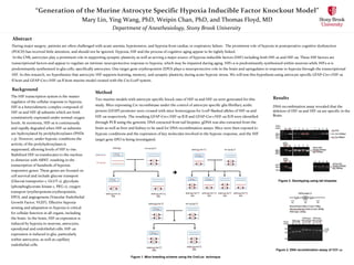
Figure 1. Mice breeding scheme using the Cre/Lox technique