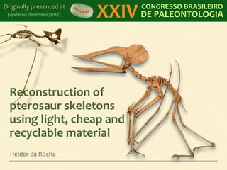
Reconstruction of Pterosaurs using XPS
•
2 likes•555 views

Presented at the 24th Brazilian Congress of Paleontology (2015). Originally in Portuguese. Translated and updated.

Recommended
Recommended
More Related Content
More from Helder da Rocha
More from Helder da Rocha (20)
Recently uploaded
Recently uploaded (20)
Reconstruction of Pterosaurs using XPS
- 6. Anhanguera piscator (2012) Based on publication by Kellner & Tomida, 2000
- 7. Tupandactylus imperator (2013) Based on specimen from Museu de Ciências da Terra (Rio de Janeiro)
- 12. Tapejara wellnhoferi (2014) Museu de Dinossauros in Peirópolis, in Brazil Instituto Geológico de Lisboa, in Portugal
- 14. Tropeognathus mesembrinus (2016) Giant 8 meter wingspan pterosaur (3,5kg) - Museu de São José dos Campos (São Paulo), Brazil It was installed in December 2016, but exhibit has not opened yet
- 19. Techniques Plaster, resin, fiberglass Ideal for casts Wax and electrotyping Wood, paper and card Also recyclable; safer construction process Rock (subtractive sculpture), metal (aditive), etc. Digital 3D sculpture + 3D printing in metal, resin, PVC ... Mixed techniques Kai-Xiang Xhong Philippe Pasqua Chicago O´Hare by Field Museum (fiber-glass) (recycled cardboard) (chrome) Jeffrie Parker (3D printer) Other
- 31. + protection Liquid epoxi + matte varnish It's optionall! Pros: increases material resistance Cons: loss of detail and reflective effect
- 32. Examples 1. Construction of Tupuxuara leonardii 2. Construction of Tapejara wellnhoferi (Download the complete presentations and read detailed descriptions of each process at imaginosaurus.wordpress.com)
- 33. Research: Tupuxuara Photos of IMCF 1502 (IWAKI, JP): Everything except pelvis, carpals, tarsals, fingers, dorsal vertebrae and pre-pubis Other sources: publications and specialized opinions Details are limited by precision of photos This is an excerpt from the presentation: Making of Tupuxuara leonardii See more in http://www.slideshare.net/helderdarocha/the-imaginary-pterosaur-makingof-tupuxuara-leonardii
- 37. Cut out
- 43. Compare
- 46. Notarium
- 50. Bone ends
- 51. Ulna: roll
- 52. Research: Tapejara 1. SMNK PAL 1137 Tapejara wellnhoferi (Germany) 2. AMNH 24440 Tapejara wellnhoferi (USA) 3. IMCF 1061 Tapejara wellnhoferi (Japan) 4. MN 6595-V Tapejara wellnhoferi (holotype) + 8 sources osseous labyrinth is well defined, leaving a deep depression on the endocast around the floccular lobes. The preserva- tion of a small portion of the lateral semicircular canal suggests that this structure would have completely sur- rounded the flocculus. Quadrate The right quadrate is complete but lies unfused to the other elements of the skull. The bone is formed by two branches, orientated dorsoventrally and mediolaterally, and con- nected by a thin diagonal laminae of bone to give the element an L-shaped appearance in its posterior aspect (Fig. 2; plate 3). The dorsoventrally directed branch is 2.4 times the length of the horizontal branch; the dorsal ter- mination of the former being smooth and well rounded in posterior view and preserving an oval shaped cross section. The ventrolateral margin of the bone forms the articular facet for the mandible where a pronounced sulcus runs in an anteromedial direction. A left quadrate of a comparable size to that described above is also present in the concretion but the vertical branch is broken only just dorsal to its base. Mandible The mandible is edentulous and preserves a short sym- physis only 44 mm in length, formed by the completely co-ossified contralateral rami. The dorsal face of the symphysis is transversely concave and is directed antero- ventrally at an angle of 18°, starting at a point 45 mm posterior of the rostral tip (Fig. 2; plate 4B). The ventral margin is almost straight but forms a sagittal crest reaching its maximum depth at the symphysis. In its dorsal aspect the bone appears as an elongate triangle, three times as long as it is wide (Fig. 2; plate 4) while the posterior section containing the articular facet is missing. Cervical vertebrae Four procoelous vertebra, identified as elements of the cervical series, are observed in various states of preserva- tion (Fig. 4). Two of these are attributed to the middle cervical column (Fig. 4f–o) while a third is identified as the 7th cervical. The remaining element represents the isolated axis (Fig. 4a–e). Fig. 4 Cervical elements of Tapejara wellnhoferi, SMNK PAL 1137, where: A–E axis in lateral (A), anterior (B), posterior (C), dorsal (D) and ventral view (E); F–J cervical vertebra in lateral (F), anterior (G), posterior (H), dorsal (I) and ventral view (J); K–O, cervical vertebra in lateral (K), anterior (L), posterior (M), dorsal (N) and ventral view (O); P–T, 7th cervical vertebra in lateral (P), anterior (Q), posterior (R), dorsal (S) and ventral view (T). f foramen, nc neural canal, ns neural spine, pe postexapophysis, pre preexapoph- ysis, pz postzygapophysis, prz prezygapophysis, vc vertebral condyle K. Eck et al. This is part of the presentation: Making of Tapejara wellnhoferi See more at http://www.slideshare.net/helderdarocha/the-imaginary-pterosaur-making-of-tapejara-wellnhoferi
- 53. Scaling Replica is 25% larger than reference size of young individual 53
- 54. Bone construction Fully documented at http://imaginosaurus.wordpress.com/2013/08/28/imaginary-pterosaur-7-finished/ 54
- 55. SMNK PAL 1137 limestone slab Approximate reconstruction (using bones of one pterosaur) 55
- 56. Neurocranium version 1 replica of SMNK PAL 1137 http://imaginosaurus.wordpress.com/2013/07/06/unfinished-tapejara-skull/ 56
- 57. Cervical vertebrae Sources: 4 specimens (SMNK, IMCF Tapejara and Tupuxuara, AMNH) 57
- 58. Cervical vertebrae http://imaginosaurus.wordpress.com/2013/07/15/tapejara-cervical-vertebrae/ 58
- 63. Workshop During the 24th. Brazilian Congress of Paleontology using the techniques described in this presentation 12 hours (3 days) - 15 participants XPS replicas made by the students during the workshop
- 65. helder.darocha@gmail.com +55 11 992 910 567 Thank you!