BIOMIMESYS® Adipose tissue
•
0 likes•66 views

BIOMIMESYS® Adipose tissue represents a new generation of mimetic Hydroscaffold™ for 3D culture of adipocyte and adipocyte-like cells. Available in a ready-to-use format, it enables the culture of adipocytes and adipocyte-like cells under physiological conditions that are representative of the microenvironment found in adipose tissue. The highly porous nature of the scaffold allows the rapid uptake of nutrients, oxygen, etc. into the cells to create a reproducible study model for all downstream analyses used with 3D adipocyte cultures.

Recommended
Recommended
More Related Content
What's hot
What's hot (19)
Similar to BIOMIMESYS® Adipose tissue
Similar to BIOMIMESYS® Adipose tissue (20)
More from HCS Pharma
More from HCS Pharma (19)
Recently uploaded
Recently uploaded (20)
BIOMIMESYS® Adipose tissue
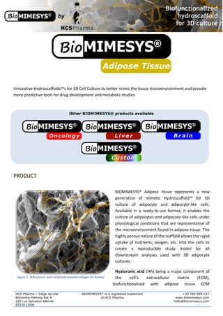
- 1. HCS Pharma – Siège de Lille Biocentre Fleming Bat A 250 rue Salvador Allende 59120 LOOS BIOMIMESYS® is a registered trademark of HCS Pharma +33 769 999 137 www.biomimesys.com hello@biomimesys.com Innovative Hydroscaffolds™s for 3D Cell Culture to better mimic the tissue microenvironment and provide more predictive tools for drug development and metabolic studies Other BIOMIMESYS® products available PRODUCT BIOMIMESYS® Adipose tissue represents a new generation of mimetic Hydroscaffold™ for 3D culture of adipocyte and adipocyte-like cells. Available in a ready-to-use format, it enables the culture of adipocytes and adipocyte-like cells under physiological conditions that are representative of the microenvironment found in adipose tissue. The highly porous nature of the scaffold allows the rapid uptake of nutrients, oxygen, etc. into the cells to create a reproducible study model for all downstream analyses used with 3D adipocyte cultures. Hyaluronic acid (HA) being a major component of the cell’s extracellular matrix (ECM), biofunctionalized with adipose tissue ECM Figure 1: SEM picture with artifically stained collagen (in brown)
- 2. HCS Pharma – Siège de Lille Biocentre Fleming Bat A 250 rue Salvador Allende 59120 LOOS BIOMIMESYS® is a registered trademark of HCS Pharma +33 769 999 137 www.biomimesys.com hello@biomimesys.com components (Collagens type I and VI and cell binding domain of Fibronectin), BIOMIMESYS® Adipose tissue Hydroscaffold™ for 3D adipocyte-like cells is thus an ideal study model because it is: Highly porous for the simple extraction of RNA and proteins Translucent for direct visualization of cells Representative of the adipocytes microenvironment in terms of cell-cell interactions, matrix composition and cell-matrix interactions HYALURONIC ACID (HA) + Collagen I & VI system, major component of the adipocyte extracellular matrix (ECM), where it plays an IMPORTANT ROLE in connective tissue. HA-based scaffolds are formed by HA-grafted RGDS + Collagen I & VI system crosslinking HA with ADH to form reticulated chains. High molecular weight HA is made from Gram+bacteria, which makes it reproducible. PHYSICOCHEMICAL FEATURES o Porosity: 70-170um o Rheology: Young’s modulus: E= 0.45 ± 0.05kPa o Swelling ratio = 60 ± 10g/g ADIPOCYTES The major role of the adipocyte is to store (lipogenesis) and release fat (lipolysis), an energy reservoir in the body. The phenomena of lipogenesis and lipolysis can be controlled, among other things, by the secretion of adipokines from adipose tissue. This secretion can have autocrine, paracrine or endocrine effects. Deregulation of lipolytic mechanisms plays a role in many diseases (obesity, diabetes, etc.). LIPOGENESIS – AN IMPORTANT ROLE OF ADIPOCYTES Lipogenesis is the process of storing the free fatty acids (FAs) in the form of triglycerides (TGs) within droplets leading to mature adipocytes. These mature adipocytes are composed of a single lipid vesicle, a nucleus present at the periphery of the cell and a very thin cytoplasm. If necessary, TGs are hydrolyzed by lipolysis and glycerol is released (1).
- 3. HCS Pharma – Siège de Lille Biocentre Fleming Bat A 250 rue Salvador Allende 59120 LOOS BIOMIMESYS® is a registered trademark of HCS Pharma +33 769 999 137 www.biomimesys.com hello@biomimesys.com Adipogenesis is a phenomenon characterized by chronological changes in the expression of many genes, in two stages: 1) Proliferation of preadipocytes, examples of early markers: Lipoprotein lipase: LPL C / EBPα PPARγ2 FABP-4 2) Differentiation: increased lipogenesis and insulin sensitivity, markers amongst others: Perilipin ATPcitratelysase Malic enzyme AcetylCoAcarboxylase LHS Glut4 … CELL CULTURE MIMICKING THE FAT CELL MICROENVIRONMENT The cell culture capable of mimicking, in vitro, the development of adipose tissue in vivo, is an experimental field for the study of physiology and function of adipocyte. This study was originally submitted to investigative challenges on animals or on isolated cells. New cell models also allow the evaluation and drug screening of therapeutic interest. Adipocytes differentiated in 2D do not express adipocyte genes in the same way as adipocytes in vivo. The confluence found in 2D and the lack of cell/cell or cell/extracellular matrix interactions results in a heterogeneous culture, with a population of undifferentiated adipocytes (fibroblast shape). This model seems less predictive in the study of diseases such as diabetes or obesity (1). Cristancho (2), Dalby (3) and Mc Beath (4) have independently shown that a spheroidal cell shape, accompanied by a physiological composition of the extracellular environment, allows to strongly induce the differentiation of preadipocytes. On this basis, HCS Pharma has developed a 3D hydroscaffold, whose composition and organization are similar to in vivo adipose tissue, by including collagens I, VI and fibronectin (RGDS pattern) in a cross-linked hyaluronic acid Hydroscaffold™.
- 4. HCS Pharma – Siège de Lille Biocentre Fleming Bat A 250 rue Salvador Allende 59120 LOOS BIOMIMESYS® is a registered trademark of HCS Pharma +33 769 999 137 www.biomimesys.com hello@biomimesys.com Figure 3: SEM picture of BIOMIMESYS® Adipose tissue (collagen chains are artificially stained in brown) In order to demonstrate the induction of adipogenesis by BIOMIMESYS® Adipose tissue, various tests were performed on two cell types: 3T3-L1 and Human White Preadipocyte (HWP) according to the following culture protocol. Protocol: as in 2D culture, seeding and differentiation steps are induced from day 0 (for HWP and 3T3-L1) until complete maturation. REFERENCES 1. F. Louis et al. A biomimetic hydrogel functionalized with adipose ECM components as la microenvironment for the 3D culture if human and murine adipocytes. Biotechnol Bioeng. 2017;114:1813-1824 2. Neels JG, Thinnes T, Loskutoff DJ. Angiogenesis in an in vivo model of adipose tissue development. FASEB J 2004;18:983–985. Figure 2: BIOMIMESYS® Adipose tissue (left) and decellularized human adipose tissue in vivo (5) (right)
- 5. HCS Pharma – Siège de Lille Biocentre Fleming Bat A 250 rue Salvador Allende 59120 LOOS BIOMIMESYS® is a registered trademark of HCS Pharma +33 769 999 137 www.biomimesys.com hello@biomimesys.com 3. Ana G. Cristancho & Mitchell A. Laza. Forming functional fat: a growing understanding of adipocyte differentiation. Nature Reviews Molecular Cell Biology 2011;12, 722-734. 4. Matthew J. Dalby, Nikolaj Gadegaard & Richard O. C. Oreffo. Harnessing nanotopography and integrin–matrix interactions to influence stem cell fate. Nature Materials 2014;13,558–569. 5. McBeath R, Pirone DM, Nelson CM, Bhadriraju K, Chen CS. Cell shape, cytoskeletal tension, and RhoA regulate stem cell lineage commitment. Dev Cell. 2004 Apr;6(4):483-95. 6. L. Wang et al. Combining decellularized human adipose tissue extracellular matrix and adipose- derived stem cells for adipose tissue engineering, Acta Biomaterialia 2013 (9):8921-8931. PROOF OF CONCEPT IN VIVO-LIKE MORPHOLOGY OF ADIPOCYTES INCREASED EXPRESSION OF DIFFERENTIATION GENES HIGHER LIPOGENESIS ACTIVITY MATURATION OF FUNCTIONAL HUMAN ADIPOCYTES APPLICATION: REGULATION OF LIPID ACCUMULATION READY TO USE BIOMIMESYS® is EASY to handle & READY-TO-USE. Upon receiving the vacuum sealed 96-well plate, open it (under a hood) and add the cells directly on top of the matrix. Changing the culture medium is easy as well. To remove medium, simply draw the medium with a pipette between the matrix and the edge of the well. To refresh the medium, place fresh medium onto the surface of the matrix. 15-50µl of cell suspension dropped on the surface of BIOMIMESYS® Culture medium is gently added on the side of the well to obtain qs. 200µl
- 6. HCS Pharma – Siège de Lille Biocentre Fleming Bat A 250 rue Salvador Allende 59120 LOOS BIOMIMESYS® is a registered trademark of HCS Pharma +33 769 999 137 www.biomimesys.com hello@biomimesys.com COMPATIBLE TECHNOLOGIES BIOMIMESYS® Hydroscaffold™ has many properties (translucent, porous, and biodegradable) that make it ideal for use with numerous downstream applications. The growing cells are easily retrieved from the matrix by a gentle and rapid procedure, this means that cells cultured in BIOMIMESYS® may be analyzed using all technologies as shown below. TRANSPARENT POROUS BIODEGRADABLE SOLID Microscopy PCRs Flow cytometry Histology Plate Reader (OD, fluorescence, luminescence Western-Blot ELISA PRICING BIOMIMESYS®Adipose tissue is sold for RESEARCH USE ONLY. 96-WELL PLATE **price per plate** Quantity of 96-well plates 1 ≥ 10 ≥ 100 ≥ 1000 BIO_ADI_96_96_transp (96 well plate with 96 Hydroscaffolds™) BIO_ADI_96_24_transp (96 well plate with 24 Hydroscaffolds™) Quantity of 96-well *BLACK*plates 1 ≥ 10 ≥ 100 ≥ 1000 BIO_ADI_96_96_black (96 well plate with 96 Hydroscaffolds™) BIO_ADI_96_24_black (96 well plate with 24 Hydroscaffolds™) Contact Information HCS Pharma / hello@biomimesys.com / http://www.biomimesys.com