Nail disorder fahad albedaiwi
•Download as PPTX, PDF•
3 likes•304 views

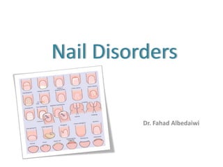
common nail disorder

Recommended
More Related Content
What's hot
What's hot (20)
Similar to Nail disorder fahad albedaiwi
Similar to Nail disorder fahad albedaiwi (20)
More from Dr Fahad Albedaiwi
Recently uploaded
Recently uploaded (20)
Nail disorder fahad albedaiwi
- 1. Nail Disorders Dr. Fahad Albedaiwi
- 2. Anatomy of the Nail Onycho = NAIL • Nail Matrix • Nail Bed • Cuticle • Nail Plate • Nail Folds • Lunula • Hyponychium
- 3. Blood Supply of Nail Lateral digital arteries supply nail through superficial , proximal & distal arcades
- 4. Nerve supply of nail through branches of Median,Ulnar & Radial nerves Nerve Supply of Nail
- 5. Nail Growth Finger nail grows at rate of 0.1 mm/ day, 3mm/ month Toe nail grows at rate of 0.033 mm/day, 1mm/month
- 6. Some nail findings with anatomic site of nail damage Nail disorders
- 7. Congenital Anonychia: • is a rare absence of some or all the nail • autosomal dominant inheritance pattern Nail patella syndrome • fingernail dysplasia with triangular lunula • absent or hypoplastic patellae • posterior iliac horns • deformation of the radial heads
- 8. Pachyonychia congenita (pachy=thick) • Hypertrophy and hyperkeratosis of the nails • palmoplanter hyperkeratosis • Warty skin lesions on the limbs • Hyperhidrosis • Scalp hair is lusterless and kinky
- 9. Traumatic Acute trauma: Hematoma (BRUISED NAIL): blood between nail plate and bed Nail biting: Is due to chronic repetitive trauma • in 60% of children, 45% of adolescents & 10% of adults • The majority of nail biters have no psychiatric disorder • Patients are susceptible to infections • Aesthetic aspect has social effects • The nails are short (no free edge is visible) • splitting of the nail into layers or a sand-papered effect, and brown longitudinal streak • Cure relies on the motivation of the patient: not to bite anymore
- 10. Onycho-gryphosis: (gryphosis: thick) • Thick, yellow and twisted great toenail in the elderly • Due to repetitive trauma by footwear Ingrowing toenail (Onychocryptosis): • The edge of the nail plate penetrates the lateral nail fold • pain, sepsis and formation of granulation tissue • Due to compression of the toe from the side due to ill-fitting footwear • Avoid tight-fitting or high-heeled shoes • Rx: Surgical removal of the ingrown toenail Pincer nail (trumpet nail): * excessive curvature of the nail * is the most painful type of ingrown nail
- 11. Onycorrhexis: • Split or brittle nails. Caused by injury or exposure to harsh chemicals Pterigium: • Abnormal winged like growth of skin (living tissue) on the nail plate and the skin is slowly stretched and dragged along the bed. • caused by severe trauma such as warts, burns & blood circulation disorders.
- 12. Nail infections Paronychia: is a soft tissue infection around a fingernail is the most common hand infection Acute paronychia • nail biting breaks down the physical barrier between the nail bed and the nail allowing the infiltration of infectious organisms • S. aureus is the most common infecting organism. • pain, tenderness, and swelling in the nail folds. • erythematous and swollen, pus collects under the skin of the nail fold. • Oral antibiotics Chronic paronychia • After 6 weeks or longer • The nail folds are swollen, erythematous, and tender with pronounced transverse ridges • Cause is a mixture of C. albicans and bacteria • Can be a complication of eczema • In housekeepers, dishwashers, and swimmers
- 13. Pseudomonas infection • It is always a complication of onycholysis or chronic paronychia • The nail plate has a characteristic bluish-black or green color due to accumulation of the pigment *pyocyanin below the nail which may remain after the organism has been removed • Treatment is as for paronychia ONYCHOMYCOSIS (TINEA UNGUIUM) • An infectious fungal disease mainly seen as white spots that can be scraped off the surface, or long yellowish streaks within the nail substance. • attacks the free edge and moves its way to the matrix. • The infected portion is thick and discoloured. WARTS • non-cancerous growths of the skin caused by infection with human papillomavirus (HPV)
- 14. Dermatoses affecting the nails Psoriasis: is common disorder affecting finger nails 10-15% of patient with psoraisis Nail changes are: • Pitting: Punctate surface depressions • Onycholysis: Separation of the nail from the nail bed either proximally or distally • Subungual hyperkeratosis: most marked distally and extends proximally • Oil drop or salmon patch: is a translucent yellow-red discoloration in the nail bed • Leukonychia (areas of white nail plate) is due to parakeratosis
- 15. Darier's disease • white and red longitudinal lines and distal notching Lichen planus (LP): • Nail involvement in 10% of individuals with disseminated LP • Nail involvement may be the only manifestation of LP • Thin nail plate and longitudinal ridging • Lunula is more elevated than the more distal portion
- 16. Alopecia areata • Affects 10-50% with alopecia areata • rough nail plate with a "hammered brass" appearance Eczema • Severe pompholyx around the nail folds may cause nail dystrophy, resulting in irregular ridges
- 17. Tumors Fibrokeratoma: periungual hyperkeratotic tip Subungual exostosis: bony outgrowth of the distal part of the toe Glomus tumour: • is painful, (pain may be spontaneous or evoked by mild trauma or temperature change) • Nail-plate changes depend on the location of the tumour: - Matrix tumours cause splitting and distortion of the nail plate. - Nail bed lesions appear as bluish or red foci of 1-5mm diameter beneath the nail
- 18. Squamous cell carcinoma: hyperkeratotic, warty changes, erosions and fissuring, macerated cuticle, periungual swelling & erythema Melanocytic nevi: longitudinal melanonychia Malignant melanoma: features suggest the possibility of malignant melanoma: • 75% will have Longitudinal melanonychia • Brown-black periungual pigmentation in a single digit in adult life • The pigmentation becomes darker and broader and has blurred edges
- 19. Nail signs in systemic disease Clubbing • bulbous uniform swelling of the soft tissue of the terminal phalanx of a digit with loss of the normal angle between the nail and the nail bed • due to vasodilation of the digit blood vessels of unknown cause Causes : 1-Primary (idiopathic) clubbing e.g. familial clubbing 2-Secondary clubbing include the following: • Pulmonary disease e.g. Lung cancer, cystic fibrosis • Cardiac disease e.g. Cyanotic congenital heart disease • GIT disease e.g. inflammatory bowel disease • Skin disease e.g. Pachydermoperiostosis • Malignancies e.g. Thyroid cancer, Hodgkin disease, leukemia • Miscellaneous conditions e.g. Acromegaly, pregnancy, and hypoxemia possibly related to long-term smoking of cannabis
- 20. Koilonychia • concave (spoon-shaped) Nails • common in infancy as a benign feature of the great toenail • The most common systemic association is with iron deficiency
- 21. Beau's line • Is a deep single horizontal ridge grooved line from side to side • caused by an infection or trauma in the nail matrix • Systemic diseases: coronary occlusion, hypocalcaemia, diabetes, certain drugs - including beta blockers Changes in nail surface
- 22. Muehrcke's lines: or leukonychia striata • are superficial white lines (not grooved as beau’s line) extend all the way across the nail and lie parallel to the lunula • are in the vascular nail bed underneath the nail plate, and so they do not move with nail growth and disappear when pressure is placed over the nail • is nonspecific, often in decreased protein synthesis (after chemotherapy) and nephrotic syndrome 'True' Leukonychia • small white spots affecting one or two nails • in young children and nail biters • In most cases disappear after around eight months
- 23. Changes in nail color Terry’s nails • The nail is proximally white and normal distally • in cirrhosis, congestive heart failure and adult-onset diabetes Yellow nail syndrome • The nails are yellow due to thickening • The lunula is obscured and there is increased transverse and longitudinal curvature and loss of cuticle • It is usually accompanied by lymphoedema and pleural effusions
- 24. Color changes due to drugs: • Chloroquine may produce blue-black pigmentation of the nail • Arsenic may produce longitudinal bands of pigment or transverse white stripes (Mees' stripes) across the nail
- 25. شكرا
Editor's Notes
- 1. Nail plate (body): * is the clear, firm & translucent portion, (hard keratin) * is created by the nail matrix * Its free edge crosses the finger * is bordered by proximal & lateral folds 2. Nail matrix : directly below the cuticle. * produces the nail plate. * contains blood vessels and nerves. * If the matrix is damaged the nail will grow deformed 3. Lunula : is the crescent shaped whitish area of the nail bed 4. Cuticle or (Eponychium): skin fold at the proximal end of the nail 5. Nail fold : hard skin overlapping the base & sides of the nail 6. Nail bed : is continuation of the matrix & the the nail plate rests on 7. Hyponychium: is under the free edge of the nail plate