
Recommended
More Related Content
Similar to Introduction GIT..ppt
Similar to Introduction GIT..ppt (20)
More from eyobkaseye
More from eyobkaseye (20)
Recently uploaded
Recently uploaded (20)
Introduction GIT..ppt
- 1. Physiology of the Digestive System
- 2. Contents • Organs of GIT • Function of the GIT • Functional structures of GIT: • Regulation of GIT • Movement of GIT • Secretions of GIT
- 3. Digestive System (GIT) • The digestive system: processes food, extracts nutrients and eliminates wastes. • It provide • Nutrients • Water and • Electrolytes - Four processes of GIT: - Digestion: mechanical or chemical - Secretion: enzymes, electrolytes (HCl, NaHCO3), mucous, and hormones - Absorption: nutrients, water & electrolytes - Motility: propulsive or mixing Digestive organs
- 4. GIT and its natural defense • GIT is hollow at both ends (mouth- to - Anus). • Harbor microorganisms in its luminal surfaces. • GI-system can protect itself by: – Mouth: Saliva contains lysozymes, IgA etc. – Stomach: HCl, Pepsin etc. have bactericidal effect – SI (e.g., Payer's patches): Immuno-competent lymph tissues – Macrophages: located in intestinal walls act to defend from bacterial invasion etc.
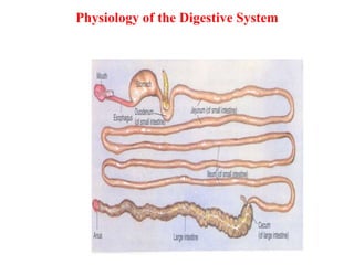
- 5. GIT-organs and its accessories 1. Mouth 2. Pharynx 3. Esophagus 4. Stomach 5. SI 6. LI 7. Rectum (Anus) Main GI-Organs Accessory Organs 1. Salivary glands 2. Pancreas 3. Liver & 4. Gallbladder Structure of the gut The alimentary canal that used to digests and absorbs food
- 6. Layers of the GIT 1. Mucosa: protection, secretion & absorption. 2. Submucosa: blood vessels, glands, lymph, nerve plexuses are found. motility + secretion 3. Muscularis externa: Circular & Longitudinal smooth muscle. – mix and propel the chyme. – 4. Serosa: outer most protective.
- 7. Regulation of GIT Neural & Hormonal Regulation A. Neural regulation of GI-activities: includes that of a. Extrinsic (Autonomic N fibers) b. Intrinsic (Enteric fibers) Myenteric (Auerbach’s) plexus: (motility of the GIT) Submucosa (Meissner’s) plexus: (secretions) c. Somatic NS: voluntary B. Hormonal regulation: Includes secretion of different hormones like Gastrin, CCK, Secretin, GIP etc. C. Paracrine Regulation: -E.g.- Histamine (stimulates parietal cells to secrete HCl -Somatostatin (inhibit secretion of gastrin by G-cells)
- 8. Receptors of the GI Tract • Receptors initiate GI-reflexes that: – Activate or inhibit GIT • Mechanoreceptors: ~ respond to distension, spastic contraction • Chemoreceptors: ~ respond to osmolarity, irritation, pH, presence of fat and protein food and end products of digested food • Thermoreceptors: respond to warm food/drinks • Pain receptors: respond to tissue injury in the GIT.
- 9. Movements of the GIT • Two basic types of movements occur in the GIT: 1. Propulsive movements: • cause food to move forward at an appropriate rate for digestion and absorption. • Peristalsis: is propulsive movement in the form of contractile rings around the gut and propels to the anal ward direction. Peristalsis and Segmentation
- 10. 2. Mixing movements : • which keep the intestinal contents thoroughly mixed at all times. • Mixing contractions are beneficial to mix the food contents with gastric juice (chyme).
- 11. Function of Saliva a. Digestion: CHO-digestion begins in saliva . • The enzyme ptyline breaks starch- to-maltose. • Lingual lipase begins fat digestion in the mouth. b. Protection: has anti-microbial actions (contains Lysozyme & thiocyanate) that kills microbes. c. Lubrication: Mucin found in saliva facilitates moistening and swallowing of food, involved in speech. d. Endocrine function: Sex steroids are found in saliva to plasma levels.
- 12. Stomach • Rugae ( a fold) increases SA. • Function: Storage up to 1.5L of food. Mixing of food to form chyme. Slow emptying the food into the SI at a rate suitable for digestion and absorption. Secretary function: HCl, mucous, pepsin, gastrin, IF • Sterilization, • Digestion: breakdown of proteins begins. • Absorption • Facilitates defecation
- 13. Secretary function of GIT * Primary secretary products of GIT are: ~ Digestive enzymes ~ GI-hormones ~ Mucous ~ Electrolytes (HCl, NaHCO3) Factors stimulating GIT secretions * Local mechanical factors: distension, irritation, pH * Nervous stimulation: ANS, ENS (submucosal plexus) Sympathetic stimulation inhibits GIT-secretions Parasympathetic stimulation increases GIT-secretions * Hormonal mechanisms: Gastrin increase HCl secretion Secretin increases NaHCO3 secretion from pancreas
- 14. Small Intestine • Runs from pyloric sphincter to the ileocecal valve • Is site for completion of digestion and absorption. • Has three subdivisions: • Duodenum, Jejunum, Ileum • The bile and pancreatic duct join duodenum. Digestive enzymes Peptidase: splits peptides into AAs 2. Disaccharidase: sucrase, maltase and lactase. 3. Intestinal lipase: splits neutral fats into glycerol & FAs. Regulation of SI secretion • Local factors: tactile, distension, irritation, PH. • Hormonal: Secretin, CCK, VIP, Glucagon, GIP • Nervous: vagal and sympathetic stimulation • Enteric reflexes: stimulation of submucosal plexus
- 15. Digestion in the Small Intestine Carbohydrates • Mouth: salivary amylase • Oesophagus & stomach: nothing happens • Duodenum: pancreatic amylase • Brush border enzymes (maltase, sucrase & lactase) Digestion of Proteins Stomach • HCl denatures or unfolds proteins • Pepsinogen pepsin • Pepsin proteins to peptides Pancreas • split peptide to AA SI • Brush border enzymes carboxypeptidases aminopeptidases dipeptidase
- 16. Dietary source of fat • Triglycerides, Cholesterol, cholesterol esters and PL Fat Emulsified fat FFA + Glycerides -Lingual lipase -Gastric lipase -Pancreas lipase Cholesterol Bile salt FFA + Glycerides Cholesterol esters Cholesterol Esterase Phospholipids-A2 Phospholipase FFA + Phospholipids Digestion of Lipids Bile salt
- 17. Absorption in the SI
- 18. Large Intestine Is subdivided into cecum, colon, rectum, and anal canal • The cecum: contains appendix Colon has distinct regions: ascending colon, transverse colon, descending colon, and sigmoid colon • The sigmoid colon joins the rectum • The anal canal
- 19. Valves and Sphincters of the Rectum and Anus Valves of the rectum stop feces from being passed with gas • The anus has two sphincters: – Internal anal sphincter composed of smooth muscle – External anal sphincter composed of skeletal muscle • Ileocecal valve (sphincter) : connects ileum -to- cecum. It is mostly in a contracted state b/s: a. Prevents bacterial penetration back to the SI. b. It permits slow flow of chyme to the LI
- 20. Large intestine • LI does not have villi, but has goblet cells that secrete mucus. Function of the large intestine 1. Absorption of drugs & water 2. Electrolyte (like NaCl) absorption 3. Mucous & HCO3 - secretion 4. Storage, transport, and evacuation of feces 5. Bacterial fermentation in the colon stimulates synthesis of vitamins K. • Movement in the LI • Two types of movements 1. Mixing movements (Haustration) 2. Propulsive movements (mass movements) • Mass movement :is initiated by local distension, gastro-colic reflex * Poor motility: causes greater absorption and constipation * Excess motility: causes less absorption and diarrhoea.
- 22. 22 Pancreas • Pancreas contains two types of secretary glands: 1. Endocrine cells (islets of Langerhans) * secrete hormones 2. Exocrine cells (acinar cells) * secrete digestive enzymes called pancreatic juice. a. Digestive enzymes: necessary to digest CHO, fat, and protein. b. Bicarbonates : to neutralize the gastric juice c. Water and electrolytes (Na+, K+ etc.)
- 23. Hormonal regulation of pancreatic secretion Secretin: causes NaHco3 bicarbonate release. GIP: increased insulin release CCK: increased digestive enzyme release • Parasympathetic impulse Stimulate secretion of pancreatic enzyme
- 24. Liver and gallbladder Liver • Is the heaviest gland. • Weighs 1.36kg. • Located below diaphragm in the abdomen • The liver is divisible into left and right lobes, separated by the falciform ligament. • • Right lobe larger • Gallbladder on right lobe
- 25. Functions of the liver 1. Metabolism: CHO glycogenesis, gluconeogenesis and glycogenolysis Lipid β-oxidation, formation of PL, LP, synthesis of cholesterol Protein • Deamination of AAs • Converts ammonia (NH3) into urea. • Synthesizes plasma proteins fibrinogen and gamma globulin. 2. Inactivation of drugs & hormones 3. Removes the waste product bilirubin 4. Releases bile salts 5. Stores: fat soluble vitamins, iron, copper and blood reservoir 6. Filtration of blood: • Old blood cells & bacteria. • Removes blood clots and toxins 7. Activates vitamin D 8. Synthesis of clotting factors (F-I, II, VII, IX, X)