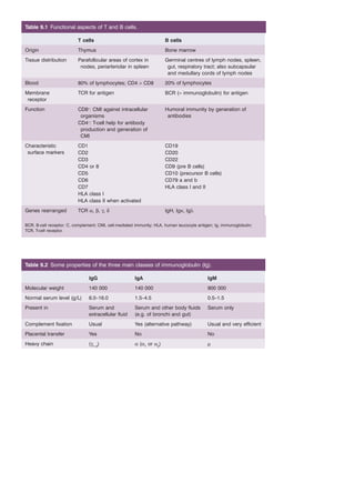
1. Table 9.1 Functional aspects of T and B cells.
T cells B cells
Origin Thymus Bone marrow
Tissue distribution Parafollicular areas of cortex in
nodes, periarteriolar in spleen
Germinal centres of lymph nodes, spleen,
gut, respiratory tract; also subcapsular
and medullary cords of lymph nodes
Blood 80% of lymphocytes; CD4 > CD8 20% of lymphocytes
Membrane
receptor
TCR for antigen BCR (= immunoglobulin) for antigen
Function CD8+: CMI against intracellular
organisms
Humoral immunity by generation of
antibodies
CD4+: T-cell help for antibody
production and generation of
CMI
Characteristic
surface markers
CD1 CD19
CD2 CD20
CD3 CD22
CD4 or 8 CD9 (pre B cells)
CD5 CD10 (precursor B cells)
CD6 CD79 a and b
CD7 HLA class I and II
HLA class I
HLA class II when activated
Genes rearranged TCR α, β, γ, δ IgH, Igκ, Igλ
BCR, B-cell receptor; C, complement; CMI, cell-mediated immunity; HLA, human leucocyte antigen; Ig, immunoglobulin;
TCR, T-cell receptor.
Table 9.2 Some properties of the three main classes of immunoglobulin (Ig).
IgG IgA IgM
Molecular weight 140 000 140 000 900 000
Normal serum level (g/L) 6.0–16.0 1.5–4.5 0.5–1.5
Present in Serum and
extracellular fluid
Serum and other body fluids
(e.g. of bronchi and gut)
Serum only
Complement fixation Usual Yes (alternative pathway) Usual and very efficient
Placental transfer Yes No No
Heavy chain (γ1–4) α (α1 or α2) μ
2. Table 9.3 Causes of lymphocytosis.
Infections
Acute: infectious mononucleosis, rubella,
pertussis, mumps, acute infectious
lymphocytosis, infectious hepatitis,
cytomegalovirus, HIV, herpes simplex or
zoster
Chronic: tuberculosis, toxoplasmosis,
brucellosis, syphilis
Chronic lymphoid leukaemias (see Chapter 17)
Acute lymphoblastic leukaemia
Non-Hodgkin lymphoma (some)
Thyrotoxicosis
HIV, human immunodeficiency virus.
Table 9.4 Classification of immunodeficiencies.
Primary
B cell (antibody deficiency) X-linked agammaglobulinaemia, acquired common variable
hypogammaglobulinaemia, selective IgA or IgG subclass deficiencies
T cell Thymic aplasia (DiGeorge’s syndrome), PNP deficiency
Mixed B and T cell Severe combined immune deficiency (as a result of ADA deficiency or
other causes); Bloom’s syndrome; ataxia-telangiectasia; Wiskott–
Aldrich syndrome
Secondary
B cell (antibody deficiency) Myeloma; nephrotic syndrome, protein-losing enteropathy, anti-CD20
(rituximab) therapy
T cell AIDS; Hodgkin lymphoma, non-Hodgkin lymphoma; drugs: steroids,
ciclosporin, azathioprine, fludarabine, etc.
T and B cell Chronic lymphocytic leukaemia, post-stem cell transplantation,
chemotherapy/radiotherapy, anti-CD52 (alemtuzumab)
ADA, adenosine deaminase; AIDS, acquired immune deficiency syndrome; Ig, immunoglobulin; PNP, purine nucleoside
phosphorylase.