
Recommended
More Related Content
What's hot
What's hot (20)
Similar to Choroidal Osteoma
Similar to Choroidal Osteoma (20)
Recently uploaded
Recently uploaded (20)
Choroidal Osteoma
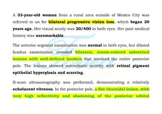
- 1. A 33-year-old woman from a rural area outside of Mexico City was referred to us for bilateral progressive vision loss, which began 20 years ago. Her visual acuity was 20/400 in both eyes. Her past medical history was unremarkable. The anterior segment examination was normal in both eyes, but dilated fundus examination revealed bilateral, cream-colored subretinal lesions with well-defined borders that involved the entire posterior pole. The lesions showed osteoclastic activity with retinal pigment epithelial hyperplasia and scarring. B-scan ultrasonography was performed, demonstrating a relatively echolucent vitreous. In the posterior pole, a flat choroidal lesion, with very high reflectivity and shadowing of the posterior orbital
- 2. contents, was observed in both eyes. Systemically, there were no other findings of familial adenomatous polyposis (Gardner syndrome). Although choroidal osteoma is benign, it usually results in poor vision over time. There is no treatment for the osteoma itself; however, it sometimes leads to choroidal neovascularization (CNV), which can be managed with anti-VEGF intravitreal injections. Since no CNV activity was identified in this case, we suggested periodic clinical follow-up.