Poster congreso vlc 2012
•
0 likes•223 views

1) The study examines the effects of polyphenols (resveratrol and pterostilbene) and cannabinoids (WIN 55,212-2) on neurons and astrocytes treated with amyloid beta (Aβ), which is implicated in Alzheimer's disease pathogenesis. 2) MTT assays showed that polyphenols and WIN 55,212-2 increased cell viability in neurons and astrocytes treated with Aβ, protecting against Aβ-induced neurodegeneration. 3) Western blot and RT-PCR analysis revealed that polyphenols and WIN 55,212-2 modulated expression of NMDA receptors and inflammation-related proteins like NF-κB and PPAR-

Recommended
More Related Content
Viewers also liked
Viewers also liked (13)
Similar to Poster congreso vlc 2012
Similar to Poster congreso vlc 2012 (9)
Recently uploaded
Recently uploaded (20)
Poster congreso vlc 2012
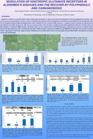
- 1. MODULATION OF IONOTROPIC GLUTAMATE RECEPTORS IN ALZHEIMER’S DISEASES AND THE RECOVER BY POLYPHENOLS AND CANNABIONOIDS Diana Aguirre-Rueda1, Patricio Paredes-Brunet1, Anna Gil-Bisquert1, Sergio García- Herreros1 and Soraya L. Valles1 1Department of Physiology, School of Medicine, University of Valencia, Spain. Introduction Alzheimer´s disease is a prevalent illness durign elderly age and actually anu drug can decrease the progression of that neurodegenerative disease. Here we use neurons and astrocytes in primary cultura to determine the protective action of polyphenols (resveratrol and pterostilbene) and cannabinoids (Win 55, 212-2) againts Amyloid β (Aβ) neurodegeneration. Usin 10 μM Win 55, 212-2 and Aβ (10 μM) during 24 h, a viability increase compared wint Aβ astrocytes was detected and similar results occur with neurons in primary culture. Also we detect a viability increase after polyphenols treatment compared with only Aβ incubation. To determine NMDA receptor expression levels, we use a western-blot and RT-PCR technique in astrocytes and neurons in primary cultura treated with Aβ, poliphenols and/or cannabinoids. We detect a different pattern expression of NMAD R-1 and NMDA R-2 in astrocytes and neurons in primary cultura, depending of which brain región we used. Polyphenols and cannabinoids increase the anti-oxidant and anti-inflammation cell response to protect neuron from the Aβ attack. Fig. 1.- Confocal microscopy. Treated cells for 24 h with 10 μM Aβ (40-1) (C), 10 μM Aβ (1-42) (Aβ), 10 μM Resveratrol (R), 10 μM Aβ (1-42) +10 μM (C ) (Aβ) (R) (Aβ + R) Resveratrol (Aβ + R), 10 μM Pterostilbene (P), 10 μM Aβ (1-42) +10 μM Pterostilbene (Aβ + P), 10 μM WIN 55, 212-2 (WIN) and 10 μM Aβ (1-42) + 10 μM WIN 55, 212-2 (Aβ + WIN). (P) (Aβ + P) (WIN) (Aβ + WIN) % of viability % of viability Fig. 2.- MTT assay on astrocytes in primary culture. Treated cells Fig. 3.- MTT assay on neurons in primary culture. Treated cells for 24 h with 10 μM for 24 h with10 μM Aβ (40-1) (C) 10 μM Aβ (1-42) (Aβ), 10 μM WIN Aβ (40-1) (C), 10 μM Aβ (1-42) (Aβ), 10 μM Resveratrol (R), 10 μM Aβ (1-42) +10 μM 55, 212-2 (WIN), 10 μM Aβ + 10 μM WIN 55, 212-2 (Aβ + WIN). Data Resveratrol (Aβ + R), 10 μM Pterostilbene (P), 10 μM Aβ (1-42) +10 μM Pterostilbene are means ± D.S. * p < 0.05 vs control cells, # p < 0,05 vs Aβ (1-42) (Aβ + P), 10 μM WIN 55, 212-2 (WIN), and 10 μM Aβ (1-42) + 10 μM WIN 55, 212-2 (Aβ treated cells. n = 3. + WIN). Data are means ± D.S. * p < 0.05 vs control cells # p < 0,05 vs Aβ treated cells. n = 3. Relative Densitometric Units Relative Densitometric Units Fig. 5.- Anti-inflamatory mediators PPAR-γ expression in astrocytes in Fig. 4.- Inflamatory mediators NFκB expression in astrocytes in primary primary culture. PPAR-γ expression were determined by western-blot in culture. NFκB expression were determined by western-blot in astrocytes astrocytes treated for 24 h with 10 μM Aβ (40-1) (C), 10 μM Aβ (1-42) (Aβ), treated for 24 h with 10 μM Aβ (40-1) (C), 10 μM Aβ (1-42) (Aβ), 10 μM 10 μM WIN 55, 212-2 (WIN) and 10 μM Aβ (1-42) + 10 μM WIN 55, 212-2 WIN 55, 212-2 (WIN) and 10 μM Aβ (1-42) + 10 μM WIN 55, 212-2 (Aβ + (Aβ + WIN). Data are means ± D.S. * p < 0.05 vs control cells # p < 0,05 vs WIN). Data are means ± D.S. * p < 0.05 vs control cells # p < 0,05 vs Aβ Aβ treated cells. n = 3. treated cells. n = 3. CONCLUSIONS MTT assay determined a viability decrease of astrocytes compared with control cells incubated with 10 μM Aβ (1-42). A viability increase of astrocytes compared with Aβ (1-42) and control cells incubated with 10 μM WIN 55, 212-2, and 10 μM Relative Densitometric Units Aβ (1-42) + 10 μM WIN 55, 212-2 (Fig 2). Similar results occur with a treatment for 24 h of Resveratrol, Pterostilbene and WIN 55, 212.-2 (10 μM), shown a viability increase of neurons (Fig 3). Inflammatory mediator NFκB, anti-inflammatory mediator PPAR-γ and oxidant SOD Cu/Zn expression in astrocytes in primary culture. NFκB, PPAR-γ and SOD Cu/Zn expression were determinated by western-blot in astrocytes treated with 10 μM Aβ (40-1) (C), 10 μM Aβ (1-42) (Aβ), 10 μM WIN 55, 212-2 (WIN) and 10 μM Aβ (1-42) + 10 μM WIN 55, 212-2 (Aβ + WIN). WIN prevents the increase of inflammatory and oxidant levels caused by Aβ in astrocytes in primary culture, suggesting that WIN Fig. 6.- Anti-oxidant SOD Cu/Zn expression in astrocytes in primary protects astrocytes against oxidative stress (Fig 4 and 6). Our findings suggest that culture. SOD Cu/Zn expression were determined by western-blot in Aβ in able to induce inflammatory mediator and oxidant in astrocytes and these astrocytes treated for 24 h with 10 μM Aβ (40-1) (C), 10 μM Aβ (1-42) effect are prevented by WIN. (Aβ), 10 μM WIN 55, 212-2 (WIN) and 10 μM Aβ (1-42) + 10 μM WIN 55, 212-2 (Aβ + WIN). Data are means ± D.S. * p < 0.05 vs control cells Our hypothesis with the results of NFkB, it is suggested that the case with NMDA # p < 0,05 vs Aβ treated cells. n = 3. receptors, are increased in the presence of Aβ and possible recovery by WIN