
Recommended
More Related Content
What's hot
What's hot (20)
Similar to Types of fractures
Similar to Types of fractures (20)
Recently uploaded
Recently uploaded (20)
Types of fractures
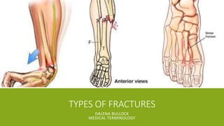
- 1. TYPES OF FRACTURES DALENA BULLOCK MEDICAL TERMINOLOGY
- 2. CONTEXT ▪Stress Fracture ▪Colles’ Fracture ▪Compound Fracture
- 3. STRESS FRACTURE STRESS FRACTURES ARE TINY CRACKS IN A BONE.
- 4. CAUSES/RISK FACTORS ▪ Stress fractures are caused by a great amount of repetitive force that causes an imbalance between the resorption and growth of bone. ▪ Example: repeatedly jumping up and down or running long distances ▪ Stress fractures often occur in people who suddenly shift from a sedentary lifestyle to an active training regimen ▪ Women who have abnormal or absent menstrual periods are at higher risk of developing stress fractures. ▪ People who have flat feet or high, rigid arches are more likely to develop stress fractures. ▪ Conditions such as osteoporosis can weaken your bones and make it easier for stress fractures to occur.
- 5. MRI SCAN OF STRESS FRACTURE Runners or people who place sports that involve running are at risk of getting stress fracture in this part of the leg.
- 6. DIAGNOSIS AND TREATMENT ▪ Bone scan, Magnetic resonance imaging (MRI), and X-rays are ways to test to see if the bone is fractured ▪ In many cases, stress fractures aren't noticeable on regular X-rays taken soon after your signs and symptoms begin. ▪ Reducing the bone's weight-bearing load until healing occurs helps, you may need to wear a walking boot or brace or use crutches. ▪ Surgery is may be necessary to ensure complete healing of some stress fractures Stress Fracture Bone Growth Imbalance Repetitive Force
- 7. COLLES’ FRACTURE A COLLES’ WRIST FRACTURE IS WHEN THE RADIUS BONE IN YOUR FOREARM BREAKS. IT IS ALSO KNOWN AS A DISTAL RADIUS FRACTURE OR A DINNER-FORK DEFORMITY OF THE WRIST.
- 8. COLLES’ FRACTURE TYPES ▪ Open fracture (if the bone broke through your skin) ▪ Comminuted fracture (if the bone broke into more than two pieces) ▪ Intra-articular fracture (if the bone broke inside your wrist joint) ▪ Extra-articular fracture (if your joint isn’t affected) COMMON CAUSES/RISK FACTORS ▪ Putting your hand out to stop yourself from falling ▪ Any type of injury that directly affects your wrist can result in a Colles’ wrist fracture. ▪ Osteoporosis ▪ Walking or other activities in snow or on ice ▪ Not enough calcium or vitamin D intake
- 9. MRI OF COLLES’ FRACTURE This type of fracture is an extra-articular fracture. The bone is broken but the joint was unharmed.
- 10. TREATMENT ▪ If the fracture isn’t severe, you can put the wrist in a splint and elevate it or your doctor might put a light cast to protect it. ▪ If your wrist is severely fractured, there’s surgery: the bones will be straightened and held together using pins, a plate and screws, or an external device that holds the pins in place. ▪ Depending on severity, there may be physical therapy to help rebuild muscle: they will have you do exercises that can help rebuild the strength and help regain normal range of motion ▪ A Colles’ wrist fracture can take a year or more to fully heal, the cast will typically be taken off about six weeks after surgery.
- 11. COMPOUND FRACTURE AN INJURY IN WHICH A BROKEN BONE PIERCES THE SKIN, CAUSING A RISK OF INFECTION.
- 12. CAUSES ▪ They can occur from direct trauma to the bone form external sources, such as a gunshot or knife wound ▪ Or from indirect trauma at a site distal to the fracture, such as rotational force ▪ Open fractures are typically caused by high-energy injuries such as car crashes, falls, or sports injuries.
- 13. CLASSIFICATIONS ▪ Grade I Open Fracture: occurs when there is a skin wound with the fracture that measures less than one centimeter ▪ Grade II Open Fracture: have larger soft-tissue injuries, measures more than one centimeter ▪ Grade III Open Fracture: the most severe injuries and include three specific sub-types of injuries: ▪ Grade IIIA: injuries that include high-energy fractures by severe bone injury and/or large, often contaminated, soft-tissue wounds ▪ Grade IIIB: significant soft-tissue damage/loss such that bone is exposed; reconstruction may require a soft-tissue transfer in order to cover the wound ▪ Grade IIIC: specifically require vascular intervention as the fracture is associated with vascular injury to the extremity
- 14. TREATMENT ▪ When a broken bone penetrates the skin there is a need for immediate treatment ▪ Irrigation and debridement is performed: ▪ The wound must be clean properly to prevent infection, irrigation is then the wound is surgically cleaning the bone-this is one of the first steps for treating an open fracture. ▪ Debridement means removing foreign material (dirt, gravel, clothing) as well as non-viable (useable) soft-tissue. ▪ Stabilizing the fractured bone helps to prevent further tissue damage: plates and screws or intramedullary rods, may not be good options if there is a high chance of bacterial contamination ▪ In many open fractures, a device called an external fixator will be used to stabilize these injuries. ▪ Antibiotics should be administered as soon as possible, even before performing the irrigation and debridement described above. The antibiotics are usually continued for 48 hours.
- 15. POST-SURGERY OF COMPOUND FRACTURE A metal rod is placed into both ends of the fractured bone to reconnect it. The rod helps to hold the bone in place so new bone can form and to stabilize it.