
Recommended
Recommended
More Related Content
Similar to ARTICLEdoi10.1038nature14098A new antibiotic kills pat.docx
Similar to ARTICLEdoi10.1038nature14098A new antibiotic kills pat.docx (20)
More from davezstarr61655
More from davezstarr61655 (20)
Recently uploaded
Recently uploaded (20)
ARTICLEdoi10.1038nature14098A new antibiotic kills pat.docx
- 1. ARTICLE doi:10.1038/nature14098 A new antibiotic kills pathogens without detectable resistance Losee L. Ling1*, Tanja Schneider2,3*, Aaron J. Peoples1, Amy L. Spoering1, Ina Engels2,3, Brian P. Conlon4, Anna Mueller2,3, Till F. Schäberle3,5, Dallas E. Hughes1, Slava Epstein6, Michael Jones7, Linos Lazarides7, Victoria A. Steadman7, Douglas R. Cohen1, Cintia R. Felix1, K. Ashley Fetterman1, William P. Millett1, Anthony G. Nitti1, Ashley M. Zullo1, Chao Chen4 & Kim Lewis4 Antibiotic resistance is spreading faster than the introduction of new compounds into clinical practice, causing a public health crisis. Most antibiotics were produced by screening soil microorganisms, but this limited resource of cultivable bacteria was overmined by the 1960s. Synthetic approaches to produce antibiotics have been unable to replace this platform. Uncultured bacteria make up approximately 99% of all species in external environments, and are an untapped source of new antibiotics. We developed several methods to grow uncultured organisms by cultivation in situ or by using specific growth factors. Here we report a new antibiotic that we term teixobactin, discovered in a screen of uncultured bacteria. Teixobactin inhibits cell wall synthesis by binding to a highly conserved motif of lipid II (precursor of peptidoglycan) and lipid III (precursor of cell wall teichoic acid). We did not obtain any mutants of Staphylococcus aureus or Mycobacterium tuberculosis resistant to teixobactin.
- 2. The properties of this compound suggest a path towards developing antibiotics that are likely to avoid development of resistance. Widespread introduction of antibiotics in the 1940s, beginning with penicillin1,2 and streptomycin3, transformed medicine, providing effec- tive cures for the most prevalent diseases of the time. Resistance develop- ment limits the useful lifespan of antibiotics and results in the requirement for a constant introduction of new compounds4,5. However, antimicro- bial drug discovery is uniquely difficult6, primarily due to poor penetra- tion of compounds into bacterial cells. Natural products evolved to breach the penetration barriers of target bacteria, and most antibiotics intro- duced into the clinic were discovered by screening cultivable soil micro- organisms. Overmining of this limited resource by the 1960s brought an end to the initial era of antibiotic discovery7. Synthetic approaches were unable to replace natural products6. Approximately 99% of all species in external environments are un- cultured (do not grow under laboratory conditions), and are a prom- ising source of new antibiotics8. We developed several methods to grow uncultured organisms by cultivation in their natural environment9,10,
- 3. or by using specific growth factors such as iron-chelating siderophores11. Uncultured organisms have recently been reported to produce interest- ing compounds with new structures/modes of action— lassomycin, an inhibitor of the essential mycobacterial protease ClpP1P2C1 (ref. 12); and diverse secondary metabolites present in a marine sponge Theonella swinhoei which are actually made by an uncultured symbiotic Entothe- onella sp.13. Here we report the discovery of a new cell wall inhibitor, teixobactin, from a screen of uncultured bacteria grown in diffusion chambers in situ. Identification of teixobactin A multichannel device, the iChip10, was used to simultaneously isolate and grow uncultured bacteria. A sample of soil is diluted so that approx- imately one bacterial cell is delivered to a given channel, after which the device is covered with two semi-permeable membranes and placed back in the soil (Extended Data Fig. 1). Diffusion of nutrients and growth factors through the chambers enables growth of uncultured bacteria in their natural environment. The growth recovery by this method ap- proaches 50%, as compared to 1% of cells from soil that will
- 4. grow on a nutrient Petri dish10. Once a colony is produced, a substantial number of uncultured isolates are able to grow in vitro14. Extracts from 10,000 isolates obtained by growth in iChips were screened for antimicrobial activity on plates overlaid with S. aureus. An extract from a new species of b-proteobacteria provisionally named Eleftheria terrae showed good activity. The genome of E. terrae was sequenced (Supplementary Dis- cussion). Based on 16S rDNA and in silico DNA/DNA hybridization, this organism belongs to a new genus related to Aquabacteria (Extended Data Fig. 2, Supplementary Discussion). This group of Gram- negative organisms is not known to produce antibiotics. A partially purified ac- tive fraction contained a compound with a molecular mass of 1,242 Da determined by mass spectrometry, which was not reported in available databases. The compound was isolated and a complete stereochemical assignment has been made based on NMR and advanced Marfey’s anal- ysis (Fig. 1, Extended Data Figs 3 and 4 and Supplementary Discussion). This molecule, which we named teixobactin, is an unusual depsipep- tide which contains enduracididine, methylphenylalanine, and four D-amino acids. The biosynthetic gene cluster (GenBank
- 5. accession num- ber KP006601) was identified using a homology search (Supplementary Discussion). It consists of two large non-ribosomal peptide synthetase (NRPS)-coding genes, which we named txo1 and txo2, respectively (Fig. 1). In accordance with the co-linearity rule, 11 modules are encoded. The in silico predicted adenylation domain specificity perfectly matches the amino acid order of teixobactin (Fig. 1), and allowed us to predict the biosynthetic pathway (Extended Data Fig. 5). Resistance and mechanism of action Teixobactin had excellent activity against Gram-positive pathogens, including drug-resistant strains (Table 1 and Extended Data Table 1). *These authors contributed equally to this work. 1NovoBiotic Pharmaceuticals, Cambridge, Massachusetts 02138, USA. 2Institute of Medical Microbiology, Immunology and Parasitology—Pharmaceutical Microbiology Section, University of Bonn, Bonn 53115, Germany. 3German Centre for Infection Research (DZIF), Partner Site Bonn-Cologne, 53115 Bonn, Germany. 4Antimicrobial Discovery Center, Northeastern University, Department of Biology, Boston, Massachusetts 02115, USA. 5Institute for Pharmaceutical Biology, University of Bonn, Bonn 53115, Germany. 6Department of Biology, Northeastern University, Boston, Massachusetts 02115, USA. 7Selcia, Ongar, Essex CM5 0GS, UK.
- 6. 2 2 J A N U A R Y 2 0 1 5 | V O L 5 1 7 | N A T U R E | 4 5 5 Macmillan Publishers Limited. All rights reserved©2015 www.nature.com/doifinder/10.1038/nature14098 Potency against most species, including difficult-to-treat enterococci and M. tuberculosis was below 1 mg ml21. Teixobactin was exception- ally active against Clostridium difficile and Bacillus anthracis (minimal inhibitory concentration (MIC) of 5 and 20 ng ml21, respectively). Tei- xobactin had excellent bactericidal activity against S. aureus (Fig. 2a), was superior to vancomycin in killing late exponential phase popula- tions (Fig. 2b), and retained bactericidal activity against intermediate resistance S. aureus (VISA) (Extended Data Fig. 6a). Note that frequent clinical failure in patients with S. aureus MRSA treated with vancomycin has been linked to the poor bactericidal activity of this compound15,16. Teixobactin was ineffective against most Gram-negative bacteria, but showed good activity against a strain of E. coli asmB1 with a defective outer membrane permeability barrier (Table 1).
- 7. We were unable to obtain mutants of S. aureus or M. tuberculosis resistant to teixobactin even when plating on media with a low dose (4 3 MIC) of the compound. Serial passage of S. aureus in the presence of sub-MIC levels of teixobactin over a period of 27 days failed to pro- duce resistant mutants as well (Fig. 2d, Supplementary Discussion). This usually points to a non-specific mode of action, with accompany- ing toxicity. However, teixobactin had no toxicity against mammalian NIH/3T3 and HepG2 cells at 100 mg ml21 (the highest dose tested). The compound showed no haemolytic activity and did not bind DNA. In order to determine specificity of action of teixobactin, we examined its effect on the rate of label incorporation into the major biosynthetic path- ways of S. aureus. Teixobactin strongly inhibited synthesis of pepti- doglycan, but had virtually no effect on label incorporation into DNA, RNA and protein (Fig. 3a). This suggested that teixobactin is a new peptidoglycan synthesis inhibitor. Resistance has not developed to this compound, suggesting that the target is not a protein. The essential lack of resistance development through mutations has been described for vancomycin which
- 8. binds lipid II, the precursor of peptidoglycan. We reasoned that teixobactin could be acting against the same target. Treatment of whole cells of S. aureus with teixobactin (1–5 3 MIC) resulted in significant accumu- lation of the soluble cell wall precursor undecaprenyl-N- acetylmuramic acid-pentapeptide (UDP-MurNAc-pentapeptide), similar to the vancomycin-treated control cells (Fig. 3b), showing that one of the membrane-associated steps of peptidoglycan biosynthesis is blocked. Teixobactin inhibited peptidoglycan biosynthesis reactions in vitro in a dose-dependent manner with either lipid I, lipid II or undecaprenyl- pyrophosphate (Fig. 3c) as a substrate. Quantitative analysis of the O O OH H N H N H N
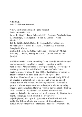
- 10. NH NH NH2 HN O NmPhe Ile Ile Ile IleSer SerGln Thr Ala End Genes Domains Amino acids a b c A T C A T C A T C A T C A T C A T C A T C A T C A T C A T C A T C txo1 txo2 NmPhe Ile Ile Ile IleSer Gln Ser Thr Ala End MT TE TE Figure 1 | The structure of teixobactin and the predicted biosynthetic gene cluster. a, The two NRPS genes, the catalytic domains they encode,
- 11. and the amino acids incorporated by the respective modules. Domains: A, adenylation; C, condensation; MT, methylation (of phenylalanine); T, thiolation (carrier); and TE, thioesterase (Ile-Thr ring closure). NmPhe, N-methylated phenylalanine. b, Schematic structure of teixobactin. The N-methylation of the first phenylalanine is catalysed by the methyltransferase domain in module 1. The ring closure between the last isoleucine and threonine is catalysed by the thioesterase domains during molecule off-loading, resulting in teixobactin. c, Teixobactin structure. 2 3 4 5 6 7 8 9 10 0 4 8 12 16 20 24 L o
- 13. Time (h) 0 4 8 12 16 20 24 Time (h) Control Oxacillin Vancomycin Teixobactin a b TeixVanOxConc 2 3 4 5 6 7 8 9 10 Control
- 14. Oxacillin Vancomycin Teixobactin 0.5 1 2 4 8 16 32 64 128 256 0 5 10 15 20 25 F o ld c h
- 15. an g e in M IC Time (days) Teixobactin Ofloxacin d Figure 2 | Time-dependent killing of pathogens by teixobactin. a, b, S. aureus were grown to early (a), and late (b) exponential phase and challenged with antibiotics. Data are representative of 3 independent experiments 6 s.d. c, Teixobactin treatment resulted in lysis. The figure is representative of 3 independent experiments. d, Resistance acquisition during serial passaging in the presence of sub-MIC levels of antimicrobials. The y axis is the highest concentration the cells grew in during passaging. For ofloxacin, 256 3 MIC was the highest concentration tested. The figure is representative of 3 independent experiments.
- 16. Table 1 | Activity of teixobactin against pathogenic microorganisms Organism and genotype Teixobactin MIC (mg ml21) S. aureus (MSSA) S. aureus 1 10% serum S. aureus (MRSA) Enterococcus faecalis (VRE) Enterococcus faecium (VRE) Streptococcus pneumoniae (penicillinR) Streptococcus pyogenes Streptococcus agalactiae Viridans group streptococci B. anthracis Clostridium difficile Propionibacterium acnes M. tuberculosis H37Rv Haemophilus influenzae Moraxella catarrhalis Escherichia coli Escherichia coli (asmB1) Pseudomonas aeruginosa Klebsiella pneumoniae 0.25 0.25 0.25 0.5 0.5 # 0.03 0.06 0.12 0.12 # 0.06 0.005 0.08
- 17. 0.125 4 2 25 2.5 .32 .32 The MIC was determined by broth microdilution. MSSA, methicillin-sensitive S. aureus; VRE, vancomycin-resistant enterococci. RESEARCH ARTICLE 4 5 6 | N A T U R E | V O L 5 1 7 | 2 2 J A N U A R Y 2 0 1 5 Macmillan Publishers Limited. All rights reserved©2015 MurG-, FemX-, and PBP2-catalysed reactions using radiolabelled sub- strates, showed an almost complete inhibition at a twofold molar excess of teixobactin with respect to the lipid substrate (Fig. 3c). The addition of purified lipid II prevented teixobactin from inhibiting growth of S. aureus (Extended Data Table 2). These experiments showed that teixobactin specifically interacts with the peptidoglycan precursor, rather than interfering with the activity of one of the enzymes. In order to evaluate the minimal motif required for high affinity binding of teix-
- 18. obactin, the direct interaction with several undecaprenyl- coupled cell envelope precursors was investigated. Purified precursors were incu- bated with teixobactin at different molar ratios, followed by extraction and subsequent thin-layer chromatography analysis (Fig. 3d). In agree- ment with the results obtained from the in vitro experiments, lipid I and lipid II were fully trapped in a stable complex that prevented extraction of the lipid from the reaction mixture in the presence of a twofold molar excess of the antibiotic, leading to the formation of a 2:1 stoichiometric complex. Teixobactin was active against vancomycin-resistant enter- ococci that have modified lipid II (lipid II-D-Ala-D-Lac or lipid II-D- Ala-D-Ser instead of lipid II-D-Ala-D-Ala)17–19. This suggested that, unlike vancomycin, teixobactin is able to bind to these modified forms of lipid II. Indeed, teixobactin bound to lipid II-D-Ala-D-Lac and lipid II-D-Ala- D-Ser (Extended Data Fig. 6b). Moreover, teixobactin efficiently bound to the wall teichoic acid (WTA) precursor undecaprenyl-PP- GlcNAc (lipid III). Although WTA is not essential per se, inhibition of late membrane-bound WTA biosynthesis steps is lethal due to accumulation of toxic intermediates20. Furthermore, teichoic acids anchor autolysins,
- 19. preventing uncontrolled hydrolysis of peptidoglycan21. Inhibition of teichoic acid synthesis by teixobactin would help liberate autolysins, contributing to the excellent lytic and killing activity of this antibiotic. Teixobactin was also able to bind undecaprenyl-pyrophosphate, but not undecaprenyl-phosphate (Fig. 3d and Extended Data Table 2). Although teixobactin efficiently binds lipid I in vitro, this is probably less significant for antimicrobial activity, as this is the intracellular form of the precursor, unlike surface-exposed lipid II and the undecaprenyl- PP-GlcNAc WTA precursors (Fig. 3e and Extended Data Fig. 7). Bind- ing to the target primarily relies on the interaction of the antibiotic with the pyrophosphate moiety, and the first sugar moiety attached to the lipid carrier, as higher concentrations of teixobactin were required to comple- tely inhibit the YbjG-catalysed monophosphorylation of undecaprenyl- pyrophosphate, involved in the recycling process of the essential lipid carrier (Fig. 3c and Extended Data Fig. 7). Corroborating this result, a tenfold higher concentration of undecaprenyl-pyrophosphate was re- quired to antagonize the antimicrobial activity of teixobactin compared to lipid II (Extended Data Table 2). The exact nature of this
- 20. first sugar is therefore not important, explaining why teixobactin is active against M. tuberculosis, where it probably binds to decaprenyl-coupled lipid intermediates of peptidoglycan and arabinogalactan. Teixobactin is also likely to bind to prenyl-PP-sugar intermediates of capsular polysacchar- ide biosynthesis which is important for virulence in staphylococci22 and whose inhibition of biosynthesis is lethal in streptococci23. In vivo efficacy Given the attractive mode of action of this compound, we investigated its potential as a therapeutic. The compound retained its potency in the presence of serum, was stable, and had good microsomal stability and low toxicity (Supplementary Discussion). The pharmacokinetic 0 20 40 60 80 100 120
- 22. ra tio n A 2 6 0 0 0.2 0.4 0.6 0.8 Untreated VAN (10× MIC) TEIX (1× MIC) TEIX (2.5× MIC) TEIX (5× MIC) 10 20 Time (min) 30 400 UDP-MurNAc-pentapeptide a b L ip
- 27. 60 70 80 90 100 P ro d u ct f o rm ed (% ) Substrate Product MurG FemX PBP2 Lipid I Lipid II Lipid II Lipid II Lipid II-Gly C55PP TEIX:substrate molar ratio
- 28. 0.25:1 0.5:1 1:1 2:1 YbjG C55PP C55P 5:1 10:1 e Figure 3 | Teixobactin binds to cell wall precursors. a, Impact of teixobactin (TEIX) on macromolecular biosyntheses in S. aureus. Incorporation of 3H-thymidine (DNA), 3H- uridine (RNA), 3H-leucine (protein), and 3H- glucosamine (peptidoglycan) was determined in cells treated with teixobactin at 1 3 MIC (grey bars). Ciprofloxacin (8 3 MIC), rifampicin (4 3 MIC), vancomycin (2 3 MIC) and erythromycin (2 3 MIC) were used as controls (white bars). Data are means of 4 independent experiments 6 s.d. b, Intracellular accumulation of the cell wall precursor UDP-MurNAc- pentapeptide after treatment of S. aureus with teixobactin. Untreated and vancomycin (VAN)- treated (10 3 MIC) cells were used as controls. UDP-MurNAc-pentapeptide was identified by mass spectrometry as indicated by the peak at m/z 1,149.675. The experiment is representative of 3 independent experiments. c, The effect of teixobactin on precursor consuming reactions. Experiments were performed in 3 biological replicates and data are presented as mean 6 s.d.
- 29. d, Complex formation of teixobactin with purified cell wall precursors. Binding of teixobactin is indicated by a reduction of the amount of lipid intermediates (visible on the thin-layer chromatogram). The figure is representative of two independent experiments. e, A model of teixobactin targeting and resistance. The teixobactin producer is a Gram-negative bacterium protected from this compound by exporting it across the outer membrane permeability barrier (upper panel). In target Gram-positive organisms lacking an outer membrane, the targets are readily accessible on the outside where teixobactin binds precursors of peptidoglycan (PG) and WTA. CM, cytoplasmic membrane; CW, cell wall; OM, outer membrane; T, teixobactin. ARTICLE RESEARCH 2 2 J A N U A R Y 2 0 1 5 | V O L 5 1 7 | N A T U R E | 4 5 7 Macmillan Publishers Limited. All rights reserved©2015 parameters determined after i.v. injection of a single 20 mg per kg dose in mice were favourable, as the level of compound in serum was main- tained above the MIC for 4 h (Extended Data Fig. 8). An animal efficacy study was then performed in a mouse septicemia model. Mice were in- fected intraperitoneally with methicillin-resistant S. aureus (MRSA) at
- 30. a dose that leads to 90% of death. One hour post-infection, teixobactin was introduced i.v. at single doses ranging from 1 to 20 mg per kg. All treated animals survived (Fig. 4a), and in a subsequent experiment the PD50 (protective dose at which half of the animals survive) was deter- mined to be 0.2 mg per kg, which compares favourably to the 2.75 mg per kg PD50 of vancomycin, the main antibiotic used to treat MRSA. Teixobactin was then tested in a thigh model of infection with S. aureus, and showed good efficacy as well (Fig. 4b). Teixobactin was also highly efficacious in mice infected with Streptococcus pneumoniae, causing a 6 log10 reduction of c.f.u. in lungs (Fig. 4c). Discussion This study, as well as previous work12,13,24 suggests that new organisms such as uncultured bacteria are likely to harbour new antimicrobials25. This is consistent with resistance mechanisms in soil bacteria being strat- ified by phylogeny, with horizontal transmission limited26 (as compared to pathogens) and the pattern of antibiotic production correlating with resistance. Exploiting uncultured bacteria is likely to revive the Waksman platform of natural product drug discovery7. Teixobactin is a promis-
- 31. ing therapeutic candidate; it is effective against drug-resistant patho- gens in a number of animal models of infection. Binding of teixobactin to WTA precursor contributes to efficient lysis and killing, due to diges- tion of the cell wall by liberated autolysins. This is akin to the action of another natural product with excellent killing ability, acyldepsipeptide, which converts the ClpP protease into a non-specific hydrolase that digests the cell27. These examples show that natural products evolved to exploit the inherent weaknesses of bacteria28, and additional com- pounds that subvert important enzymes into killing devices are likely to be discovered. Teixobactin binds to multiple targets, none of which is a protein (Fig. 3e and Extended Data Fig. 7). Polyprenyl- coupled cell envelope precursors, such as lipid II, are readily accessible on the out- side of Gram-positive bacteria and represent an ‘Achilles heel’ for antibi- otic attack28. The target of teixobactin, the pyrophosphate- sugar moiety of these molecules, is highly conserved among eubacteria. The producer is a Gram-negative bacterium, and its outer membrane will protect it from re-entry of the compound (Fig. 3e and Extended Data Fig. 7). This suggests that the producer does not employ an alternative path-
- 32. way for cell wall synthesis that would protect it from teixobactin, and which other bacteria could borrow. Resistance could eventually emerge from horizontal transmission of a resistance mechanism from some soil bacterium, and given the highly conserved teixobactin binding motif, this would likely take the form of an antibiotic modifying enzyme. How- ever, although determinants coding for enzymes attacking frequently found antibiotics such as b-lactams or aminoglycosides are common, they are unknown for the rare vancomycin. The recently discovered teixobactin is even less common than vancomycin. After its introduc- tion into the clinic, it took 30 years for vancomycin resistance to appear29. The lipid II modification pathway resulting in vancomycin resistance probably originated in the producer of vancomycin, Amycolatopsis orientalis19. It will probably take even longer for resistance to the better-protected teixobactin to emerge. Teixobactin is the first mem- ber of a new class of lipid II binding antibiotics, structurally distinct from glycopeptides, lantibiotics30,31, and defensins32. The properties of teixobactin suggest that it evolved to minimize resistance development by target microorganisms. It is likely that additional natural compounds
- 33. with similarly low susceptibility to resistance are present in nature and are waiting to be discovered. Online Content Methods, along with any additional Extended Data display items and Source Data, are available in the online version of the paper; references unique to these sections appear only in the online paper. Received 29 July; accepted 19 November 2014. Published online 7 January; corrected online 21 January 2015 (see full-text HTML version for details). 1. Fleming, A. On the antibacterial action of cultures of a penicillium, with special reference to their use in the isolation of B. influenzae. Br. J. Exp. Pathol. 10, 226–236 (1929). 2. Kardos, N. & Demain, A. L. Penicillin: the medicine with the greatest impact on therapeutic outcomes. Appl. Microbiol. Biotechnol. 92, 677–687 (2011). 3. Schatz, A., Bugie, E. & Waksman, S. A. Streptomycin, a substance exhibiting antibiotic activity against gram-positive and gram-negative bacteria. Proc. Soc. Exp. Biol. Med. 55, 66–69 (1944). 4. Spellberg, B. & Shlaes, D. Prioritized current unmet needs for antibacterial therapies. Clin. Pharmacol. Ther. 96, 151–153 (2014).
- 34. a 0. 5 5 0. 1 0. 25 0. 5 1 5 0 20 40 60 80 100 P er ce n ta g e su rv
- 35. iv al Vancomycin (mg per kg) Teixobactin (mg per kg) Vancomycin (mg per kg) Teixobactin (mg per kg) *** *** ****** * 0. 5 1. 0 2. 5 5. 0 0. 5 1. 0
- 38. 5 5 10 3 4 5 6 7 8 9 10 Teixobactin (mg per kg) b c In fe ct io n co nt ro
- 40. fe ct io n co nt ro l 2 h Am ox ici llin 10 m g pe r k g Figure 4 | Teixobactin is efficacious in three mouse models of infection. a, Single dose treatment (i.v., 1 h post-infection, 6 mice per group) with teixobactin and vancomycin in septicemia protection model using MRSA.
- 41. Survival is depicted 48 h after infection. b, Single dose (i.v., 2 h post-infection, 4 mice per group) treatment with teixobactin and vancomycin in neutropenic mouse thigh infection model using MRSA. For drug-treated animals, thigh colony-forming units (c.f.u.) were determined at 26 h post- infection. For controls, c.f.u. in thighs were determined at 2 h and 26 h post- infection. c, Two dose treatment, 5 mice per group, with teixobactin (i.v., 24 h and 36 h post- infection) and single dose treatment with amoxicillin (subcutaneous, 24 h post- infection) in immunocompetent lung infection model using S. pneumoniae. Lung c.f.u. were determined at 48 h post-infection. The c.f.u. from each mouse are plotted as individual points and error bars represent the deviation within an experimental group. *P , 0.05, ***P , 0.001 (determined by non-parametric log-rank test). RESEARCH ARTICLE 4 5 8 | N A T U R E | V O L 5 1 7 | 2 2 J A N U A R Y 2 0 1 5 Macmillan Publishers Limited. All rights reserved©2015 www.nature.com/doifinder/10.1038/nature14098 5. Bush, K. et al. Tackling antibiotic resistance. Nature Rev. Microbiol. 9, 894–896 (2011).
- 42. 6. Payne, D. J., Gwynn, M. N., Holmes, D. J. & Pompliano, D. L. Drugs for bad bugs: confronting the challenges of antibacterial discovery. Nature Rev. Drug Discov. 6, 29–40 (2007). 7. Lewis, K. Antibiotics: Recover the lost art of drug discovery. Nature 485, 439–440 (2012). 8. Lewis, K. Platforms for antibiotic discovery. Nature Rev. Drug Discov. 12, 371–387 (2013). 9. Kaeberlein, T., Lewis, K. & Epstein, S. S. Isolating ‘‘uncultivable’’ microorganisms in pure culture in a simulated natural environment. Science 296, 1127–1129 (2002). 10. Nichols, D. et al. Use of ichip for high-throughput in situ cultivation of ‘‘uncultivable’’ microbial species. Appl. Environ. Microbiol. 76, 2445–2450 (2010). 11. D’Onofrio, A. et al. Siderophores from neighboring organisms promote the growth of uncultured bacteria. Chem. Biol. 17, 254–264 (2010). 12. Gavrish, E. et al. Lassomycin, a ribosomally synthesized cyclic peptide, kills Mycobacterium tuberculosis by targeting the ATP-dependent protease ClpC1P1P2. Chem. Biol. 21, 509–518 (2014).
- 43. 13. Wilson, M. C. et al. An environmental bacterial taxon with a large and distinct metabolic repertoire. Nature 506, 58–62 (2014). 14. Nichols, D. et al. Short peptide induces an ‘‘uncultivable’’ microorganism to grow in vitro. Appl. Environ. Microbiol. 74, 4889–4897 (2008). 15. Sakoulas, G. et al. Relationship of MIC and bactericidal activity to efficacy of vancomycin for treatment of methicillin-resistant Staphylococcus aureus bacteremia. J. Clin. Microbiol. 42, 2398–2402 (2004). 16. Kollef, M. H. Limitations of vancomycin in the management of resistant staphylococcal infections. Clin. Infect. Dis. 45 (Suppl 3), S191– S195 (2007). 17. Arthur, M., Depardieu, F., Reynolds, P. & Courvalin, P. Quantitative analysis of the metabolism of soluble cytoplasmic peptidoglycan precursors of glycopeptide- resistant enterococci. Mol. Microbiol. 21, 33–44 (1996). 18. Bugg, T. D. et al. Molecular basis for vancomycin resistance in Enterococcus faecium BM4147: biosynthesis of a depsipeptide peptidoglycan precursor by vancomycin resistance proteins VanH and VanA. Biochemistry 30, 10408–10415 (1991). 19. Marshall, C. G., Broadhead, G., Leskiw, B. K. & Wright, G. D. D-Ala-D-Ala ligases from glycopeptide antibiotic-producing organisms are highly
- 44. homologous to the enterococcal vancomycin-resistance ligases VanA and VanB. Proc. Natl Acad. Sci. USA 94, 6480–6483 (1997). 20. D’Elia, M. A. et al. Lesions in teichoic acid biosynthesis in Staphylococcus aureus lead to a lethal gain of function in the otherwise dispensable pathway. J. Bacteriol. 188, 4183–4189 (2006). 21. Bierbaum, G. & Sahl, H. G. Induction of autolysis of staphylococci by the basic peptide antibiotics Pep 5 and nisin and their influence on the activity of autolytic enzymes. Arch. Microbiol. 141, 249–254 (1985). 22. O’Riordan, K. & Lee, J. C. Staphylococcus aureus capsular polysaccharides. Clin. Microbiol. Rev. 17, 218–234 (2004). 23. Xayarath, B. & Yother, J. Mutations blocking side chain assembly, polymerization, or transport of a Wzy-dependent Streptococcus pneumoniae capsule are lethal in the absence of suppressor mutations and can affect polymer transfer to the cell wall. J. Bacteriol. 189, 3369–3381 (2007). 24. Degen, D. et al. Transcription inhibition by the depsipeptide antibiotic salinamide A. eLife 3, e02451 (2014). 25. Doroghazi, J. R. et al. A roadmap for natural product discovery based on large-scale genomics and metabolomics. Nature Chem. Biol. 10, 963–968
- 45. (2014). 26. Forsberg, K. J. et al. Bacterial phylogeny structures soil resistomes across habitats. Nature 509, 612–616 (2014). 27. Conlon, B. P. et al. Activated ClpP kills persisters and eradicates a chronic biofilm infection. Nature 503, 365–370 (2013). 28. Schneider, T. & Sahl, H. G. An oldie but a goodie—cell wall biosynthesis as antibiotic target pathway. Int. J. Med. Microbiol. 300, 161–169 (2010). 29. Leclercq, R., Derlot, E., Duval, J. & Courvalin, P. Plasmid- mediated resistance to vancomycin and teicoplanin in Enterococcus faecium. N. Engl. J. Med. 319, 157–161 (1988). 30. Wiedemann, I. et al. Specific binding of nisin to the peptidoglycan precursor lipid II combines pore formation and inhibition of cell wall biosynthesis for potent antibiotic activity. J. Biol. Chem. 276, 1772–1779 (2001). 31. Hasper, H. E. et al. An alternative bactericidal mechanism of action for lantibiotic peptides that target lipid II. Science 313, 1636–1637 (2006). 32. Schneider, T. et al. Plectasin, a fungal defensin, targets the bacterial cell wall precursor Lipid II. Science 328, 1168–1172 (2010). Supplementary Information is available in the online version of
- 46. the paper. Acknowledgements This work was supported by NIH grant T- RO1 AI085585 to K.L., by NIH grant AI085612 to A.L.S., by the Charles A. King Trust to B.P.C., and by the German Research Foundation (DFG; SCHN1284/1-2) and the German Center for Infection Research (DZIF) to T.S. and I.E. The NRS strains were provided by the Network on Antimicrobial Resistance in Staphylococcus aureus for distribution by BEI Resources, NIAID, NIH. Preclinical Services offered by NIAID are gratefully acknowledged. We thank H. G. Sahl for reading the manuscript and making comments, A. Makriyannis for suggestions, P. Muller, B. Berdy and S. Kaluziak for taxonomy analysis, and M. Josten for performing mass spectrometry analysis. Author Contributions K.L. and T.S. designed the study, analysed results, and wrote the paper. L.L.L. designed the study and analysed results. A.J.P. designed the study, performed compound isolation and structure determination and analysed data. B.P.C. designed the study, performed susceptibility experiments and wrote the paper. D.E.H. oversaw preclinical work including designing studies and analysing data. S.E. designed cultivation experiments and analysed data. M.J., L.L. and V.A.S. designed and performed experiments on structure determination and analysed data. I.E. and A.M. designed and performed experiments on mechanism of action. A.L.S., D.R.C., C.R.F.,
- 47. K.A.F., W.P.M., A.G.N., A.M.Z. and C.C. performed experiments on compound production, isolation, susceptibility testing and data analysis. T.F.S. identified the biosynthetic cluster. Author Information The biosynthetic gene cluster for teixobactin has been deposited with GenBank under accession number KP006601. Reprints and permissions information is available at www.nature.com/reprints. The authors declare competing financial interests: details are available in the online version of the paper. Readers are welcome to comment on the online version of the paper. Correspondence and requests for materials should be addressed to K.L. ([email protected]). ARTICLE RESEARCH 2 2 J A N U A R Y 2 0 1 5 | V O L 5 1 7 | N A T U R E | 4 5 9 Macmillan Publishers Limited. All rights reserved©2015 www.nature.com/doifinder/10.1038/nature14098 http://www.ncbi.nlm.nih.gov/nuccore/?term=KP006601 www.nature.com/reprints www.nature.com/doifinder/10.1038/nature14098 www.nature.com/doifinder/10.1038/nature14098 mailto:[email protected] METHODS Isolation and cultivation of producing strains. A sample of 1 g of soil sample
- 48. collected from a grassy field in Maine was agitated vigorously in 10 ml of deionized H2O for 10 min. After letting the soil particulates settle for 10 min, the supernatant was diluted in molten SMS media (0.125 g casein, 0.1 g potato starch, 1 g casamino acids, 20 g bacto-agar in 1 litre of water) to achieve an average concentration of one cell per 20 ml of medium. Then 20 ml aliquots were then dispensed into the wells of an iChip. The iChip was placed in direct contact with the soil. After one month of incubation, the iChips were disassembled and individual colonies were streaked onto SMS agar to test for the ability to propagate outside the iChip and for colony purification. Extract preparation and screening for activity. Isolates that grew well outside the iChip were cultured in seed broth (15 g glucose, 10 g malt extract, 10 g soluble starch, 2.5 g yeast extract, 5 g casamino acids, and 0.2 g CaCl2N2H2O per 1 litre of deionized H2O, pH 7.0) to increase biomass, followed by 1:20 dilution into 4 dif- ferent fermentation broths. After 11 days of agitation at 29 uC, the fermentations were dried and resuspended in an equal volume of 100% DMSO. Then 5 ml of ex- tracts were spotted onto a lawn of growing S. aureus NCTC8325-4 cells in Mueller- Hinton agar (MHA) plates. After 20 h of incubation at 37 uC, visible clearing zones indicated antibacterial activity. The extract from this isolate, which was provision- ally named Eleftheria terrae sp., produced a large clearing zone.
- 49. Although E. terrae sp. produced antibacterial activity under several growth conditions, the best activity (that is, largest clearing zone) was seen with R4 fermentation broth (10 g glucose, 1 g yeast extract, 0.1 g casamino acids, 3 g proline, 10 g MgCl2-6H2O, 4 g CaCl2- 2H2O, 0.2 g K2SO4, 5.6 g TES free acid (2-[[1,3-dihydroxy-2- (hydroxymethyl) propan-2-yl]amino]ethanesulfonic acid) per 1 litre of deionized H2O, pH 7). Sequencing of the strain. Genomic DNA of E. terrae was isolated. Sequencing was performed at the Tufts University Core Facility (Boston, MA). A paired-end library with an insert size of approximately 800 bases was generated and sequenced using Illumina technology. The read length was 251 bases per read. Strain identification. A suspension of cells was disrupted by vigorous agitation with glass beads (106 nm or smaller) and the supernatant used as template to am- plify the 16S rRNA gene, using GoTaq Green Master Mix (Promega M7122), and the universal primers E8F and U1510R33. The thermocycler parameters included 30 cycles of 95 uC for 30 s, 45 u C for 30 s and 72 uC for 105 s. The amplified DNA fragment was sequenced by Macrogen USA (Cambridge, MA), and the sequence compared by BLAST to cultured isolates in the Ribosomal Database Project. The assembled genome for E. terrae was submitted to the RAST genome anno-
- 50. tation server at (http://rast.nmpdr.org/)34 which produced a list of closest relatives with published genomes. These are Alicycliphilus denitrificans, Leptothrix cholodnii, Methylibium petroleiphilum, and Rubrivivax gelatinosus, and their genomes were downloaded from the NCBI ftp site (ftp://ftp.ncbi.nih.gov/genomes/ASSEMBLY_ BACTERIA/). DNA–DNA hybridization (DDH) values of these genomes to E. terrae were then predicted by the Genome-to-Genome Distance calculator 2.0, formula 2, (http://ggdc.dsmz.de/)35–37. Note that M. petroleiphilum and R. gelatinosus are pres- ent on the phylogeny tree of E. terrae (Extended Data Fig. 2). Biosynthetic gene cluster identification. By screening the draft genome of E. terrae, obtained by Illumina sequencing, many gene fragments putatively belonging to NRPS coding genes were identified. The assembly was manually edited and gap closure PCRs were performed. Sanger sequencing of the resulting fragments allowed the closure of the gene locus corresponding to the teixobactin biosynthetic gene cluster. The specificity of the adenylation domains was determined using the on- line tool NRPSpredictor2 (ref. 38). Strain fermentation and purification of teixobactin. Homogenized colonies were first grown with agitation in seed broth. After 4 days at 28 uC, the culture was diluted 5% (v/v) into the R4 fermentation media, and production monitored with analytical HPLC. For scale-up isolation and purification of teixobactin, 40 litres of
- 51. cells were grown in a Sartorius Biostat Cultibag STR 50/200 Bioreactor for about 7 days. The culture was centrifuged and the pellet extracted with 10 litres of 50% aqueous acetonitrile and the suspension again centrifuged for 30 min. The acet- onitrile was removed from the supernatant by rotary evaporation under reduced pressure until only water remained. The mixture was then extracted twice with 5 litres of n-BuOH. The organic layer was transferred to a round bottom flask and the n-BuOH removed by rotary evaporation under reduced pressure. The result- ing yellow solid was dissolved in DMSO and subjected to preparatory HPLC (SP: C18, MP: H2O/MeCN/0.1% TFA). The fractions containing teixobactin were then pooled and the acetonitrile removed by rotary evaporation under reduced pres- sure. The remaining aqueous mixture was then lyophilized to leave a white pow- der (trifluoroacetate salt). Teixobactin was then converted to a hydrochloride salt, and endotoxin removed as follows. 100 mg of teixobactin (TFA salt) was dissolved in 100 ml of H2O and 5 g of Dowex (1 3 4 Cl 2 form) was added and the mixture incubated for 20 min with occasional shaking. A 10 g Dowex (134 Cl2 form) col- umn was prepared and the mixture was then poured onto the prepared column and the solution was allowed to elute slowly. This solution was then poured over a
- 52. fresh 10 g Dowex (1x4 Cl- form) column and the resulting solution filtered through a Pall 3K Molecular Weight Centrifugal filter. The clear solution was then lyophilized to leave a white powder. Minimum inhibitory concentration (MIC). MIC was determined by broth micro- dilution according to CLSI guidelines. The test medium for most species was cation- adjusted Mueller-Hinton broth (MHB). The same test medium was supplemented with 3% lysed horse blood (Cleveland Scientific, Bath, OH) for growing Strepto- cocci. Haemophilus Test Medium was used for H. influenzae (Teknova, Hollister, CA), Middlebrook 7H9 broth (Difco) was used for mycobacteria, Schaedler- anaerobe broth (Oxoid) was used for C. difficile, and fetal bovine serum (ATCC) was added to MHB (1:10) to test the effect of serum. All test media were supple- mented with 0.002% polysorbate 80 to prevent drug binding to plastic surfaces39, and cell concentration was adjusted to approximately 5 3 105 cells per ml. After 20 h of incubation at 37 uC (2 days for M. smegmatis, and 7 days for M. tuberculosis), the MIC was defined as the lowest concentration of antibiotic with no visible growth. Expanded panel antibacterial spectrum of teixobactin was tested at Micromyx, Kal- amazoo, MI, in broth assays. Experiments were performed with biological replicates. Minimum bactericidal concentration (MBC). S. aureus NCTC8325-4 cells from the wells from an MIC microbroth plate that had been incubated
- 53. for 20 h at 37 uC were pelleted. An aliquot of the initial inoculum for the MIC plate was similarly processed. The cells were resuspended in fresh media, plated onto MHA, and the colonies enumerated after incubating for 24 h at 37 uC. The MBC is defined as the first drug dilution which resulted in a 99.9% decrease from the initial bacterial titre of the starting inoculum, and was determined to be 2 3 MIC for teixobactin. Ex- periments were performed with biological replicates. Time-dependent killing. An overnight culture of cells (S. aureus HG003; vanco- mycin intermediate S. aureus SA1287) was diluted 1:10,000 in MHB and incubated at 37 uC with aeration at 225 r.p.m. for 2 h (early exponential) or 5 h (late exponen- tial). Bacteria were then challenged with antibiotics at 10 3 MIC (a desirable con- centration at the site of infection), oxacillin (1.5 mg ml21), vancomycin (10 mg ml21) or teixobactin (3 mg ml21) in culture tubes at 37 uC and 225 r.p.m. At intervals, 100 ml aliquots were removed, centrifuged at 10,000g for 1 min and resuspended in 100 ml of sterile phosphate buffered saline (PBS). Tenfold serially diluted suspen- sions were plated on MHA plates and incubated at 37 uC overnight. Colonies were counted and c.f.u. per ml was calculated. For analysis of lysis, 12.5 ml of culture at A600 nm (OD600) of 1.0 was treated with 10 3 MIC of antibiotics for 24 h, after which, 2 ml of each culture was added to glass test tubes and photographed. Experiments
- 54. were performed with biological replicates. Resistance studies. For single step resistance, S. aureus NCTC8325-4 at 1010 c.f.u. were plated onto MHA containing 23, 43, and 10 3 MIC of teixobactin40. After 48 h of incubation at 37 uC, no resistant colonies were detected, giving the calcu- lated frequency of resistance to teixobactin of , 10210. For M. tuberculosis, cells were cultured in 7H9 medium and plated at 109 cells per ml on 10 plates and in- cubated for 3 weeks at 37 uC for colony counts. No colonies were detected. For resistance development by sequential passaging40,41, S. aureus ATCC 29213 cells at exponential phase were diluted to an A600 nm (OD600) of 0.01 in 1 ml of MHB supplemented with 0.002% polysorbate 80 containing teixobactin or oflox- acin. Cells were incubated at 37 uC with agitation, and passaged at 24 h intervals in the presence of teixobactin or ofloxacin at subinhibitory concentration (see Sup- plementary Discussion for details). The MIC was determined by broth microdi- lution. Experiments were performed with biological replicates. Mammalian cytotoxicity. The CellTiter 96 AQueous One Solution Cell Prolif- eration Assay (Promega) was used to determine the cytotoxicity
- 55. of teixobactin. Ex- ponentially growing NIH/3T3 mouse embryonic fibroblast (ATCC CRL-1658, in Dulbecco’s Modified Eagle’s medium supplemented with 10% bovine calf serum), and HepG2 cells (ATCC HB-8065, in Dulbecco’s Modified Eagle’s medium sup- plemented with 10% fetal calf serum) were seeded into a 96- well flat bottom plate, and incubated at 37 uC. After 24 h, the medium was replaced with fresh medium containing test compounds (0.5 ml of a twofold serial dilution in DMSO to 99.5 ml of media). After 48 h of incubation at 37 uC, reporter solution was added to the cells and after 2 h, the A490nm (OD490) was measured using a Spectramax Plus Spectro- photometer. Experiments were performed with biological replicates. Haemolytic activity. Fresh human red blood cells were washed with PBS until the upper phase was clear after centrifugation. The pellet was resuspended to an A600nm (OD600) of 24 in PBS, and added to the wells of a 96-well U- bottom plate. Tei-
- 56. xobactin was serially diluted twofold in water and added to the wells resulting in a final concentration ranging from 0.003 to 200 mg ml21. After one hour at 37 uC, cells were centrifuged at 1,000g. The supernatant was diluted and A450nm (OD450) measured using a Spectramax Plus Spectrophotometer. Experiments were per- formed with biological replicates. Macromolecular synthesis. S. aureus NCTC8325-4 cells were cultured in minimal medium (0.02 M HEPES, 0.002 M MgSO4, 0.0001 M CaCl2, 0.4% succinic acid, RESEARCH ARTICLE Macmillan Publishers Limited. All rights reserved©2015 http://rast.nmpdr.org ftp://ftp.ncbi.nih.gov/genomes/ASSEMBLY_BACTERIA ftp://ftp.ncbi.nih.gov/genomes/ASSEMBLY_BACTERIA http://ggdc.dsmz.de 0.043 M NaCl2, 0.5% (NH4)2 SO4) supplemented with 5%
- 57. tryptic soy broth (TSB). Cells were pelleted and resuspended into fresh minimal medium supplemented with 5% TSB containing test compounds and radioactive precursors to a density of 108 cells per ml. The radioactive precursors were glucosamine hydrochloride, D-[6-3H(N)] (1 mCi ml21), leucine, L-[3,4,5-3H(N)] (1 mCi ml21), uridine, [5-3H] (1 mCi ml21), or thymidine, [methyl-3H] (0.25 mCi ml21) to measure cell wall, protein, RNA, and DNA synthesis, respectively. After 20 min of incubation at 37 uC, aliquots were removed, added to ice cold 25% trichloroacetic acid (TCA), and fil- tered using Multiscreen Filter plates (Millipore Cat. MSDVN6B50). The filters were washed twice with ice cold 25% TCA, twice with ice-cold water, dried and counted with scintillation fluid using Perkin Elmer MicroBeta TriLux Microplate Scintil- lation and Luminescence counter. Experiments were performed with biological replicates. Intracellular accumulation of UDP-N-acetyl-muramic acid
- 58. pentapeptide. Ana- lysis of the cytoplasmic peptidoglycan nucleotide precursor pool was examined using S. aureus ATCC 29213 grown in 25 ml MHB. Cells were grown to an A600nm (OD600) of 0.6 and incubated with 130 mg ml 21 of chloramphenicol for 15 min. Teixobactin was added at 1, 2.5 and 5 3 MIC and incubated for another 60 min. Vancomycin (VAN; 10 3 MIC), known to form a complex with lipid II, was used as positive control. Cells were collected and extracted with boiling water. The cell extract was then centrifuged and the supernatant lyophilized42. UDP-linked cell wall precursors were analysed by RP18-HPLC43 and confirmed by MALDI-ToF44 mass spectrometry. Experiments were performed with biological replicates. Cloning, overexpression and purification of S. aureus UppS and YbjG as His6- tag fusions. S. aureus N315 uppS (SA1103) and ybjB (SA0415) were amplified using forward and reverse primers uppS_FW-59-
- 59. TCGGAGGAAAGCATATGT TTAAAAAGC-39, uppS_RV-59- ATACTCTCGAGCTCCTCACTC-39, SA0415_ FW-59- GCGCGGGATCCATGATAGATAAAAAATTAACATCAC-39 and SA0415_ RV-59-GCGCGCTCGAGAACGCGTTGTCGTCGATGAT-39, respectively and cloned into a modified pET20 vector44 using restriction enzymes NdeI (uppS) or BamHI (ybjG) and XhoI, to generate C-terminal His6-fusion proteins. Recombi- nant UppS-His6 enzyme was overexpressed and purified as described for MurG 32. For overexpression and purification of YbjG-His6 E. coli BL21(DE3) C43 cells trans- formed with the appropriate recombinant plasmid were grown in 2YT-medium (50 mg ml21 ampicillin) at 25 uC. At an A600nm (OD600) of 0.6, IPTG was added at a concentration of 1 mM to induce expression of the recombinant proteins. After 16 h, cells were harvested and resuspended in buffer A (25 mM
- 60. Tris/HCl, pH 7.5, 150 mM NaCl, 2 mM b-mercaptoethanol, 30% glycerol, and 1 mM MgCl2). 2 mg ml 21 lyso- zyme, 75 mg ml21 DNase and 75 mg ml21 RNase were added; cells were incubated for 1 h on ice, sonicated and the resulting suspension was centrifuged (20,000g, 30 min, 4 uC). Pelleted bacterial membranes were washed three times to remove remaining cytoplasmic content. Membrane proteins were solubilized in two suc- cessive steps with buffer A containing 17.6 mM n-dodecyl-b-D- maltoside (DDM). Solubilized proteins were separated from cell debris by centrifugation (20,000g, 30 min, 4 uC) and the supernatant containing recombinant proteins was mixed with Talon-agarose (Clontech) and purification was performed42. Purity was controlled by SDS–PAGE and protein concentration was determined using Bradford protein assay (Biorad). In vitro peptidoglycan synthesis reactions. In vitro
- 61. peptidoglycan biosynthesis reactions were performed as described using purified enzymes and substrates32,45. The MurG activity assay was performed in a final volume of 30 ml containing 2.5 nmol purifed lipid I, 25 nmol UDP-N-acetyl glucosamine (UDP-GlcNAc) in 200 mM Tris-HCl, 5.7 mM MgCl2, pH 7.5, and 0.8% Triton X- 100 in the presence of 0.45 mg of purified, recombinant MurG-His6 enzyme. Reaction mixtures were incubated for 60 min at 30 uC. For quantitative analysis 0.5 nmol of [14C]-UDP- GlcNAc (9.25 GBq mmol21; ARC) was added to the reaction mixtures. The assay for synthesis of lipid II–Gly1 catalysed by FemX was performed as described pre- viously without any modifications32,45. Enzymatic activity of S. aureus PBP2 was determined by incubating 2 nmol [14C]-lipid II in 100 mM MES, 10 mM MgCl2, pH 5.5 in a total volume of 50 ml. The reaction was initiated by the addition of 5 mg PBP2–His6 and incubated for 2.5 h at 30 uC. Monophosphorylation of C55-PP was
- 62. carried out using purified S. aureus YbjG–His6 enzyme as described previously for E. coli pyrophosphatase46, with modifications. 0.5 nmol [14C]- C55-PP (1.017 kBq) was incubated with 0.6 mg YbjG-His6 in 20 mM Tris/HCl, pH 7.5, 60 mM NaCl, 0.8% Triton X-100 for 10 min at 30 uC. In all in vitro assays teixobactin was added in molar ratios ranging from 0.25 to 8 with respect to the amount of [14C]-C55-PP, lipid I or lipid II and [ 14C]-lipid II, respectively. Synthesized lipid intermediates were extracted from the reaction mix- tures with n-butanol/pyridine acetate, pH 4.2 (2:1; vol/vol) after supplementing the reaction mixture with 1 M NaCl and analysed by thin-layer chromatography (TLC). Quantification was carried out using phosphoimaging (Storm imaging sys- tem, GE Healthcare) as described32,45. Experiments were performed with biological replicates.
- 63. Synthesis and purification of lipid intermediates. Large scale synthesis and pu- rification of the peptidoglycan precursors lipid I and II was performed45. Radio- labelled lipid II was synthesized using [14C]-UDP-GlcNAc (9.25 GBq mmol21; ARC) as substrate. For synthesis of the lipid II variant with a terminal D-Lac res- idue, UDP-MurNAc-depsipeptide (Ala-Glu-Lys-Ala-Lac) was purified from Lac- tobacillus casei ATCC393 . Briefly, L. casei was grown in MRS broth to an A600nm (OD600) of 0.6 and incubated with 65 mg ml 21 of chloramphenicol for 15 min. In- tracellular accumulation was achieved by incubation with Bacitracin (10 3 MIC, 40 mg ml21) in the presence of 1.25 mM zinc for another 60 min. For synthesis of lipid II ending D-Ala-D-Ser the UDP-MurNAc-pentapeptide (Ala-Glu-Lys-Ala-Ser) was used. The wall teichoic acid precursor lipid III (undecaprenyl-PP-GlcNAc) was prepared using purified TarO enzyme44. In short, purified
- 64. recombinant TarO protein was incubated in the presence of 250 nmol C55-P, 2.5 mmol of UDP-GlcNAc in 83 mM Tris-HCl (pH 8.0), 6.7 mM MgCl2, 8.3% (v/v) dimethyl sulfoxide, and 10 mM N-lauroylsarcosine. The reaction was initiated by the addition of 150 mg of TarO-His6 and incubated for 3 h at 30 uC. Lipid intermediates were extracted from the reaction mixtures with n-butanol/ pyridine acetate (pH 4.2) (2:1; vol/vol), ana- lysed by TLC and purified. C55-P and C55-PP were purchased from Larodan Fine Chemicals, Sweden. [14C]-C55-PP was synthesized using purified S. aureus UppS enzyme based on a protocol elaborated for E. coli undecaprenyl pyrophosphate synthase47. Synthesis was performed using 0.5 nmol [14C]- farnesyl pyrophosphate (ARC; 2.035 GBq mmol21), 5 nmol isopentenyl pyrophosphate (Sigma–Aldrich) and 5 mg UppS enzyme in 100 mM HEPES, pH 7.6, 50 mM KCl, 5 mM MgCl2, and 0.1% Triton X-100. After 3 h of incubation at 30 uC radiolabelled C55-PP was ex-
- 65. tracted from the reaction mixture with BuOH and dried under vacuum. Product identity was confirmed by TLC analysis. Experiments were performed with bio- logical replicates. Antagonization assays. Antagonization of the antibiotic activity of teixobactin by potential target molecules was performed by an MIC-based setup in microtitre plates. Teixobactin (8 3 MIC) was mixed with potential HPLC- purified antagonists (C55-P, farnesyl-PP [C15-PP; Sigma Aldrich], C55-PP, UDP- MurNAc-pentapeptide, UDP-GlcNAc [Sigma Aldrich], lipid I, lipid II, and lipid III) at a fixed molar ratio (fivefold molar excess) or at increasing concentrations with respect to the antibiotic, and the lowest ratio leading to complete antagonization of teixobactin activity was determined. S. aureus ATCC 29213 (5 3 105 c.f.u. per ml) were added and samples were examined for visible bacterial growth after overnight incubation. Vancomycin (8 3 MIC) was used as a control. Experiments were performed with biological
- 66. replicates. Complex formation of teixobactin. Binding of teixobactin to C55-P, C55-PP, lipid I, lipid II, lipid II-D-Ala-D-Ser, lipid II-D-Ala-D-Lac and lipid III was anal- ysed by incubating 2 nmol of each purified precursor with 2 to 4 nmoles of teixo- bactin in 50 mM Tris/HCl, pH 7.5, for 30 min at room temperature. Complex formation was analysed by extracting unbound precursors from the reaction mix- ture with n-butanol/pyridine acetate (pH 4.2) (2:1; vol/vol) followed by TLC ana- lysis using chloroform/methanol/water/ammonia (88:48:10:1, v/v/v/v) as the solvent and detection of lipid-containing precursors by phosphomolybdic acid staining48. Experiments were performed with biological replicates. hERG inhibition testing. Teixobactin was tested for inhibition of hERG activity using an IonWorksTM HT instrument (Molecular Devices Corporation), which performs electrophysiology measurements in a 384-well plate (PatchPlate). Chinese hamster ovary (CHO) cells stably transfected with hERG (cell-
- 67. line obtained from Cytomyx, UK) were prepared as a single-cell suspension in extracellular solution (Dulbecco’s phosphate buffered saline with calcium and magnesium pH 7), and aliquots added to each well of the plate. The cells were positioned over a small hole at the bottom of each well by applying a vacuum beneath the plate to form an elec- trical seal. The resistance of each seal was measured via a common ground-electrode in the intracellular compartment and individual electrodes placed into each of the upper wells. Experiments were performed with three biological replicates. Cytochrome P450 inhibition. Teixobactin and control compounds were incu- bated with human liver microsomes at 37 uC to determine their effect on five major human cytochromes P450s (CYP). The assay included probe substrates (midazolam for Cyp3A4, testosterone for Cyp3A4, tolbutamide for Cyp2C9, dextro-methorphan for Cyp2D6, S-mephenytoin for Cyp2C19, and phenacetin for Cyp1A2, 2 mM
- 68. NADPH, 3 mM MgCl2 in 50 mM potassium phosphate buffer, pH 7.4. The final microsomal concentration was 0.5 mg ml21. NADPH was added last to start the assay. After ten minutes of incubation, the amount of probe metabolite in the super- natant was determined by LC/MS/MS using an Agilent 6410 mass spectrometer coupled with an Agilent 1200 HPLC and a CTC PAL chilled autosampler, all con- trolled by MassHunter software (Agilent). Experiments were performed with three biological replicates. In vitro genotoxicity. Teixobactin was tested in an in vitro micronucleus test that employs fluorescent cell imaging to assess cytotoxicity and quantify micronuclei. The assay was performed with CHO-K1 cells in the presence or absence of Aroclor ARTICLE RESEARCH Macmillan Publishers Limited. All rights reserved©2015
- 69. (to induce CYP activity)-treated rat liver S9 fraction (contains phase I and phase II metabolizing enzymes) to determine if any genotoxic metabolites are produced. No evidence of genotoxicity was observed with teixobactin up to 125 mg ml21 (the highest concentration tested) under either condition. Experiments were performed with three biological replicates. DNA binding. Compounds were serially diluted and mixed with sheared salmon sperm DNA (6.6 mg ml21 final concentration). An aliquot was spotted onto a lawn of growing S. aureus NCTC 8325-4 cells, and the zones of growth inhibition measured after 20 h of growth at 37 uC. A reduction in the inhibition zone size in the presence of DNA would indicate loss of antibacterial activity due to binding to the DNA. Experiments were performed with three biological replicates. Plasma protein binding. Protein binding of teixobactin in rat plasma was deter- mined using a Rapid Equilibrium Dialysis (RED) kit (Pierce)
- 70. with LC–MS/MS ana- lysis. Teixobactin (10 mg ml21) and rat plasma in 5% dextrose containing 0.005% polysorbate 80 were added to one side of the single-use RED plate dialysis chamber having an 8kD MW cutoff membrane. Following four hours of dialysis the samples from both sides were processed and analysed by LC/MS/MS. The teixobactin con- centration was determined, and the percentage of compound bound to protein was calculated. Teixobactin exhibited 84% plasma protein binding. Experiments were performed with three biological replicates. Microsomal stability. The metabolic stability of teixobactin was measured in rat liver microsomes (Invitrogen/Life Technologies, CA) using NADPH Regeneration System (Promega) by monitoring the disappearance of the compound over an incubation period of two hours. Teixobactin (60 mg ml21) or verapamil (5 mM) serving as positive control were added to 1 mg ml21 microsomes at 37 uC. Aliquots were removed at 0 h, 0.5 h, 1 h and 2 h, and the reactions
- 71. stopped by addition of 3 volumes of ice-cold acetonitrile. Samples were analysed by LC/MS/MS. Experi- ments were performed with three biological replicates. Animal studies. All animal studies were carried out at Vivisource Laboratories, (Waltham, MA), and University of North Texas Health Science Center (Fort Worth, TX), and conformed to institutional animal care and use policies. Neither random- ization nor blinding was deemed necessary for the animal infection models, and all animals were used. All animal studies were performed with female CD-1 mice, 6–8-weeks old. Pharmacokinetic analysis. CD-1 female mice were injected intravenously with a single dose of 20 mg per kg in water and showed no adverse effects. Plasma sam- ples were taken from 3 mice per time point (5, 15, 30 min; 1, 2, 4, 8 and 24 h post- dose). An aliquot of plasma sample or calibration sample was mixed with three volumes of methanol containing internal standard, incubated on ice for 5 min, and
- 72. centrifuged. The protein-free supernatant was analysed by LC/MS/MS using an Agilent 6410 mass spectrometer coupled with an Agilent 1200 HPLC and a CTC PAL chilled autosampler, all controlled by MassHunter software (Agilent). After separation on a C18 reverse phase HPLC column (Agilent) using an acetonitrile- water gradient system, peaks were analysed by mass spectrometry using ESI ion- ization in MRM mode. The product m/z analysed was 134.1D, which provided a low limit of quantification of 1 ng ml21. The mean plasma concentration and the standard deviation from all 3 animals within each time point were calculated. PK parameters of test agent were calculated with a non- compartmental analysis model based on WinNonlin. The mean plasma concentrations from all 3 mice at each time point were used in the calculation. Mouse sepsis protection model. Teixobactin was tested against clinical isolate S. aureus MRSA ATCC33591 in a mouse septicemia protection assay to assess its
- 73. in vivo bioavailability and PD50 (protective dose resulting in 50% survival of in- fected mice after 48 h). CD-1 female mice were infected with 0.5 ml of bacterial suspension (3.28 3 107 c.f.u. per mouse) via intraperitoneal injection, a concentra- tion that achieves at least 90% mortality within 48 h after infection. At one hour post-infection, mice (6 per group) were treated with teixobactin at single intraven- ous doses of 20, 10, 5, 2.5, and 1 mg per kg. Infection control mice were dosed with vehicle or vancomycin. Survival is observed 48 h after infection and the probability determined by non-parametric log-rank test. To obtain the PD50, the experiment was repeated at lower doses 5, 1, 0.5, 0.25, and 0.1 mg per kg. Mouse thigh infection model. Teixobactin was tested against MRSA ATCC33591 in a neutropenic mouse thigh infection model. Female CD-1 mice were rendered neutropenic by cyclophosphamide (two consecutive doses of 150 and 100 mg per kg delivered on 4 and 1 days before infection). Bacteria were
- 74. resuspended in sterile saline, adjusted to an A625nm (OD625) of 0.1, and a 0.1 ml inoculum (2.8 3 10 5 c.f.u. per mouse) injected into the right thighs of mice. At 2 h post- infection, mice re- ceived treatment with teixobactin at 1, 2.5, 5, 10 or 20 mg per kg administered in a single dose, intravenous injection (four mice per group). One group of infected mice was euthanized and thighs processed for c.f.u. to serve as the time of treatment controls. At 26 h post-infection mice were euthanized by CO2 inhalation. The right thighs were aseptically removed, weighed, homogenized, serially diluted, and pla- ted on trypticase soy agar for c.f.u. titres. Mouse lung infection model. Teixobactin was tested against Streptococcus pneu- moniae ATCC 6301 (UNT012-2) in an immunocompetent mouse pneumonia model to determine the compound’s potential to treat acute respiratory infections. CD-1 mice were infected intranasally (1.5 3 106 c.f.u. per
- 75. mouse). The compound was delivered intravenously at 24 and 36 h post-infection, whereas amoxicillin was delivered subcutaneously at a single concentration to serve as positive control. Teix- obactin was delivered at doses ranging from 0.5 to 10 mg per kg per dose (5 mice per dose). At 48 h post-infection, treated mice were euthanized, lungs aseptically re- moved and processed for c.f.u. counts. 33. Baker, G. C., Smith, J. J. & Cowan, D. A. Review and re- analysis of domain-specific 16S primers. J. Microbiol. Methods 55, 541–555 (2003). 34. Aziz, R. K. et al. The RAST Server: rapid annotations using subsystems technology. BMC Genomics 9, 75 (2008). 35. Auch, A. F., Klenk, H. P. & Goker, M. Standard operating procedure for calculating genome-to-genome distances based on high-scoring segment pairs. Stand. Genomic Sci. 2, 142–148 (2010).
- 76. 36. Auch, A. F., von Jan, M., Klenk, H. P. & Goker, M. Digital DNA–DNA hybridization for microbial species delineation by means of genome-to-genome sequence comparison. Stand. Genomic Sci. 2, 117–134 (2010). 37. Meier-Kolthoff, J. P., Auch, A. F., Klenk, H. P. & Goker, M. Genome sequence-based species delimitation with confidence intervals and improved distance functions. BMC Bioinformatics 14, 60 (2013). 38. Röttig, M. et al. NRPSpredictor2–a web server for predicting NRPS adenylation domain specificity. Nucleic Acids Res. 39, W362–W367 (2011). 39. Arhin, F. F. et al. Effect of polysorbate 80 on oritavancin binding to plastic surfaces: implications for susceptibility testing. Antimicrob. Agents Chemother. 52, 1597–1603 (2008). 40. Bogdanovich, T., Ednie, L. M., Shapiro, S. & Appelbaum, P. C. Antistaphylococcal activity of ceftobiprole, a new broad-spectrum cephalosporin.
- 77. Antimicrob. Agents Chemother. 49, 4210–4219 (2005). 41. Metzler, K., Drlica, K. & Blondeau, J. M. Minimal inhibitory and mutant prevention concentrations of azithromycin, clarithromycin and erythromycin for clinical isolates of Streptococcus pneumoniae. J. Antimicrob. Chemother. 68, 631–635 (2013). 42. Schneider, T. et al. The lipopeptide antibiotic Friulimicin B inhibits cell wall biosynthesis through complex formation with bactoprenol phosphate. Antimicrob. Agents Chemother. 53, 1610–1618 (2009). 43. Brötz, H., Bierbaum, G., Reynolds, P. E. & Sahl, H. G. The lantibiotic mersacidin inhibits peptidoglycan biosynthesis at the level of transglycosylation. Eur. J. Biochem. 246, 193–199 (1997). 44. Müller, A., Ulm, H., Reder-Christ, K., Sahl, H. G. & Schneider, T. Interaction of type A
- 78. lantibiotics with undecaprenol-bound cell envelope precursors. Microb. Drug Resist. 18, 261–270 (2012). 45. Schneider, T. et al. In vitro assembly of a complete, pentaglycine interpeptide bridge containing cell wall precursor (lipid II-Gly5) of Staphylococcus aureus. Mol. Microbiol. 53, 675–685 (2004). 46. El Ghachi, M., Derbise, A., Bouhss, A. & Mengin–Lecreulx, D. Identification of multiple genes encoding membrane proteins with undecaprenyl pyrophosphate phosphatase (UppP) activity in Escherichia coli. J. Biol. Chem. 280, 18689–18695 (2005). 47. El Ghachi, M., Bouhss, A., Blanot, D. & Mengin–Lecreulx, D. The bacA gene of Escherichia coli encodes an undecaprenyl pyrophosphate phosphatase activity. J. Biol. Chem. 279, 30106–30113 (2004). 48. Sham, L. T. et al. Bacterial cell wall. MurJ is the flippase of
- 79. lipid-linked precursors for peptidoglycan biogenesis. Science 345, 220–222 (2014). 49. Mohammadi, T. et al. Identification of FtsW as a transporter of lipid-linked cell wall precursors across the membrane. EMBO J. 30, 1425–1432 (2011). 50. Lazarevic, V. & Karamata, D. The tagGH operon of Bacillus subtilis 168 encodes a two-component ABC transporter involved in the metabolism of two wall teichoic acids. Mol. Microbiol. 16, 345–355 (1995). RESEARCH ARTICLE Macmillan Publishers Limited. All rights reserved©2015 Extended Data Figure 1 | The iChip. a–c, The iChip (a) consists of a central plate (b) which houses growing microorganisms, semi- permeable membranes on each side of the plate, which separate the plate from the
- 80. environment, and two supporting side panels (c). The central plate and side panels have multiple matching through-holes. When the central plate is dipped into suspension of cells in molten agar, the through-holes capture small volumes of this suspension, which solidify in the form of small agar plugs. Alternatively, molten agar can be dispensed into the chambers. The membranes are attached and the iChip is then placed in soil from which the sample originated. ARTICLE RESEARCH Macmillan Publishers Limited. All rights reserved©2015 Extended Data Figure 2 | 16S rRNA gene phylogeny of Eleftheria terrae. a, The phylogenetic position of E. terrae within the class b- proteobacteria. The
- 81. 16S rRNA gene sequences were downloaded from Entrez at NCBI using accession numbers retrieved from peer-reviewed publications. b, The phylogenetic position of E. terrae among its closest known relatives. The sequences were downloaded from NCBI using accession numbers retrieved from the RDP Classifier Database. For both trees, multiple sequence alignments (MSA) were constructed using ClustalW2, implementing a default Cost Matrix, the Neighbour-Joining (NJ) clustering algorithm, as well as optimized gap penalties. Resulting alignments were manually curated and phylogenetic trees were constructed leveraging PhyML 3.0 with a TN93 substitution model and 500 Bootstrap iterations of branch support. Topology search optimization was conducted using the Subtree–Pruning–Regrafting (SPR) algorithm with an estimated Transition–Transversion ratio and gamma distribution
- 82. parameters as well as fixed proportions of invariable sites. RESEARCH ARTICLE Macmillan Publishers Limited. All rights reserved©2015 Extended Data Figure 3 | NMR assignment of teixobactin. a, 13C-NMR of teixobactin (125 mHz, d in p.p.m.). b, Structure of teixobactin with the NMR assignments. ARTICLE RESEARCH Macmillan Publishers Limited. All rights reserved©2015 Extended Data Figure 4 | NMR spectra of teixobactin. a, 13C NMR spectrum of teixobactin. b, 1H NMR spectrum. c, HMBC NMR spectrum. d, HSQC NMR spectrum. e, COSY NMR spectrum. RESEARCH ARTICLE
- 83. Macmillan Publishers Limited. All rights reserved©2015 Extended Data Figure 5 | Hypothetical biosynthesis pathway of teixobactin. The eleven modules of the non-ribosomal peptide synthetases Txo1 and Txo2 are depicted with the growing chain attached. Each module is responsible for the incorporation of one specific amino acid in the nascent peptide chain. The N-methylation of the first amino acid phenylalanine is catalysed by the methyltransferase domain in module 1. The ring closure (marked by a dashed arrow) between the last isoleucine and threonine is catalysed by the thioesterase domains during molecule off-loading, resulting in teixobactin. ARTICLE RESEARCH
- 84. Macmillan Publishers Limited. All rights reserved©2015 Extended Data Figure 6 | Teixobactin activity against vancomycin-resistant strains. a, Vancomycin intermediate S. aureus (VISA) were grown to late exponential phase and challenged with vancomycin or teixobactin. Cell numbers were determined by plating for colony counts. Data are representative of 3 independent experiments 6 s.d. b, Complex formation of teixobactin with cell wall precursor variants as formed by vancomycin- resistant strains. Purified lipid intermediates with altered stem peptides were incubated with teixobactin at a molar ratio of 2:1 (TEIX:lipid II variant). Reaction mixtures were extracted with BuOH/PyrAc and binding of teixobactin to lipid II variants is indicated by its absence on the thin-layer chromatogram. Migration
- 85. behaviour of unmodified lipid II is used for comparison. The figure is representative of 3 independent experiments. RESEARCH ARTICLE Macmillan Publishers Limited. All rights reserved©2015 Extended Data Figure 7 | Model for the mechanism of action of teixobactin. Inhibition of cell wall synthesis by teixobactin. Lipid II, precursor of peptidoglycan, is synthesized in the cytoplasm and flipped to the surface of the inner membrane by MurJ48 or FtsW49. Lipid III, a precursor of wall teichoic acid (WTA), is similarly formed inside the cell and WTA lipid- bound precursors are translocated across the cytoplasmic membrane by the ABC- transporter TarGH50. Teixobactin (TEIX) forms a stoichiometric complex with cell wall precursors, lipid II and lipid III. Abduction of these
- 86. building blocks simultaneously interrupts peptidoglycan (right), WTA (left) biosynthesis as well as precursor recycling. Binding to multiple targets within the cell wall pathways obstructs the formation of a functional cell envelope. Left panel, teixobactin targeting and resistance. The producer of teixobactin is a Gram- negative bacterium which is protected from this compound by exporting it outside of its outer membrane permeability barrier. The target Gram-positive organisms do not have an outer membrane. CM, cytoplasmic membrane; CW, cell wall; OM, outer membrane; LTA, lipoteichoic acid; WTA, wall teichoic acid. ARTICLE RESEARCH Macmillan Publishers Limited. All rights reserved©2015
- 87. Extended Data Figure 8 | Pharmacokinetic analysis of teixobactin. a, The mean plasma concentrations of teixobactin after a single i.v. injection of 20 mg per kg teixobactin (3 mice per time point). Data are the mean of plasma concentration, and error bars represent the standard deviation from 3 animals in each time point. b, Pharmacokinetic parameters of teixobactin calculated with a non-compartmental analysis model based on WinNonlin. RESEARCH ARTICLE Macmillan Publishers Limited. All rights reserved©2015 Extended Data Table 1 | Antibacterial spectrum of teixobactin a, Antibacterial spectrum of teixobactin. MIC was determined by broth microdilution. aB. anthracis BB resources isolates are from NIH Biodefense and Emerging Infections Research
- 88. Resources repository. b, Antibacterial activity of teixobactin and known drugs against contemporary clinical isolates. 1In the Viridans Group Streptococci, one isolate of each of the following was tested S. sanguis, S. mitis, S. anginosus, S. intermedius and S. salivarius. PISP, penicillin-intermediate S. pneumoniae; PRSP, penicillin-resistant S. pneumoniae; PSSP, penicillin-sensitive S. pneumoniae. ARTICLE RESEARCH Macmillan Publishers Limited. All rights reserved©2015 Extended Data Table 2 | Antagonization of the antimicrobial activity of teixobactin by cell wall precursors a, S. aureus ATCC 29213 was incubated with teixobactin and vancomycin at 8 3 MIC in nutrient broth in a microtitre plate, and growth was measured after a 24 h incubation at 37 uC. Putative HPLC-purified antagonists (undecaprenyl-phosphate [C55-P], farnesyl- pyrophosphate [C15-PP], undecaprenyl-pyrophosphate [C55- PP], UDP-MurNAc-pentapeptide, UDP-GlcNAc, lipid I, lipid II,
- 89. and lipid III) were added in a fivefold molar excess with respect to the antibiotic. b, Teixobactin at 8 3 MIC was exposed to increasing concentrations of putative antagonistic lipid intermediates. Experiments were performed with biological replicates. RESEARCH ARTICLE Macmillan Publishers Limited. All rights reserved©2015 TitleAuthorsAbstractIdentification of teixobactinResistance and mechanism of actionIn vivo efficacyDiscussionReferencesMethodsIsolation and cultivation of producing strainsExtract preparation and screening for activitySequencing of the strainStrain identificationBiosynthetic gene cluster identificationStrain fermentation and purification of teixobactinMinimum inhibitory concentration (MIC)Minimum bactericidal concentration (MBC)Time- dependent killingResistance studiesMammalian cytotoxicityHaemolytic activityMacromolecular synthesisIntracellular accumulation of UDP-N-acetyl-muramic acid pentapeptideCloning, overexpression and purification of S. aureus UppS and YbjG as His6-tag fusionsIn vitro peptidoglycan synthesis reactionsSynthesis and purification of lipid intermediatesAntagonization assaysComplex formation of
- 90. teixobactinhERG inhibition testingCytochrome P450 inhibitionIn vitro genotoxicityDNA bindingPlasma protein bindingMicrosomal stabilityAnimal studiesPharmacokinetic analysisMouse sepsis protection modelMouse thigh infection modelMouse lung infection modelMethods ReferencesFigure 1 The structure of teixobactin and the predicted biosynthetic gene cluster.Figure 2 Time-dependent killing of pathogens by teixobactin.Figure 3 Teixobactin binds to cell wall precursors.Figure 4 Teixobactin is efficacious in three mouse models of infection.Table 1 Activity of teixobactin against pathogenic microorganismsExtended Data Figure 1 The iChip.Extended Data Figure 2 16S rRNA gene phylogeny of Eleftheria terraeExtended Data Figure 3 NMR assignment of teixobactin.Extended Data Figure 4 NMR spectra of teixobactin.Extended Data Figure 5 Hypothetical biosynthesis pathway of teixobactin.Extended Data Figure 6 Teixobactin activity against vancomycin-resistant strains.Extended Data Figure 7 Model for the mechanism of action of teixobactin.Extended Data Figure 8 Pharmacokinetic analysis of teixobactin.Extended Data Table 1 Antibacterial spectrum of teixobactinExtended Data Table 2 Antagonization of the antimicrobial activity of teixobactin by cell wall precursors