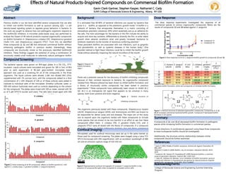
Study of alterations of bacterial membrane proteins involved in β lactam sens...
Natural Compounds Impact Oral Biofilms
1. Abstract:
Effects of Natural Products-Inspired Compounds on Commensal Biofilm Formation
Previous studies in our lab have identified several compounds that are able
to inhibit oral biofilm formation as well as quorum sensing (QS) - a cell
density-based signaling system to regulate group behaviors in bacteria. In
this work we sought to observe how non-pathogenic organisms respond to
the biofilm/QS inhibitors. A microtiter plate-based assay was performed to
analyze the impact of 48 natural products-inspired organosulfur compounds
on biofilm formation in Streptococcus cristatus CR3, Streptococcus gordonii
Challis CH1, and Streptococcus sanguinis VT517. From this, we identified
three compounds (2, 12, and 16) that enhanced commensal biofilm without
enhancing pathogenic biofilm in previous studies. Interestingly, these
compounds are structurally similar to the previously identified biofilm/QS
inhibitors. These findings suggest the potential of using a combination of
structurally related small molecules to selectively reduce pathogenic biofilm
formation while retaining commensal organisms.
Abstract:
Gavin Clark-Gartner, Stephen Kasper, Nathaniel C. Cady
SUNY College of Nanoscale Science & Engineering, Albany, NY USA
Background:
Summary:
•Compounds 2, 12, and 16 all decreased biofilm formation in pathogenic
species while increasing biofilm formation in commensal species.
•Future directions: A combinatorial approach using these three compounds
to treat multispecies biofilm should be investigated.
•Additionally, the structure-activity relationships between similar
compounds should be further examined.
B C
A
C
Acknowledgments:
It is estimated that 60-80% of bacterial infections are caused by bacteria that
grow in a biofilm, as opposed to the planktonic growth mode.1 A biofilm is a
network of bacteria that encapsulate themselves in a matrix consisting of
extracellular polymeric substance, (EPS) which essentially acts as an adhesive for
the cells. The main advantages for the bacteria in this film include the ability to
acquire and store key nutrients, differentiate to diversify the film, to enter latent
growth when adverse conditions arise, and greatly increased resistance to
antibiotic treatment via the transfer of resistance conferring plasmids.2
These persistent biofilms are associated with oral infections, such as gingivitis
and periodontitis, as well as systemic diseases in the human body.3 One
possible method to fight these infections could be to inhibit the biofilm growth
mode without adversely impacting the natural microflora of the mouth.
Plants are a potential avenue for the discovery of biofilm inhibiting compounds
because of their constant exposure to bacteria. An organosulfur compound
isolated from Petiveria alliacea has been shown to inhibit biofilm formation, and
a library of structurally similar compounds has been tested in previous
experiments.4 These compounds have additionally been shown to inhibit AI-2
QS. AI-2 is an interspecies QS signal that appears to be universal in many
species, both Gram-positive and Gram-negative.
Figure 3: General structure of
biofilm
inhibiting compounds.
The organisms previously tested with these compounds, Streptococcus mutans
UA159, Streptococcus sanguis 10556, and Actinomyces oris MG1 are known to
be responsible for dental caries and oral diseases. The major aim of this study
was to expand upon the organisms tested with these compounds to include
some species that are known to be non-harmful in an effort to see how these
compounds affect them. S. cristatus CR3, S. gordonii Challis CH1, and S.
sanguinis VT517 were all tested with the library of compounds. If a compound
could be shown to inhibit pathogenic bacteria, but not these commensal
bacteria, it may be a good candidate for oral health applications.
Compound Screening:
The bacterial species were grown on BHI-agar plates in a 5% CO2, 37ºC
incubator. Liquid cultures were inoculated and grown for 18h in 5mL of BHI
prior to each experiment. A 96-well glass-bottom microplate based
approach was used as a screening for all of the compounds in the three
organisms. The liquid cultures were diluted 1:100 into diluted BHI (25%)
supplemented with 2% sucrose (w/v), with the compounds added to a final
concentration of 1 mM. Aliquots of 100 µL of these cultures were added in
triplicate to individual wells of the microplate. Dimethyl sulfoxide, water, or
300 mM sodium hydroxide were used as controls depending on the solvent
for the compound. The plates were rinsed with 100 µL water, stained with 50
µL of 5 µM SYTO-9 (nucleic acid stain). The cells were rinsed again with 100
µL of water and fluorescence measured in a Tecan M-200 plate reader.
The dose response experiments investigated the response of all
commensal species to various organosulfur compounds. Below are the
results of compounds 12 and 16 tested against S. sanguinis.
Figure 6: Heat map of responses to organosulfur compounds. Green corresponds to
increased biofilm formation, while red corresponds to inhibited biofilm formation.
1 Lewis K (2001) Riddle of biofilm resistance. Antimicrob Agents Chemother 45:
999–1007
2Watnick P, Kotler R (2000) Biofilm, city of microbes. J Bacteriol 182(10): 2675–
2679
3 Costerton JW, Stewart PS, Greenberg EP (1999) Bacterial biofilms: a common
cause of persistent infections. Science 284: 1318–1322
4 Cady NC, McKean KA, Behnke J. et al. Inhibition of biofilm bormation, quorum
sensing and infection in Pseudomonas aeruginosa by natural products-inspired
organosulfur compounds. PLoS ONE. 2012;7:e38492
5Aaron Mosier, Ph.D.
References:
Microplates used for confocal microscopy were set up in the same manner as
those used for compound screening. The plates were imaged using a Leica TCS
SP5 II microscope with 20X magnification, with an excitation wavelength of 488
nm and an emission capture range of 510-550 nm.
Confocal Imaging:
Figure 1: Initial screening of all 48 compounds’ effects on biofilm growth at 1 mM
against S. cristatus (top), S. gordonii (middle), S. sanguinis (bottom).
Figure 2: Diagram of
biofilm formation
steps.5
Figure 4: Representative confocal micrographs of increased S. sanguinis biofilm in the
presence of compound N (right) when compared to untreated control (left).
Dose Response:
Figure 5: Dose response curve of S. sanguinis biofilm growth to compounds 12 and 16.
College of Nanoscale Science and Engineering Summer Internship
Program.
Figure 7: Chemical structure of selected organosulfur compounds. Compounds 2, 3, 12,
16 all increased commensal bacterial biofilm growth. Compound 7 decreased biofilm
formation in all species.
S. mutans
UA159
S. sanguis
10556
A. oris
MG1
S. cristatus
CR3
S.
gordonii
Challis
CH1
S.
sanguinis
VT517
7 12.38 57.86 0.3 8.99 22.91 38.98
12 31.27 64.86 44.06 29.27 44.02 147.67
3 69.05 72.81 55.54 88.22 97.17 153.06
2 76.02 87.9 103.08 153.14 139.77 117.14
16 80.77 92.03 71.73 42.59 60.88 201.76
2 3 7
12 16