
Recommended
More Related Content
What's hot
What's hot (20)
Similar to Biología molecular
Similar to Biología molecular (20)
Biología molecular
- 1. LUNG CANCER 0272–5231/02 $15.00 .00 MOLECULAR BIOLOGY OF LUNG CANCER: CLINICAL IMPLICATIONS Kwun M. Fong, MBBS, PhD, and John D. Minna, MD Lung cancer accounts for the most cancer-related MOLECULAR EPIDEMIOLOGY: deaths in men and women in the United States, INHERITED LUNG CANCER causing about 29% of all cancer deaths, more than SUSCEPTIBILITY prostate, colorectal, and breast cancers combined.59 There is intense effort now looking at the screening The major classes of carcinogens in tobacco and early detection of lung cancer, with the in- smoke are the polycyclic hydrocarbons (such as creasingly used low-dose spiral CT scanning tech- benzo (a) pyrene), the nitrosamines, and the aro- nology. In addition, there has been a flurry of new, matic amines. Tobacco smoke carcinogens may be biologically based therapy designed from knowl- activated enzymatically to chemically reactive elec- edge of molecular and biologic changes in lung trophiles that form carcinogen DNA adducts. Al- cancer cells. This review discusses the relevance of though most lung cancer cases are linked to smok- recent molecular data on lung cancer pathogenesis ing, only a minority of heavy smokers develop to clinical practice. The challenge is to translate lung cancer, leading to the notion that there may discoveries regarding how lung cancers achieve be genetic factors that affect individual susceptibil- uncontrolled growth, proliferation, and metastatic ity to develop lung cancer. Familial aggregation behavior by disruption of key cell-cycle regulators (clustering of cases) was described some time ago, and signal transduction cascades into improved with the observation of more lung cancer in rela- clinical outcomes. The molecular basis of lung car- tives of lung cancer cases.129, 191 Segregation analy- cinogenesis, essentially by genetically or epigenet- ses have suggested a mendelian codominant pat- ically altering oncogenes and tumor suppressor tern of inheritance 161 perhaps most relevant to genes, must be understood more fully and ex- early onset, never-smoking lung cancer cases.158 In ploited to enhance survival in the presence of this addition, there has been much interest in identi- highly lethal cancer. The molecular epidemiology fying the more common genetic variants or poly- of individual susceptibility to tobacco smoke car- morphisms that are hypothesized to affect lung cinogens may help in focusing on the highest risk cancer risk, particularly focusing on molecules as- group for screening technologies, which are now sociated with carcinogen handling and DNA re- capable of detecting very small lung nodules.63 pair. An individual’s susceptibility to cancer may Moreover, clinicians need new clinical strategies to be affected partially by the balance between the complement surgery, radiotherapy, and chemo- capacity to activate inhaled procarcinogens (phase therapy, and to assist in primary and secondary I enzymes) and the capacity to detoxify carcino- prevention efforts. gens (phase II enzymes), for example.178 It is recog- nized increasingly that genetic polymorphisms common in the population can affect each of these We are grateful to Dr Maree Colosimo for reviewing processes, leading to the notion that an individu- this manuscript. Supported by Lung Cancer SPORE al’s lung cancer susceptibility could be affected Grant P50 CA70907 by genetic polymorphisms, modified by tobacco From the Prince Charles Hospital, Chermside, Brisbane, Australia (KMF); and the Hamon Center for Therapeutic Oncology Research, University of Texas Southwestern Medical Center, Dallas, Texas (JDM) CLINICS IN CHEST MEDICINE VOLUME 23 • NUMBER 1 • MARCH 2002 83
- 2. 84 FONG & MINNA smoking. The genetic variations thought to be im- MOLECULAR CHANGES IN LUNG portant in lung cancer include polymorphisms at CARCINOGENESIS: THERAPEUTIC the P-450 gene loci, such as CYP1A1, CYP2D6 and IMPLICATIONS the glutathione S-transferases gene cluster.77, 148, 209 Molecular changes in lung cancer and associated Some studies have yielded conflicting results, preneoplastic cells increasingly are being identi- however, perhaps limited by the infrequency of fied, particularly with the advent of tools (such certain polymorphisms, ethnic confounding, and as the fluorescent bronchoscope) that improve the differences in smoking consumption—that is, risk detection and sampling of bronchial intraepithelial and tumor heterogeneity. Different metabolic en- lesions, and tissue microdissection techniques (in- zymes may be associated with susceptibility of cluding the rapid laser capture microdissection tumor subtypes to various tobacco smoke carcino- system) that allow for careful analysis of specific gens, for instance.96 Furthermore, the possibility lung epithelial cell populations. There is an intense of gene–gene interactions means that large, well- effort to translate these data into surrogate bio- designed studies are needed to understand fully markers for lung cancer risk assessment, for the role of these genetic variations. achieving early diagnosis, for monitoring chemo- Another area of study is the individual variabil- prevention studies, and for developing new thera- ity in DNA repair, which may be hypothesized to pies, making an understanding of the major molec- be linked to different rates of DNA repair and ular abnormalities acquired by bronchial cells therefore, accumulation of DNA mutations. ‘‘Mu- during their transformation to lung cancer an es- tagen sensitivity,’’ based on the number of chromo- sential resource for clinicians dealing with lung some breaks in peripheral blood lymphocytes after cancer. they were exposed in vitro to benzo (a) pyrene Lung cancer is the end stage of multistep carci- diol-epoxide (BPDE), a carcinogenic derivative of nogenesis, in most cases driven by genetic and benzopyrene, has been linked to increased lung epigenetic damage caused by chronic exposure to cancer risk.8 In fact, more chromosomal 3p21.3 de- tobacco smoke carcinogens and tumor promoters. letions were found by fluorescent in situ hybridiza- The genetic instability in human cancers seems tion (FISH) in peripheral blood lymphocytes of to exist at two levels—at the chromosomal level, lung cancer cases than controls after BPDE expo- including large-scale losses and gains, and at the sure, and increased rates of 3p21.3 deletions after nucleotide level, including single or several base BPDE exposure were associated with approxi- changes and DNA promoter-region methylation.97 mately a 14-fold increased risk for having lung Lung cancers are characterized by genomic imbal- cancer.210 This finding is of interest because the ances, consisting of many numerical chromosome exact same 3p21.3 region also is lost in respiratory abnormalities (aneuploidy) and structural cytoge- epithelial cells in smokers.204, 205 Studies of other netic abnormalities, including deletions, amplifi- DNA repair genes (ERCC1, XPD, XPF, XRCC3, and cations, and nonreciprocal translocations. Chromo- XRCC1) are reported increasingly and are promis- somal instability leading to aneuploidy can occur ing for at least some of these molecules.24, 142 because of the loss of function of a mitotic check- The major clinical implication lies in the poten- point, but the precise mechanism in lung cancer is tial for these studies to provide new genetic epide- not known. In addition to karyotyping, chromo- miology tools to help identify the highest risk pop- somal copy number aberrations can be mapped ulation for developing lung cancer. These more finely using newer molecular cytogenetic individuals then can be targeted for education, techniques.188 These techniques include compara- novel screening methods, such as low-dose helical tive genomic hybridization, which has demon- CT scan, and medical surveillance (Table 1). strated multiple abnormalities in lung cancer,99, 137 Table 1. MOLECULAR BIOLOGY OF LUNG CANCER: CLINICAL IMPLICATIONS Issue Possible Approaches (in Conjunction with Traditional Methods) Molecular Risk stratify according to individual susceptibility for prevention and screening epidemiology Early detection Detection of lung cancer–specific mutations, other genetic alterations, and methylation in sputum, blood, bronchoscopy specimens Develop intermediate biomarkers to gauge chemoprevention response Diagnosis Adjunct to morphologic and histologic diagnosis Assist in subtype differentiation Therapy Biologically based approaches, such as monoclonal antibodies and inhibitors against oncogenes, growth factors and receptors, tyrosine kinase inhibitors, replacement gene therapy (e.g., p53), matrix metalloproteinase inhibitors, cyclin-dependent kinase inhibitors, farnesyltransferase inhibitors, cyclooxygenase inhibitors, protein phosphatase and kinase modulators, antisense molecules, apoptosis modulators, angiogenesis inhibitors, immunotherapy, vaccination, antibody– or ligand–toxin conjugates Prognosis Predict metastatic disease, prognosis, response, and side effects of chemotherapy and radiotherapy
- 3. MOLECULAR BIOLOGY OF LUNG CANCER: CLINICAL IMPLICATIONS 85 Table 2. MAJOR MOLECULAR ALTERATIONS IN LUNG CANCER SCLC (%) NSCLC (%) Tumor suppressor genes p53 abnormalities mutation with 17p13 LOH 75–100 50 abnormal p53 expression (IHC) 40–70 40–60 p16-cyclin D1-CDK4-RB pathway lesions p16 mutation or DNA methylation with 9p21 LOH 1 10–40 absent p16 expression (IHC2) 0–10 30–70 absent RB expression with 13q14 LOH 90 15–30 APC (5q21 LOH) DNA methylation 26 46 Chromosome 3p LOH (several sites) 100 90 RAR- 3p24 (DNA methylation) 76 40 RASSF1A 3p21.3 (DNA methylation) 90 30–40 FHIT 3p14.2 (DNA methylation and del) 64 50 CDH13 (HCAD) (DNA methylation) 20 45 Proto-oncogenes and growth stimulation Putative autocrine loops GRP/GRP receptor HGF/MET SCF/KIT NDF/ERBB RAS mutation 1 15–20 MYC amplification* 15–30 5–10 Other molecular changes BCL-2 expression 75–95 10–35 Telomerase activity 100 80–85 Microsatellite instability 35 22 Promoter hypermethylation marker dependent marker dependent SCLC Small cell lung cancer; NSCLC non–small cell lung cancer; IHC immunohistochemistry; GRP gastrin releasing peptide; SCF stem cell factor; HGF hepatocyte growth factor; MET metatasis oncogene; NGF new differentiation factor; ERBB epidermal growth factor receptor. *Overexpression without amplification is observed in other cases. SCLC amplifications include MYC, MYCN, and MYCL. and FISH, which allows the examination of in- type. Microsatellite alterations have been reported terphase cells from tissue sections or cell suspen- in approximately 35% of small cell lung cancers sion, and therefore is potentially applicable for the (SCLC) and 22% of non–small cell lung cancers analysis of preneoplasia.184 Chromosomal losses of- (NSCLC).159 Microsatellite alterations seem to be ten are observed at sites that house known or associated with younger age,153 reduced survival,147 suspected tumor suppressor genes (TSGs; e.g., 3p, and advanced tumor stage.2 Finally, other DNA 5q, 17p), which have a fundamental tumorigenic repair pathways may be implicated in lung cancer, role. Tumor suppressor genes classically are inacti- given that there are reports of occasional mutations vated by structural deletion (loss) of one parental in the OGG1 gene involved in repairing oxidative allele combined with a point or small mutation, or DNA damage31 and inactivation of the DNA repair epigenetic methylation inactivation of the re- gene O6-methylguanine-DNA methyltransferase maining allele (Table 2). expression by the epigenetic mechanism of pro- At least two classes of cellular genes other than moter hypermethylation.43 These changes can be TSGs are involved in tumorigenesis—proto-onco- detected by sensitive PCR-based tests in bronchial genes and DNA repair genes. Oncogene activation washings, biopsies, and sputum and therefore are often occurs through point mutation, gene ampli- being tested as molecular markers for risk assess- fication, or rearrangement. Additionally, dysregu- ment, early detection, and monitoring prevention lated (increased or decreased) gene expression can studies. occur by other, as yet unknown, mechanisms. Studies in lung cancer have not confirmed a prom- Tumor Suppressor Genes inent role for somatically acquired abnormalities of DNA repair genes, but there are alterations in The p53 TSG maintains genomic integrity in the DNA repeat sequences that are reminiscent of the context of DNA damage from or ultraviolet irra- replication error repair (RER ) phenotype seen diation and carcinogens. DNA damage or hypoxia in colorectal and other tumors characterized by up-regulates p53, which acts as a sequence-specific mutations in DNA mismatch repair genes. None- transcription factor regulating downstream genes, theless, the phenotype in lung cancers seems dis- including p21, MDM2, GADD45, and BAX, tinct, affecting fewer markers and causing a single thereby helping to regulate the G1/S cell cycle ‘‘shift’’ of individual allelic bands, in contrast to transition, G2/M DNA damage checkpoint, and ‘‘RER laddering’’ prompting the term microsatel- apoptosis or programmed cell death. Dysfunction lite alteration to distinguish this lung cancer pheno- of p53 allows the inappropriate survival of geneti-
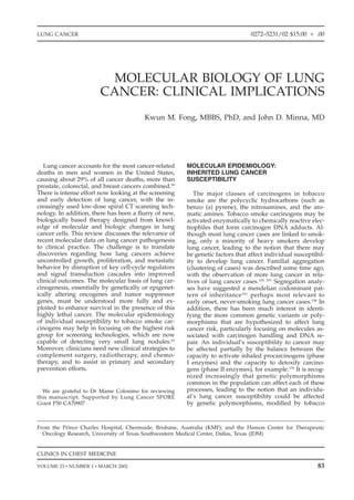
- 4. 86 FONG & MINNA Figure 1. The p53 tumor suppressor gene pathway. p53 is situated on the short arm of chromosome 17 (17p). It helps maintain genomic stability, inhibits the cell cycle at G1, and causes apoptosis if DNA is damaged beyond repair. These functions are lost because of mutation in p53 in 90% of small cell lung cancer (SCLCs) and more than 50% of non–small cell lung cancer (NSCLCs). p53 activity is antagonized by MDM2 (which thus functions as an oncogene), with the two forming a negative feedback loop. MDM2 is not frequently abnormal in lung cancers. In addition, p19 alternate reading frame (ARF) is an alternatively spliced transcript from the same chromosome 9p21 locus that gives rise to p16. It antagonizes MDM2, thus function- ing as a tumor suppressor. p19ARF mutations are apparently not common in lung cancer. p53 is inactivated by damage to both alleles, often by allelic loss and somatic missense mutations. Tumor suppressor genes also can be inactivated by promoter hypermethylation (see later) but this is apparently not common for p53. Gene therapy with wild-type p53 replacement is clinically successful in some NSCLCs. cally damaged cells, setting the stage for the accu- p53, which inhibits cyclin and cyclin-dependent mulation of multiple mutations and the subse- kinase (CDK) complexes at the G1 phase. Al- quent evolution of a cancer cell (Fig. 1). p53 plays though not somatically mutated in lung cancer, a critical role in lung cancer; its chromosome 17p13 p21 was overexpressed in 65% to 75% of NSCLCs, locus frequently is deleted hemizygously and mu- especially in well-differentiated tumors. 106 One tational inactivation of the remaining allele occurs NSCLC study reported that p21 expression was in 75% or more of SCLCs and approximately 50% linked to a favorable outcome27; another suggested of NSCLCs.17, 58, 185 Mutations of p53 in lung tumors that concordant expression of p21 and tumor correlate with cigarette smoking and are primarily growth fact or- 1 (TGF- 1) predicts better chance of the guanine to thymine (G-T) transversions ex- survival than discordant expression.16 The MDM2 pected of tobacco smoke carcinogens.58 Further- oncogene product inhibits p53 function by more, benzo (a) pyrene selectively forms adducts blocking its regulation of target genes and en- at the major p53 mutational hot spots in bronchial hances proteasome-dependent degradation of p53. epithelial cells.39 Missense p53 mutations can pro- Conversely, p53 regulates (increases) the expres- long the protein half-life, mutant p53 protein lead- sion of MDM2 by directly binding and activating ing to easily detected by immunohistochemistry the MDM2 promoter. Because the investigator (IHC).28 Other types of p53 mutations do not corre- would expect p21 expression to be lost with p53 late with IHC staining. Studies have shown abnor- loss of function, the overexpression indicates there mal p53 expression by IHC in 40% to 70% of must be other mechanisms stimulating p21. In ad- SCLCs and 40% to 60% of NSCLCs (squamous cell dition, the fact that a lung cancer develops in the carcinomas higher than adenocarcinomas).21, 56, 120 face of p21 overexpression indicates there are other Mutations of p53 have been linked to response to cellular defects that can bypass this negative cis-platinum–based chemotherapy or radiotherapy growth regulator. The MDM2 protein is overex- in NSCLC.79, 107 pressed in 25% of NSCLCs65 and MDM2 expres- Two proteins homologous to p53 include p51 sion without abnormal p53 expression has been and p73, both of which can induce growth sup- reported to be a favorable prognostic factor. Differ- pression and apoptosis. Mutations of p51 and p73 ent ways of inactivating the p53 pathway therefore appear infrequently in lung cancer, however.122, 217 may have different clinical outcomes. The expression of the gene p21 is up-regulated by Therapeutic implications: The in vitro reintro-
- 5. MOLECULAR BIOLOGY OF LUNG CANCER: CLINICAL IMPLICATIONS 87 duction of wild-type p53 into lung cancer cells that in lung cancers, with protein abnormalities de- have various other genetic abnormalities besides tected in approximately 90% of SCLCs and 15% to p53 blocks tumor cell growth by inducing 30% of NSCLCs.25, 40, 56, 143 Functional RB loss can apoptosis.1 Direct injection of a retroviral vector include deletion, nonsense mutations, or splicing containing wild-type p53 into tumors in nine pa- abnormalities, frequently leading to a truncated tients with NSCLC who had failed conventional RB protein. Whether absent RB expression is asso- treatment led to tumor regression in one third of ciated with poor prognosis in NSCLCs is contro- the patients.149, 150 Other promising strategies for versial.40, 56, 92, 212 Functionally, in vitro reintroduc- delivering p53 gene therapy include liposome–p53 tion into tumor cells of a wild-type RB suppresses complexes delivered endobronchially and adeno- SCLC growth.130 Although the inherited germline virus-mediated p53 transfer given locally, endo- form of mutant RB is associated with the develop- bronchially, or even systemically.222 Intratumoral ment of the childhood disease retinoblastoma, rela- injection of adenoviral p53 gene therapy, however, tives of patients with retinoblastoma carrying seemed to provide no additional benefit in patients germline RB mutation are about 15 times more receiving first-line chemotherapy for advanced likely to die from lung cancer than the general NSCLC.157 In addition, vaccine trials with mutant population.154 As a result, in this rare inherited p53 peptides are ongoing. Finally, because most disorder, there seems to be an example of genetic p53 mutations are missense, small molecules are predisposition to lung cancer. Two RB-related being developed to try to reactivate p53 wild-type genes also have been implicated in lung cancer, function by binding to and changing the conforma- including p107 and pRB2/p130; decreased expres- tion of the mutant p53 protein. sion of these proteins is associated with more ag- The p16–cyclin D1–cyclin-dependent kinase 4 gressive histologic behavior.110 (CDK4)–RB pathway is central to controlling the Therapeutic implications: Experiments in cell G1 to S transition of the cell cycle, and its compo- culture and transgenic lung cancer models demon- nents are altered functionally or mutated in many strate inhibition of tumor-cell growth in vivo after cancers (Fig. 2). Inactivation of both alleles of the transfecting in wild-type pRB2/p130, but human RB TSG at chromosome region 13q14 is common studies have not yet been reported.33, 119, 182 Figure 2. The p16-cyclin D1-CDK4-RB pathway. The product of the retinoblastome (RB) tumor suppressor gene binds to the E2F transcription factor during the resting (G0/G1) phase of the cell cycle. When complexed to RB, E2F cannot activate the genes needed to initiate the S phase. Moreover, the RB-E2F complex also represses the transcription of other target genes. RB is phosphorylated at the end of G1 by cyclin/cyclin dependent kinase (CDK) complexes, for example cyclin D/CDK4-6, and dephosphorylated at the end of mitosis (M). Phosphorylation of RB releases E2F, which initiates the S phase, overcoming the block to the cell cycle. In quiescent cells, RB is unphosphorylated, cyclin D levels are low, and CDK inhibitors, for example p16, p21 and p27, inhibit the cyclin/CDK complexes. In lung cancers, acquired loss of RB function allows continued cell cycling. This pathway can be turned on by mutations inactivating RB, inactivating p16, overexpression of cyclin D1, or overexpression of CDK4. Mutations inactivating p53 function also can impinge on this pathway. p21 abnormalities have not been reported. Drugs that replace RB or p16 function or that would inhibit cyclin D1 or CDK4 would represent new therapeutics. In SCLC the abnormality is usually in RB and in NSCLC the abnormality is usually in p16.
- 6. 88 FONG & MINNA Cyclin D1 inhibits the activity of RB by stimulat- somal regions in lung cancers. In the classical TSG ing its phosphorylation by CDK4. Cyclin D1 over- paradigm, the presence of underlying TSGs is sug- expression therefore is an alternative mechanism gested as the target of these genetic losses. The for disrupting the p16–cyclin D1–CDK4–RB path- chromosomal regions showing hemizygous dele- way. Cyclin D1 was overexpressed in 25% to 47% tions include 1p, 1q, 2q, 3p, 4p, 4q, 5q, 6p, 6q, 8p, of primary NSCLCs and may be associated with 8q, 11p, 11q, 14q, 17q, 18q, and 22q.98, 123, 125, 131, 155, poor prognosis. 18, 26 Transfection of a cyclin-D1 163, 194, 206 Although several of these chromosomal antisense construct into lung cancer cell lines in- arms contain known or candidate TSGs (such as duces destabilization of RB and retards growth.41 adenomatous polyposis coli [APC] at 5q21, Wilm’s It is not clear whether amplification of CDK4, tumor (WT1) at 11p13, neurofibromatosis (NF2) at which can occur in other malignancies, plays a 22q12), these genes are not known to be mutated role in lung cancer. in lung cancer. Some, however, such as APC, can Therapeutic implications: A CDK inhibitor, fla- be inactivated by tumor-acquired promoter hyper- vopiridol, is in clinical trials against lung and methylation (discussed subsequently). Hemizy- other cancers. gous loss of chromosome 3p regions, including p16 regulates RB function by inhibiting CDK4 3p25-26, 3p21.3-22, and 3p14-cen, often occurs in and CDK6 kinase activity. p16 (p16INK4/CDKN2) lung cancer, consistent with the presence of TSGs.64 is situated at chromosome 9p21 and frequently The notion of critical 3p TSGs also is supported undergoes allele loss and mutation in lung can- by the observation of homozygously deleted re- cer.159 Homozygous deletion or point mutations gions at 3p12-13, 3p14.2, and 3p21 in lung cancer occur in 10% to 40% of NSCLCs.105, 152 An alterna- cell lines.159 One candidate is fragile histidine triad tive abnormality down-regulating p16 expression (FHIT) gene at 3p14.2, which undergoes hemizy- in 30% to 40% of cases is promoter hypermethyl- gous and, occasionally, homozygous deletion in ation.109, 133 Thus, p16 inactivation represents a fur- lung cancer cells and encodes a dinucleoside hy- ther mechanism for disrupting the p16-cyclin D1– drolase. Lung cancer cells frequently (40%–80%) CDK4–RB pathway, particularly in NSCLC, func- express abnormal mRNA transcripts of FHIT but tionally analogous to the preferential RB nearly always also express wild-type FHIT tran- inactivation in SCLC.82, 86, 92, 117, 127, 162 The end result scripts.48, 177 Unlike classical TSG inactivation, FHIT from all of these mechanisms is that 30% to 50% point mutations are rare, and abnormal transcripts of early stage primary NSCLCs do not express can be found in normal lung tissue.190 Notwith- p16. Coinactivation of RB and p16 in any one standing, FHIT is expressed in normal lung but tumor is rare but cyclin D1 overexpression can FHIT protein expression loss occurs in primary coexist with these abnormalities in the same tu- lung tumors. This loss frequently is caused by mor.162 Notably, 10% to 30% of NSCLCs seem nor- promoter hypermethylation.221 Moreover, FHIT al- mal for RB and p16, indicating involvement of lele loss is more common in smokers than non- cyclin D1, CDK4, or other pathway members in smokers176 and may be associated with a poorer these cases. p16 alteration and loss of function in likelihood of survival in NSCLC.23 Furthermore, lung cancer may be associated with tumor progres- reintroduction of exogenous wild-type FHIT sup- sion, clinical stage, and survival, although not all pressed tumorigenicity of a lung cancer cell line in studies concur.56, 86, 92, 183 nude mice,74, 170 whereas others have reported that The p16 locus also encodes a second alternative FHIT transfection does not suppress tumor growth reading frame (ARF) protein, p19ARF, which over- of human cancer cell lines.132 laps with p16, and that also may be important in The 3p21.3 region has been examined exten- growth regulation. p19ARF sometimes called p14ARF sively for putative TSGs, particularly at homozy- binds to the MDM2-p53 complex and prevents gously deleted regions.89, 90, 201, 213 The RASSF1A p53 degradation, resulting in p53 activation. IHC isoform of RASSF1 undergoes promoter hyper- analysis suggests more frequent loss of p19ARF pro- methylation in approximately 30% of primary tein expression in tumors with neuroendocrine fea- NSCLCs and more than 90% of SCLCs, causing tures.54 One genetic locus at 9p21 therefore pro- loss of RASSF1 A expression. This promoter hyper- duces two products, p16 and p19ARF, both of which methylation is correlated with poorer survival in play a critical role in growth regulation; p16 with resected patients with NSCLC. Transfection and the RB pathway, and p19ARF with the p53 pathway. re-expression of wild-type RASSF1 A result in sup- Reduced expression of another CDK-inhibitor pression of lung cancer tumorigenicity. 22, 37 The gene, p27KIP1, correlates with poor prognosis in RARB2 isoform also undergoes promoter hyper- NSCLC.42, 216 In contrast, most SCLCs exhibit in- methylation in approximately 60% to 70% of creased p27KIP1 staining, suggesting a possible link NSCLCs and SCLCs. This occurrence leads to loss with neuronal differentiation.216 p 57KIP2 at chromo- of RARB2 expression, which occurs in most lung some region 11p15 is imprinted with maternal ex- tumors and in some preneoplastic lesions. This pression and p57KIP2 expression is down-regulated situation could account for the retinoid resistance by selective loss of the maternal alleles in some seen in lung cancers.55, 195 lung cancers.91 Other candidate TSGs include the PTEN gene at Other candidate tumor suppressor genes are im- chromosome 10q23, which encodes a phospha- plied by the finding of many hemizygous and tase.100 Protein tyrosine phosphatases are able to some homozygous deletions at multiple chromo- antagonize the growth-promoting protein kinases,
- 7. MOLECULAR BIOLOGY OF LUNG CANCER: CLINICAL IMPLICATIONS 89 and the PTEN gene is mutated in a subset of lung vated earlier in women in response to tobacco cancers and homozygously deleted in several lung exposure.164 cancer cell lines.52 Another candidate at 10q, region Therapeutic implications: The in vitro formation 25.3-26.1, is DBMT1, which frequently is down- of soft agar clones and the in vitro growth of nude regulated and occasionally homozygously deleted mouse xenografts of SCLC cell lines are inhibited in lung cancer.210 There also seem to be important by a neutralizing monoclonal antibody directed chromosomal deletions at 11q23-24, including the against GRP/BN and by antagonists of BN.35, 60 A locus for PPP2RIB (beta isoform of the A subunit clinical trial of the anti-BN monoclonal antibody of the human protein phosphatase-2A) gene, in has shown some antitumor activity in previously which mutations have been described in lung and treated patients with SCLC, with phase I results colon cancers, suggesting a TSG function.198, 199 published recently.29, 81 The development of peptide Clinical implications: The increasing discovery antagonists by new small-molecule therapeutics is of TSGs important in lung cancer pathogenesis, another attractive strategy. particularly those at 3p, which occur early in lung cancer pathogenesis, is likely to lead to the oppor- tunity to develop new diagnostic and therapeutic ERBB Family strategies. These strategies would include monitor- ing target tissue for 3p allele loss and promoter Non–small cell lung cancers, rather than SCLCs, hypermethylation, treatment with demethylation often demonstrate abnormalities of the neuregulin agents, and replacement gene therapy for 3p TSGs. receptors, ERBB2 and ERBB1, which are part of a family of transmembrane receptor tyrosine ki- nases. On ligand binding, ERBB receptors homo- Proto-oncogenes and Growth Stimulation or heterodimerize, thereby inducing intrinsic ki- nase activities that initiate intracellular signal Autocrine and paracrine growth stimulatory transduction cascades, including the MAP kinase loops exist in lung cancers as a consequence of the pathway. ERBB2 (also called HER2/neu) is highly expression of growth factors, regulatory peptides, expressed in approximately 30% of NSCLCs, espe- and their receptors by the cancerous or adjacent cially adenocarcinomas.141, 202 Transfection experi- normal cells. Several but not all components of ments suggest that ERBB2 overexpression contri- these stimulatory pathways are proto-oncogene butes to tumorigenicity in immortalized human products. bronchial epithelial cells.121 High ERBB2 levels are The gastrin-releasing peptide/bombesin (GRP/ associated with the multiple-drug-resistance phe- BN) growth stimulatory loop has a role in lung notype 192 and increased metastatic potential in development and repair.179 Immunohistochemical NSCLC,219 which may help explain the poor clini- studies show that approximately 20% to 60% of cal outcome linked to ERBB2 overexpression re- SCLCs express GRP/BN, whereas NSCLCs express ported by some investigators.83, 139 GRP/BN less frequently.144 The human GRP/BN Therapeutic implications: In preclinical studies receptor subtypes belong to the G-protein–coupled with tumor cell lines, trastuzumab (Herceptin), a receptor superfamily and include receptors for monoclonal antibody against the ERBB2 receptor, GRP, neuromedin B, and bombesin subtype-3; all was found to have additive and synergistic effects of these can be expressed in SCLC, NSCLC, and with some chemotherapeutic agents. Clinical trials in some bronchial epithelial biopsies from smok- investigating combination chemotherapy with ers.45, 166 Why these embryonic regulatory loops trastuzumab and a variety of chemotherapeutic become ‘‘reactivated’’ in lung cancers is uncertain agents are in progress in lung cancer,4 and a com- because mutations of GRP/BN or its receptor are mercial US Food and Drug Administration (FDA)- seemingly absent. Nonetheless, the GRP/BN auto- approved diagnostic kit is now available for detec- crine loop is an important growth-stimulatory loop tion of ERBB2 (HER-2/neu) expression. in lung cancer, particularly SCLC. It may play an ERBB1 (also called EGF receptor) regulates epi- early pathogenic role because there is an increased thelial proliferation and differentiation and usually likelihood of expression of the GRP receptor is activated in lung cancer cells by overexpression mRNA in the respiratory epithelium of some indi- by an unknown mechanism. The production of viduals with a history of prolonged tobacco expo- ERBB1 ligands such as EGF and TGF- by lung sure, and expression of the GRP receptor mRNA cancer cells expressing cognate receptors indicates is accompanied by in vitro responsiveness of these an autocrine loop.141, 151 More common in NSCLC, respiratory epithelial cells to the mitogenic effects ERBB1 activation may be related to tumor stage of bombesin-like peptides. 166 In addition, there and differentiation.38, 187 may be gender-specific GRP abnormalities, inter- Clinical implications: Monoclonal antibodies esting because of possible differences in lung can- against the ERBB1 receptor (C225, ImClone) are cer risk between the genders. A recent study, for entering clinical trials combined with chemother- instance, found that the GRP receptor gene situ- apy, with early phase I results available.14 In addi- ated on the X chromosome is expressed more fre- tion, several tyrosine kinase inhibitors that have quently in women than in men in the absence of some selectivity as EGF receptor (ERBB1) blockers smoking and that expression of this gene is acti- (CP358774, ZD1839-Iressa, OS1774) also are being
- 8. 90 FONG & MINNA tested in clinical trials, with a major advantage PDGF tyrosine kinases) and has promising activity that most are orally active and able to be given in SCLC cell lines.200 with conventional chemotherapy (Fig. 3). Members of the RAS proto-oncogene family Other membrane tyrosine kinases include the (KRAS, HRAS, and NRAS) encode plasma mem- hepatocyte growth factor (HGF), which normally brane proteins and are activated in some lung can- stimulates epithelial cell proliferation, mobility, cers by point mutations, resulting in inappropriate and differentiation programs. During fetal lung signaling for continued cell division (Fig. 4). KRAS development, HGF acts as a mesenchyme-derived is the most frequently activated RAS gene in lung morphogenic factor. The constitutively low levels cancer, usually by mutations at codon 12 but, occa- of HGF increase in response to lung or distant sionally, codons 13 and 61. KRAS mutations affect injury.214 Hepatocyte growth factor stimulates mi- approximately 20% to 30% of lung adenocarcino- togenesis or motogenesis of human bronchial epi- mas and 15% to 20% of all NSCLCs, but rarely thelial, alveolar type II, and SCLC cells in vitro. SCLCs.144 There is subtype heterogeneity—for ex- The receptor for HGF is the product of the metasta- ample, KRAS mutations are present in parenchy- sis (MET) proto-oncogene. It generally is expressed mal but not bronchial adenocarcinomas34 and gob- in normal lung and SCLC and NSCLC; HGF, how- let-cell subtypes of adenocarcinoma have the highest frequency of KRAS mutations. 193 KRAS ever, is expressed mainly in NSCLCs, suggesting mutations correlate with smoking,171 often being an autocrine loop specific for NSCLC.62, 128 Clini- the guanine to thymine (G-T) transversions associ- cally, high HGF levels are associated with a poor ated with polycyclic hydrocarbons and nitrosa- outcome in resectable patients with NSCLC.168 The mines.58 KRAS mutations may imply a poor prog- KIT proto-oncogene encodes the tyrosine kinase nosis in NSCLC, although this observation is receptor, CD117. It is coexpressed together with debated.57, 113, 146, 167, 172 Neither chemotherapy sensi- its ligand, stem cell factor, in many SCLCs,93, 160 tivity nor survival is correlated with KRAS muta- representing another autocrine loop that may pro- tions in a prospective study of advanced lung ade- vide a growth advantage or mediate chemoattrac- nocarcinoma.145 Moreover, KRAS mutations were tion. Other putative loops involve insulin growth not associated with in vitro resistance against a factor (IGF)-I, IGF-2, and type I IGF receptors, range of chemotherapeutic agents in NSCLC cell which frequently are coexpressed in SCLC and lines.192 NSCLC140 as are PDGF and its receptor.11 Therapeutic implications: To be active in the cell, Therapeutic implications: These growth loops Ras has to undergo lipid modification (farnesyla- provide additional avenues for the development tion), a process regulated by the farnesyltransfer- of novel, biologically based therapies. The tyrosine ase enzyme. This process potentially can be kinase inhibitor, STI 571, for instance, can inhibit blocked by specific farnesyltransferase inhibitors the KIT tyrosine kinase (in addition to bcr-Abl and (FTIs), several of which are in clinical trials against Figure 3. Epidermal growth factor receptor (EGFR) tyrosine kinase inhibition. After ligand binding, the EGFR receptor dimerizes and signals downstream mole- cules through the activity of a specific tyrosine kinase, which can be inhibited with specificity by drugs with specificity for the EGFR.
- 9. MOLECULAR BIOLOGY OF LUNG CANCER: CLINICAL IMPLICATIONS 91 Figure 4. Mutant KRAS pathway. RAS is active when bound to guanosine triphos- phate (GTP) and inactive when bound to guanosine diphosphate (GDP). The intrinsic GTPase activity of RAS is stimulated by GAP resulting in inactive RAS. Oncogenic RAS mutations inhibit GTPase activity causing it to remain permanently activated with ensuing positive signaling to downstream molecules. Drugs that could interfere with the localization of mutant KRAS in the membrane (and thus inactivate it) or interfere with its expression such as antisense compounds, or with components of the downstream signaling cascade such as mitogen stimulated extracellular regu- lated kinase (MEK) inhibitors all have therapeutic potential. lung cancer (e.g., BMS214662, RII5777, SCH 66336). growth inhibition of an SCLC cell line by all-trans- Trials of vaccination with mutant KRAS peptides retinoic acid is associated with increased neuroen- also are underway. Although tumor responses docrine differentiation, increased MYCL, and de- have been seen, KRAS the commonly mutated creased MYC expression.134 Gene therapy in the form in lung cancer, probably is not blocked by form of antisense technology, although encourag- FTIs. Other experimental approaches to block ing in cell culture systems, has not yet been re- KRAS have included antisense treatment, ribo- ported in clinical trials. zymes to block protein expression, and signal Many new therapeutic strategies therefore are transduction interference further down the KRAS- being tested to exploit the molecular abnormalities activated pathway. observed in lung cancer cells. Developments in antiangiogenic and antimetastatic agents are de- scribed later, and these new therapeutic ap- MYC Proto-oncogenes proaches based on specific tumor biology offer much potential promise. RAS signaling ultimately activates nuclear proto-oncogene products such as MYC, which, on heterodimerization, transcriptionally activates MOLECULAR CHANGES IN LUNG downstream genes that drive cells to grow. The CARCINOGENESIS: EARLY DETECTION myc family comprises MYC, MYCN, and MYCL AND DIAGNOSTIC IMPLICATIONS (originally isolated from a lung cancer118). MYC is activated most frequently and affects SCLC and Rapid advances in molecular biology, radiology, NSCLC, whereas the others usually only affect bronchoscopy, and other biopsy methods have oc- SCLC. Activation occurs by gene amplification curred since the classic tests of chest radiography ( 20–100 copies per cell) or by transcriptional dys- and sputum cytology, neither of which seemed to regulation, both of which lead to protein overex- alter outcome when used as a screening modality. pression. Richardson et al144 concluded from 17 Knowledge of the molecular evolution is crucial if studies that 18% to 31% of SCLCs had amplifica- tumor characteristics are to be exploited as a tool tion of one MYC family member. Conversely, only for early detection and diagnosis. 8% to 20% of NSCLCs were affected.144 Cancers arise from precursor lesions in a Therapeutic implications: MYC amplification multistep fashion, with preneoplastic cells somati- seems to occur more frequently in chemotherapy- cally acquiring genetic alterations, ultimately treated patients, and the ‘‘variant’’ SCLC subtype,76 evolving into invasive cancer by clonal expansion. and may correlate with adverse survival. In vitro Morphologically distinct preneoplastic changes
- 10. 92 FONG & MINNA (hyperplasia, metaplasia, dysplasia, and carcinoma cently.69 There is the increasing ability to obtain in situ) can be observed in bronchial epithelium cells and cellular products such as DNA and RNA before overt, invasive cancer develops. The se- from clinical specimens such as sputum, bronchial quential morphologic changes that occur in pre- biopsies, brushings, lavage fluids, and blood to neoplastic lesions are best described for squamous use as substrates for these new techniques. A cell carcinomas. Other potential premalignant le- promising monoclonal antibody that recognizes sions now have been recognized, however, includ- hnRNP A2/B1 seems to have good sensitivity for ing atypical adenomatous hyperplasia as a precur- detecting lung cancers, with prospective study re- sor for peripheral adenocarcinomas; angiogenic sults awaited.47, 189 Microsatellite alterations, detect- squamous dysplasia, in which dysplastic lesions able by PCR, have been suggested as useful mark- occur with an angiogenic response; and diffuse ers of lung cancer and can be found in tumors and idiopathic neuroendocrine cell hyperplasia for car- corresponding sputum samples.111 The finding of cinoids. Some reversal of morphology can occur microsatellite alterations in bodily fluids from pa- after smoking cessation even though the elevated tients with chronic obstructive pulmonary disease risk for lung cancer does not return completely and individuals without clinical lung cancer, how- to baseline, raising the possibility of irreversibly ever, raises an important issue.46, 165 Any potential genetically damaged areas of at-risk bronchial epi- biomarker needs to be detectable easily in bodily thelium. These preneoplastic cells and even macro- fluids (i.e., is sensitive) but must be specific for scopically normal bronchial epithelium adjacent to neoplastic transformation and not just reflect cancers can contain genetic abnormalities identical smoking-related lung damage. The ease of ob- to those in invasive cancer cells. These abnormali- taining peripheral blood has led to attempts to ties include allele loss at several loci (3p, 9p, 8p, identify blood-based biomarkers, especially given 17p), MYC and RAS up-regulation, cyclin D1 ex- that tumor DNA is released into plasma. In this pression, p53 immunoreactivity, BCL2 overexpres- regard, microsatellite alterations have been re- sion, and DNA aneuploidy.19, 20, 67, 101, 156 Examina- ported in plasma DNA,30 but recent efforts have tion of microdissected, preneoplastic lesions focused on detecting methylation changes in circu- suggests that the earliest change is allele loss at lating DNA in blood samples. chromosome region 3p, then 9p, 8p, 17p (with p53 mutation), and 5q, followed by RAS mutations.72, 87, 180, 207 There are data suggesting that KRAS acti- Tumor-acquired Promoter vation also can occur at early stages, and muta- Hypermethylation as a Common Method tions can be found in atypical alveolar hyperpla- of Inactivating Gene Expression in sia.34, 101, 203 The genetic changes found in invasive Tumors cancers and preneoplasia also can be identified in Methylation of CpG islands in the promoter re- morphologically normal-appearing bronchial epi- gions of genes is a mechanism for controlling gene thelium from current or former smokers.104, 208 The expression. Abnormalities of DNA methylation oc- earliest change appearing in histologically normal cur consistently in human neoplasia and promoter- epithelium seems to be allele loss at 3p, in about region hypermethylation may inactivate genes 50% of specimens from current or former smokers. transcriptionally, including TSGs (Fig. 5). In There is also a progressive increase in the fre- NSCLCs, hypermethylation contributes to down- quency and size of the areas undergoing allele loss regulation of p16 expression, occurs at an early as they progress from normal to hyperplasia or stage in lung carcinogenesis,15, 109, 133 and correlates metaplasia (representing mildly abnormal lesions) with smoking.85 An increasing number of genes to dysplasia, to carcinoma in situ.207, 208 These ob- have been observed to undergo promoter methyla- servations are consistent with ‘‘field canceriza- tion in lung cancer but not in associated normal tion,’’ whereby the whole tissue region is exposed lung, including p16, DAPK, GSTPI, MGMT, FHIT, repeatedly to tobacco smoke and is at risk for RARB, APC, CDHI3(HCAD), and RASSFIA22, 43, 195, developing multiple, separate, clonally unrelated 220, 222 . Other regional sites of hypermethylation foci of neoplasia, a notion supported by the wide- have been found in lung cancer, including sites at spread presence of aneuploidy in the respiratory 3p, 4q34, 10q26, and 17p13, although the precise tree of smokers.173 A case report of a smoker with- gene targets at these sites are uncertain and the out invasive lung cancer with an identical somatic significance not yet apparent.88, 103 p53 gene mutation at geographically dispersed preneoplastic bronchial lesions is consistent with the clonal expansion of a single progenitor clone Clinical Implications throughout the respiratory tree.53 Hypermethylation of certain gene promoters also may have relevance in predicting clinical out- Diagnostic Implications come for patients with lung cancer.22, 85 Because The considerable inroads made in developing methylated DNA sequences can be found even in new tools for the early detection of lung cancer, the setting of a high background of constitution- including antibodies for immunostaining, poly- ally unmethylated normal DNA, they are attractive merase chain reaction (PCR) techniques, and com- candidates for early molecular detection tools and puterized image analysis, have been reviewed re- for following chemoprevention studies. A small
- 11. MOLECULAR BIOLOGY OF LUNG CANCER: CLINICAL IMPLICATIONS 93 Figure 5. Tumor acquired promoter hypermethylation leads to silencing of gene expression. Schematic diagram of how hypermethylation of cytosine (C) in CpG islands in the promoter region of a gene in a tumor cell may down-regulate gene expression. Active gene expression often is associated with unmethylated promoter CpG islands. In lung cancers, hypermethylation of promoter CpG is- lands is associated with down-regulation of gene expression and thus is a mechanism for inactivating tumor suppressor genes. These methylated DNA products can be detected by methylation specific polymerase chain reaction (PCR) in tissue or body fluid specimens, and the expression of these genes can be restored by treatment with methylation inhibitors such as 5 aza-cytidine, which is augmented by treatment with histone deacetylase inhibitors. study reported the detection of aberrant methyla- have telomerase activity that is able to compensate tion of p16 or O6-MGMT in DNA from sputum in for telomere shortening by replacing the hexameric patients with squamous cell lung cancer up to 3 repeats, providing cellular ‘‘immortality.’’ Three years before clinical diagnosis.135 In addition, ab- major components of human telomerase—RNA normal identically methylated DNA can be de- component, telomerase-associated protein, and ca- tected in serum from cases in which the tumor talytic subunit—frequently are overexpressed in was methylated.44 Apart from potential diagnostic lung cancers, and activity can be determined by use, the ability for methylation to be reversed with measuring telomerase enzyme activity (TRAP drugs such as 5-aza-2 deoxycytidine leads to po- assay). 13 Nearly all SCLCs and 80% to 85% of tential therapeutic developments. Retinoic acid, for NSCLCs have high levels of telomerase activity.6, 70 instance, plays an important role in lung develop- High telomerase activity is associated with in- ment and differentiation, acting primarily through creased cell proliferation rates and advanced stage nuclear receptors encoded by the retinoic acid re- in NSCLCs. Additionally, telomerase activity and ceptor- (RAR- ) gene. RAR- often is hypermeth- dysregulated RNA expression frequently are ylated in lung cancers, particularly SCLC,195 and found in carcinoma in situ lesions, implicating chemical demethylation may provide an avenue involvement early in lung cancer development.215 for the re-expression of RAR- . Clinical Implications Telomerase Activation The widespread presence of dysregulated te- Another potential biomarker in lung cancer is lomerase in lung cancers has prompted examina- telomerase activation. Human telomeres are struc- tion of its role as a molecular marker. Telomerase tures located at the ends of chromosomes compris- positivity can be found in bronchoalveolar lavage ing TTAGGG tandem DNA repeats bound to spe- fluid and may provide a useful adjunct to tradi- cific proteins. During normal cell division, the tional cytology.211 On the other hand, it seems to absence of telomerase activity is associated with have limited value in the molecular staging of progressive telomere shortening, perhaps repre- established NSCLC. 5 The development of te- senting an intrinsic ‘‘cellular clock,’’ leading to nor- lomerase inhibitors is being pursued in new thera- mal cell ‘‘senescence’’ and ‘‘mortality.’’ In contrast, peutic drug development. germ cells, some stem cells, and most cancer cells Biomarkers based on other molecular mutations
- 12. 94 FONG & MINNA also hold promise for early detection, although sion), BCL-xL antisense in NSCLC, and a bispecific sensitivity remains an issue. Some 15% to 25% BCL-2–BCLxL antisense to target SCLC and of patients with lung cancer develop antibodies NSCLC. against the p53 protein, for instance, indicating that mutant p53 protein overexpression can lead to a humoral immune response. Whether serum Angiogenesis p53 antibodies can be used as a diagnostic marker Tumors cannot exceed 1 to 2 mm3 volume with- is presently unclear.95, 102, 114 Additionally, p53 muta- out developing new blood vessels and therefore tions can be detected in DNA from tumor cells in require angiogenic factors early in their pathogene- plasma.169 sis. Microvessel density may be regarded as a mor- phologic measure of tumor angiogenesis and has been linked variably to metastases and impaired MOLECULAR CHANGES IN LUNG survival in lung cancer.10, 49, 124, 136 Tumor angiogen- CARCINOGENESIS: PROGNOSTIC esis is complex and controlled by a diverse family IMPLICATIONS of inducers and inhibitors governing angiogenesis and regulating endothelial cell proliferation and Refining the staging of lung cancer with reliable migration. 61 Vascular endothelial growth factor and robust prognostic markers may allow the indi- (VEGF) and basic fibroblast growth factor are im- vidualization of therapy. Molecular abnormalities, portant angiogenesis inducers. Vascular endothe- being a fundamental step in tumorigenesis, may lial growth factor expression is associated with prove to be useful in this context.36, 94, 115 Before intratumoral microvessel density and tumor-cell adoption into routine clinical use, however, large, proliferation in squamous lung cancer.7, 51, 108 Vascu- high-quality, prospective studies of potential bio- lar endothelial growth factor expression is higher markers are required to validate their role because in lung cancers with nodal metastasis than those most studies have been retrospective, relatively without126 and is associated with poor prognosis small in size, possibly exposed to bias such as in NSCLC.51, 196 In squamous cell carcinomas, the selection bias, examined markers with different VEGF receptor, Flt-1, is expressed frequently, sug- techniques, and came to different conclusions. gesting a possible autocrine role of VEGF.196 Mu- Meta-analyses of p53 as a potential prognostic bio- tant p53 acts synergistically with hypoxia to in- marker have been reported and serve to highlight duce VEGF expression,84 whereas wild-type p53 the heterogeneity of the studies to date.71, 112 Apart up-regulates expression of the antiangiogenic pro- from the prognostic relationships of the various tein, TSP-1.116 Fibroblast growth factor is expressed biomarkers discussed earlier, other potentially im- in approximately 70% of NSCLCs, but its prognos- portant biomarkers of lung cancer progression and tic significance is controversial.186, 197 outcome are described herewith. Therapeutic implications: There are multiple new therapeutic strategies in lung cancer directed against angiogenesis in general and the VEGF sys- Apoptosis tem in particular. There currently are clinical trials testing rhuMAbVEGF, an antiangiogenic human- Tumor cells often escape the normal physiologic ized monoclonal antibody. Other potential agents response termed programmed cell death or include a monoclonal antibody against VEGF re- apoptosis when challenged by cellular and DNA ceptor 2 (KDR/FLK1, ImCone), a tyrosine kinase damage. A key player in apoptosis is the product inhibitor (SU5416, Sugen), and a ribozyme directed of the breakpoint cluster lymphoma (BCL2) antia- against the VEGF receptor 1 (Flt-1) mRNA (Angio- poptotic proto-oncogene. Its expression is higher zyme). in SCLCs (75%–95%) than in NSCLCs,75, 78 in which expression is greater for squamous cell carcinoma than adenocarcinoma.12, 66, 75, 138 The former finding Molecules Involved in Metastatic is interesting because SCLCs are more responsive Behavior to chemotherapy, a modality generally resulting in tumor apoptosis. Clinically, there is a trend toward Tumor metastasis is a complex, multistep, host- longer survival in SCLCs that express BCL-2,78 but dependent process. Cell adhesion molecules have this relationship is controversial in NSCLC.12, 50, 66, 138 been implicated in epithelial differentiation, carci- BAX counteracts BCL-2 and promotes apoptosis nogenesis, and metastasis. E-cadherin is responsi- and is a downstream transcription target of p53. ble for the organization, maintenance, and mor- Expression of BAX and BCL-2 is related inversely phogenesis of epithelial tissues, and reduced in neuroendocrine cancers; most typical and atypi- expression has been associated with tumor dedif- cal carcinoids have low BCL-2 and high BAX ex- ferentiation, increased lymphogenous metastasis, pression, with the reverse in most SCLCs and large and poor survival rates in NSCLC.181 The integrins cell neuroendocrine cancers.21 Therapeutic Implica- that bind to collagen and laminin in basement tions: Among the anti-apoptosis strategies in pre- membranes are down-regulated in NSCLC.174 Re- clinical trials are studies of antisense BCL-2 in duced integrin- 3 expression is linked with poor SCLC (to down-regulate BCL-2 protein expres- prognosis in adenocarcinomas.3 Others have re-
- 13. MOLECULAR BIOLOGY OF LUNG CANCER: CLINICAL IMPLICATIONS 95 ported that the v6 isoform of the CD44 lymphocyte 3. Adachi M, Taki T, Huang C, et al: Reduced integrin homing receptor correlates with adverse prognosis 3 expression as a factor of poor prognosis of pa- in NSCLC.68 The degradation of the extracellular tients with adenocarcinoma of the lung. J Clin Oncol matrix and basement membrane, particularly type 16:1060–1067, 1998 4. Agus DB, Bunn PA Jr, Franklin W, et al: HER-2/neu IV collagen, by tumor cells is essential for invasion as a therapeutic target in non–small-cell lung cancer, and metastasis. Type IV collagen expression of ad- prostate cancer, and ovarian cancer. Semin Oncol enocarcinomas is lost progressively with increas- 27:53–63; discussion 92–100, 2000 ing tumor dedifferentiation.32 5. Ahrendt SA, Yang SC, Wu L, et al: Comparison of Therapeutic implications: The matrix metallo- oncogene mutation detection and telomerase activ- proteinase enzymes that degrade stroma and ity for the molecular staging of non-small cell lung therefore could lead to tumor invasion and metas- cancer. Clin Cancer Res 3:1207–1214, 1997 tasis include collagenases, gelatinases, stromely- 6. Albanell J, Lonardo F, Rusch V, et al: High te- sins, membrane-type matrix metalloproteinases, lomerase activity in primary lung cancers: Associa- matrilysin, and metalloelastase. Gelatinase A, for tion with increased cell proliferation rates and ad- vanced pathologic stage. J Natl Cancer Inst 89: instance, is expressed in SCLCs ( 50%) and 1609–1615, 1997 NSCLCs ( 65%).80 Stromelysin-3 has been shown 7. Ambs S, Bennett WP, Merriam WG, et al: Vascular to be overexpressed by stromal elements in pri- endothelial growth factor and nitric oxide synthase mary NSCLCs compared with adjacent normal expression in human lung cancer and the relation lung specimens.9 Several matrix metalloproteinase to p53. Br J Cancer 78:233–239, 1998 inhibitors (Prinomastat, Marimastat, and BAY 8. Amos CI, Xu W, Spitz MR: Is there a genetic basis 129566) are in clinical trials. for lung cancer susceptibility? Recent Results Can- cer Res 151:3–12, 1999 9. Anderson IC, Sugarbaker DJ, Ganju RK, et al: Stro- SUMMARY melysin-3 is overexpressed by stromal elements in primary non-small cell lung cancers and regulated This review summarizes the rapidly expanding by retinoic acid in pulmonary fibroblasts. Cancer Res 55:4120–4126, 1995 knowledge of the molecular pathogenesis of lung 10. Angeletti CA, Lucchi M, Fontanini G, et al: Prognos- cancer. It is clear that respiratory epithelial cells tic significance of tumoral angiogenesis in com- require many genetic alterations to become inva- pletely resected late stage lung carcinoma (stage sive and metastatic cancer. Much more is to be IIIA-N2). Impact of adjuvant therapies in a subset learned, but with modern technology. Clinicians of patients at high risk of recurrence. Cancer 78: can detect ‘‘field cancerized’’ regions and preneo- 409–415, 1996 plastic and malignant cells, therefore offering the 11. Antoniades HN, Galanopoulos T, Neville-Golden J, opportunity to intercede with biomarker-moni- et al: Malignant epithelial cells in primary human tored prevention and early detection efforts. Such lung carcinomas coexpress in vivo platelet-derived molecular screening and detection efforts will growth factor (PDGF) and PDGF receptor mRNAs and their protein products. Proc Natl Acad Sci U S likely be coupled to advances in low-dose com- A 89:3942–3946, 1992 puted tomographic imaging, positron emission to- 12. Apolinario RM, van der Valk P, de Jong JS, et al: mography scans, and other imaging modalities.63, Prognostic value of the expression of p53, bcl-2, and 73, 175, 218 Although this molecular marker approach bax oncoproteins, and neovascularization in pa- has great potential, there is not yet a molecular tients with radically resected non-small-cell lung marker validated in large prospective trials that cancer. J Clin Oncol 15:2456–2466, 1997 has major independent predictive prognostic 13. Arinaga M, Shimizu S, Gotoh K, et al: Expression value. There is an urgent need for large, ade- of human telomerase subunit genes in primary lung quately powered, carefully designed prospective cancer and its clinical significance. Ann Thorac Surg studies to identify clinically useful new biomark- 70:401–405; discussion 405–406, 2000 ers. Finally, new therapeutic strategies with genetic 14. Baselga J, Pfister D, Cooper MR, et al: Phase I stud- ies of anti-epidermal growth factor receptor chim- manipulation, small molecules, antibodies, vac- eric antibody C225 alone and in combination with cines, and, particularly, new drugs targeting spe- cisplatin. J Clin Oncol 18:904–914, 2000 cific biologic pathways found to be abnormal in 15. Belinsky SA, Nikula KJ, Palmisano WA, et al: Aber- lung provide for future optimism. Researchers rant methylation of p16(INK4a) is an early event in need to define their individual value, especially lung cancer and a potential biomarker for early when integrated with standard therapies. diagnosis. Proc Natl Acad Sci U S A 95:11891– 11896, 1998 16. Bennett WP, el-Deiry WS, Rush WL, et al: p21waf1/ References cip1 and transforming growth factor beta 1 protein expression correlate with survival in non-small cell 1. Adachi J, Ookawa K, Shiseki M, et al: Induction of lung cancer. Clin Cancer Res 4:1499–1506, 1998 apoptosis but not G1 arrest by expression of the 17. Bennett WP, Hussain SP, Vahakangas KH, et al: Mo- wild-type p53 gene in small cell lung carcinoma. lecular epidemiology of human cancer risk: Gene- Cell Growth Differ 7:879–886, 1996 environment interactions and p53 mutation spec- 2. Adachi J, Shiseki M, Okazaki T, et al: Microsatellite trum in human lung cancer. J Pathol 187:8–18, 1999 instability in primary and metastatic lung carcino- 18. Betticher DC, Heighway J, Hasleton PS, et al: Prog- mas. Genes Chromosomes Cancer 14:301–306, 1995 nostic significance of CCND1 (cyclin D1) overex-
- 14. 96 FONG & MINNA pression in primary resected non-small-cell lung tors in human small-cell lung cancer. Nature 316: cancer. Br J Cancer 73:294–300, 1996 823–826, 1985 19. Betticher DC, Heighway J, Thatcher N, et al: Abnor- 36. D’Amico TA, Aloia TA, Moore MB, et al: Molecular mal expression of CCND1 and RB1 in resection biologic substaging of stage I lung cancer according margin epithelia of lung cancer patients. Br J Cancer to gender and histology. Ann Thorac Surg 69:882– 75:1761–1768, 1997 886, 2000 20. Brambilla E, Gazzeri S, Lantuejoul S, et al: p53 mu- 37. Dammann R, Li C, Yoon JH, et al: Epigenetic inacti- tant immunophenotype and deregulation of p53 vation of a RAS association domain family protein transcription pathway (Bcl2, Bax, and Waf1) in pre- from the lung tumour suppressor locus 3p21.3. Nat cursor bronchial lesions of lung cancer. Clin Cancer Genet 25:315–319, 2000 Res 4:1609–1618, 1998 38. Damstrup L, Rygaard K, Spang-Thomsen M, et al: 21. Brambilla E, Negoescu A, Gazzeri S, et al: Expression of the epidermal growth factor receptor Apoptosis-related factors p53, Bcl2, and Bax in neu- in human small cell lung cancer cell lines. Cancer roendocrine lung tumors. Am J Pathol 149:1941– Res 52:3089–3093, 1992 1952, 1996 39. Denissenko MF, Pao A, Tang M, et al: Preferential 22. Burbee DG, Forgacs E, Zochbauer-Muller S, et al: formation of benzo[a]pyreme adducts at lung cancer Epigenetic inactivation of RASSF1A in lung and mutational hotspots in P53. Science 274:430–432, breast cancers and malignant phenotype suppres- 1996 sion. J Natl Cancer Inst 93:691–699, 2001 40. Dosaka-Akita H, Hu S-X, Fujino M, et al: Altered 23. Burke L, Khan M, Freedman A, et al: Allelic deletion retinoblastoma protein expression in nonsmall cell analysis of the FHIT gene predicts poor survival in lung cancer: Its synergistic effects with altered ras non-small cell lung cancer. Cancer Res 58:2533– and p53 protein status on prognosis. Cancer 79: 2536, 1998 1329–1337, 1997 24. Butkiewicz D, Rusin M, Enewold L, et al: Genetic 41. Driscoll B, Wu L, Buckley S, et al: Cyclin D1 anti- polymorphisms in DNA repair genes and risk of sense RNA destabilizes pRb and retards lung cancer lung cancer. Carcinogenesis 22:593–597, 2001 cell growth. Am J Physiol 273:941–949, 1997 25. Cagle PT, El-Naggar AK, Xu H-J, et al: Differential 42. Esposito V, Baldi A, Deluca A, et al: Prognostic role retinoblastoma protein expression in neuroendo- of the cyclin-dependent kinase inhibitor p27 in non- crine tumors of the lung. Potential diagnostic impli- small cell lung cancer. Cancer Res. 57:3381–3385, cations. Am J Pathol 150:393–400, 1997 1997 26. Caputi M, De Luca L, Papaccio G, et al: Prognostic 43. Esteller M, Hamilton SR, Burger PC, et al: Inactiva- role of cyclin D1 in non small cell lung cancer: An tion of the DNA repair gene O6-methylguanine- immunohistochemical analysis. Eur J Histochem 41: DNA methyltransferase by promoter hypermethyla- 133–138, 1997 tion is a common event in primary human neopla- 27. Caputi M, Esposito V, Baldi A, et al: p21waf1/cip1- sia. Cancer Res 59:793–797, 1999 mda-6 expression in non-small-cell lung cancer: Re- 44. Esteller M, Sanchez-Cespedes M, Rosell R, et al: lationship to survival. Am J Respir Cell Mol Biol 18: Detection of aberrant promoter hypermethylation of 213–217, 1998 tumor suppressor genes in serum DNA from non- 28. Casey G, Lopez ME, Ramos JC, et al: DNA sequence small cell lung cancer patients. Cancer Res 59:67– analysis of exons 2 through 11 and immunohisto- 70, 1999 chemical staining are required to detect all known 45. Fathi Z, Way JW, Corjay MH, et al: Bombesin recep- p53 alterations in human malignancies. Oncogene tor structure and expression in human lung carci- 13:1971–1981, 1996 noma cell lines. J Cell Biochem Suppl 24:237–246, 29. Chaudhry A, Carrasquillo JA, Avis IL, et al: Phase I 1996 and imaging trial of a monoclonal antibody directed 46. Field JK, Liloglou T, Xinarianos G, et al: Genetic against gastrin-releasing peptide in patients with alterations in bronchial lavage as a potential marker lung cancer. Clin Cancer Res 5:3385–3393, 1999 for individuals with a high risk of developing lung 30. Chen XQ, Stroun M, Magnenat JL, et al: Microsatel- cancer. Cancer Res 59:2690–2695, 1999 lite alterations in plasma DNA of small cell lung 47. Fielding P, Turnbull L, Prime W, et al: Heteroge- cancer patients. Nature Medicine 2:1033–1035, 1996 neous nuclear ribonucleoprotein A2/B1 up-regula- 31. Chevillard S, Radicella JP, Levalois C, et al: Muta- tion in bronchial lavage specimens: A clinical tions in OGG1, a gene involved in the repair of marker of early lung cancer detection. Clin Cancer oxidative DNA damage, are found in human lung Res 5:4048–4052, 1999 and kidney tumours. Oncogene 16:3083–3086, 1998 48. Fong KM, Biesterveld EJ, Virmani A, et al: FHIT 32. Clarke MR, Landreneau RJ, Finkelstein SD, et al: and FRA3B 3p14.2 allele loss are common in lung Extracellular matrix expression in metastasizing and cancer and preneoplastic bronchial lesions and are nonmetastasizing adenocarcinomas of the lung. associated with cancer-related FHIT cDNA splicing Hum Pathol 28:54–59, 1997 aberrations. Cancer Res 57:2256–2267, 1997 33. Claudio PP, Howard CM, Pacilio C, et al: Mutations 49. Fontanini G, Lucchi M, Vignati S, et al: Angiogen- in the retinoblastoma-related gene RB2/p130 in esis as a prognostic indicator of survival in non- lung tumors and suppression of tumor growth in small-cell lung carcinoma: A prospective study. J vivo by retrovirus-mediated gene transfer. Cancer Natl Cancer Inst 89:881–886, 1997 Res 60:372–382, 2000 50. Fontanini G, Vignati S, Bigini D, et al: Bcl-2 protein: 34. Cooper CA, Carby FA, Bubb VJ, et al: The pattern A prognostic factor inversely correlated to p53 in of K-ras mutation in pulmonary adenocarcinoma non-small-cell lung cancer. Br J Cancer 71:1003– defines a new pathway of tumour development in 1007, 1995 the human lung. J Pathol 181:401–404, 1997 51. Fontanini G, Vignati S, Lucchi M, et al: Neoangio- 35. Cuttitta F, Carney DN, Mulshine J, et al: Bombesin- genesis and p53 protein in lung cancer: Their prog- like peptides can function as autocrine growth fac- nostic role and their relation with vascular endothe-
- 15. MOLECULAR BIOLOGY OF LUNG CANCER: CLINICAL IMPLICATIONS 97 lial growth factor (VEGF) expression. Br J Cancer 69. Hirsch FR, Franklin WA, Gazdar AF, et al: Early 75:1295–1301, 1997 detection of lung cancer: Clinical perspectives of 52. Forgacs E, Biesterveld EJ, Sekido Y, et al: Mutation recent advances in biology and radiology. Clin Can- analysis of the PTEN/MMAC1 gene in lung cancer. cer Res 7:5–22, 2001 Oncogene 17:1557–1565, 1998 70. Hiyama K, Hiyama E, Ishioka S, et al: Telomerase 53. Franklin WA, Gazdar AF, Haney J, et al: Widely activity in small-cell and non-small-cell lung can- dispersed p53 mutation in respiratory epithelium. J cers. J Natl Cancer Inst 87:895–902, 1995 Clin Invest 100:2133–2137, 1997 71. Huncharek M, Kupelnick B, Geschwind JF, et al: 54. Gazzeri S, Gouyer V, Vour’ch C, et al: Mechanisms Prognostic significance of p53 mutations in non- of p16INK4A inactivation in non small-cell lung small cell lung cancer: A meta-analysis of 829 cases cancers. Oncogene 16:497–504, 1998 from eight published studies. Cancer Lett 153:219– 55. Geradts J, Chen JY, Russell EK, et al: Human lung 226, 2000 cancer cell lines exhibit resistance to retinoic acid 72. Hung J, Kishimoto Y, Sugio K, et al: Allele-specific treatment. Cell Growth Differ 4:799–809, 1993 chromosome 3p deletions occur at an early stage 56. Geradts J, Fong KM, Zimmerman PV, et al: Correla- in the pathogenesis of lung carcinoma. JAMA 273: tion of abnormal RB, p16ink4a, and p53 expression 558–563, 1995 with 3p loss of heterozygosity, other genetic abnor- 73. Ito T, Noguchi Y, Satoh S, et al: Expression of facili- malities, and clinical features in 103 primary non- tative glucose transporter isoforms in lung carcino- small cell lung cancers [JG and KMF contributed mas: Its relation to histologic type, differentiation equally to this work]. Clin Cancer Res 5:791–800, grade, and tumor stage [see comments]. Mod Pathol 1999 11:437–443, 1998 57. Graziano SL, Gamble GP, Newman NB, et al: Prog- 74. Ji L, Fang B, Yen N, et al: Induction of apoptosis nostic significance of K-ras codon 12 mutations in and inhibition of tumorigenicity and tumor growth patients with resected stage I and II non-small-cell by adenovirus vector-mediated fragile histidine lung cancer. J Clin Oncol 17:668–675, 1999 triad (FHIT) gene overexpression. Cancer Res 59: 58. Greenblatt MS, Bennett WP, Hollstein M, et al: Mu- 3333–3339, 1999 tations in the p53 tumor suppressor gene: Clues to 75. Jiang SX, Kameya T, Sato Y, et al: Bcl-2 protein cancer etiology and molecular pathogenesis. Cancer expression in lung cancer and close correlation with Res 54:4855–4878, 1994 neuroendocrine differentiation. Am J Pathol 148: 59. Greenlee RT, Murray T, Bolden S, et al: Cancer sta- 837–846, 1996 tistics, 2000. CA Cancer J Clin 50:7–33, 2000 76. Johnson BE, Russell E, Simmons AM, et al: MYC 60. Halmos G, Schally AV: Reduction in receptors for family DNA amplification in 126 tumor cell lines bombesin and epidermal growth factor in xeno- from patients with small cell lung cancer. J Cell grafts of human small-cell lung cancer after treat- Biochem Suppl 24:210–217, 1996 ment with bombesin antagonist RC-3095. Proc Natl 77. Jourenkova-Mironova N, Wikman H, Bouchardy C, Acad Sci USA 94:956–960, 1997 et al: Role of glutathione S-transferase GSTM1, 61. Hanahan D, Folkman J: Patterns and emerging GSTM3, GSTP1 and GSTT1 genotypes in modulat- mechanisms of the angiogenic switch during tumor- ing susceptibility to smoking-related lung cancer. igenesis. Cell 86:353–364, 1996 Pharmacogenetics 8:495–502, 1998 62. Harvey P, Warn A, Newman P, et al: Immunoreac- 78. Kaiser U, Schilli M, Haag U, et al: Expression of bcl- tivity for hepatocyte growth factor/scatter factor 2–protein in small cell lung cancer. Lung Cancer 15: and its receptor, met, in human lung carcinomas 31–40, 1996 and malignant mesotheliomas. J Pathol 180:389– 79. Kandioler-Eckersberger D, Kappel S, Mittlbock M,¨ 394, 1996 et al: The TP53 genotype but not immunohisto- 63. Henschke CI, McCauley DI, Yankelevitz DF, et al: chemical result is predictive of response to cisplatin- Early Lung Cancer Action Project: Overall design based neoadjuvant therapy in stage III non-small and findings from baseline screening. Lancet 354: cell lung cancer. J Thorac Cardiovasc Surg 117:744– 99–105, 1999 750, 1999 64. Hibi K, Takahashi T, Yamakawa K, et al: Three dis- 80. Kawano N, Osawa H, Ito T, et al: Expression of tinct regions involved in 3p deletion in human lung gelatinase A, tissue inhibitor of metalloproteinases- cancer. Oncogene 7:445–449, 1992 2, matrilysin, and trypsin(ogen) in lung neoplasms: 65. Higashiyama M, Doi O, Kodama K, et al: MDM2 An immunohistochemical study. Hum Pathol 28: gene amplification and expression in non-small-cell 613–622, 1997 lung cancer: Immunohistochemical expression of its 81. Kelley MJ, Linnoila RI, Avis IL, et al: Antitumor protein is a favourable prognostic marker in patients activity of a monoclonal antibody directed against without p53 protein accumulation. Br J Cancer 75: gastrin-releasing peptide in patients with small cell 1302–1308, 1997 lung cancer. Chest 112:256–261, 1997 66. Higashiyama M, Doi O, Kodama K, et al: bcl-2 82. Kelley MJ, Nakagawa K, Steinberg SM, et al: Differ- oncoprotein in surgically resected non-small cell ential inactivation of CDKN2 and Rb protein in non- lung cancer: Possibly favorable prognostic factor in small-cell and small-cell lung cancer cell lines. J Natl association with low incidence of distant metastasis. Cancer Inst 87:756–761, 1995 J Surg Oncol 64:48–54, 1997 83. Kern JA, Slebos RJ, Top B, et al: C-erbB-2 expression 67. ´ Hirano T, Franzen B, Kato H, et al: Genesis of squa- and codon 12 K-ras mutations both predict short- mous cell lung carcinoma. Sequential changes of ened survival for patients with pulmonary adeno- proliferation, DNA ploidy, and p53 expression. Am carcinomas. J Clin Invest 93:516–520, 1994 J Pathol 144:296–302, 1994 84. Kieser A, Weich HA, Brandner G, et al: Mutant p53 68. Hirata T, Fukuse T, Naiki H, et al: Expression of potentiates protein kinase C induction of vascular CD44 variant exon 6 in stage I non-small cell lung endothelial growth factor expression. Oncogene 9: carcinoma as a prognostic factor. Cancer Res 58: 963–969, 1994 1108–1110, 1998 85. Kim DH, Nelson HH, Wiencke JK, et al: Promoter
- 16. 98 FONG & MINNA methylation of DAP-kinase: Association with ad- some loci in human lung and colon cancer. Proc vanced stage in non-small cell lung cancer. Onco- Natl Acad Sci U S A 89:1929–1933, 1992 gene 20:1765–1770, 2001 104. Mao L, Lee JS, Kurie JM, et al: Clonal genetic alter- 86. Kinoshita I, Dosaka-Akita H, Mishina T, et al: Al- ations in the lungs of current and former smokers tered p16INK4 and retinoblastoma protein status in [see comments]. J Natl Cancer Inst 89:857–862, 1997 non-small cell lung cancer: Potential synergistic ef- 105. Marchetti A, Buttitta F, Pellegrini S, et al: Alterations fect with altered p53 protein on proliferative activ- of P16 (MTS1) in node-positive non-small cell lung ity. Cancer Res 56:5557–5562, 1996 carcinomas. J Pathol 181:178–182, 1997 87. Kishimoto Y, Sugio K, Hung JY, et al: Allele-specific 106. Marchetti A, Doglioni C, Barbareschi M, et al: p21 loss in chromosome 9p loci in preneoplastic lesions RNA and protein expression in non-small cell lung accompanying non-small-cell lung cancers [see com- carcinomas: Evidence of p53-independent expres- ments]. J Natl Cancer Inst 87:1224–1229, 1995 sion and association with tumoral differentiation. 88. Kohno T, Kawanishi M, Inazawa J, et al: Identifica- Oncogene 12:1319–1324, 1996 tion of CpG islands hypermethylated in human 107. Matsuzoe D, Hideshima T, Kimura A, et al: p53 lung cancer by the arbitrarily primed-PCR method. mutations predict non-small cell lung carcinoma re- Hum Genet 102:258–264, 1998 sponse to radiotherapy. Cancer Lett 135:189–194, 89. Kok K, Osinga J, Carritt B, et al: Deletion of a DNA 1999 sequence at the chromosomal region 3p21 in all 108. Mattern J, Koomagi R, Volm M: Association of vas- major types of lung cancer. Nature 330:578–581, 1987 cular endothelial growth factor expression with in- 90. Kok K, van den Berg A, Veldhuis PM, et al: A tratumoral microvessel density and tumour cell pro- homozygous deletion in a small cell lung cancer cell liferation in human epidermoid lung carcinoma. Br line involving a 3p21 region with a marked instabil- J Cancer 73:931–934, 1996 ity in yeast artificial chromosomes. Cancer Res 54: 109. Merlo A, Herman JG, Mao L, et al: 5’CpG island 4183–4187, 1994 methylation is associated with transcriptional silenc- 91. Kondo M, Matsuoka S, Uchida K, et al: Selective ing of the tumour suppressor p16/CDKN2/MTS1 maternal-allele loss in human lung cancers of the in human cancers. Nature Medicine 1:686–692, 1995 maternally expressed p57KIP2 gene at 11p15.5. On- 110. Minimo C, Bibbo M, Claudio PP, et al: The role of cogene 12:1365–1368, 1996 pRb2/p130 protein in diagnosing lung carcinoma 92. Kratzke RA, Greatens TM, Rubins JB, et al: Rb and on fine needle aspiration biopsies. Pathol Res Pract p16INK4a expression in resected non-small cell lung 195:67–70, 1999 tumors. Cancer Res 56:3415–3420, 1996 111. Miozzo M, Sozzi G, Musso K, et al: Microsatellite 93. Krystal GW, Hines SJ, Organ CP: Autocrine growth alterations in bronchial and sputum specimens of of small cell lung cancer mediated by coexpression lung cancer patients. Cancer Res 56:2285–2288, 1996 of c-kit and stem cell factor. Cancer Res 56:370–376, 112. Mitsudomi T, Hamajima N, Ogawa M, et al: Prog- 1996 nostic significance of p53 alterations in patients with 94. Kwiatkowski DJ, Harpole DH Jr, Godleski J, et al: non-small cell lung cancer: A meta-analysis. Clin Molecular pathologic substaging in 244 stage I non- Cancer Res 6:4055–4063, 2000 small-cell lung cancer patients: Clinical implications. 113. Mitsudomi T, Steinberg SM, Oie HK, et al: ras gene J Clin Oncol 16:2468–2477, 1998 mutations in non-small cell lung cancers are associ- 95. Lai CL, Tsai CM, Tsai TT, et al: Presence of serum ated with shortened survival irrespective of treat- anti-p53 antibodies is associated with pleural effu- ment intent. Cancer Res 51:4999–5002, 1991 sion and poor prognosis in lung cancer patients. 114. Mitsudomi T, Suzuki S, Yatabe Y, et al: Clinical Clin Cancer Res 4:3025–3030, 1998 implications of p53 autoantibodies in the sera of 96. Le Marchand L, Sivaraman L, Pierce L, et al: Associ- patients with non-small-cell lung cancer. J Natl Can- ations of CYP1A1, GSTM1, and CYP2E1 polymor- cer Inst 90:1563–1568, 1998 phisms with lung cancer suggest cell type specificit- 115. Miyake M, Adachi M, Huang C, et al: A novel ies to tobacco carcinogens. Cancer Res 58: molecular staging protocol for non-small cell lung 4858–4863, 1998 cancer. Oncogene 18:2397–2404, 1999 97. Lengauer C, Kinzler KW, Vogelstein B: Genetic in- stabilities in human cancers. Nature 396:643–649, 116. Mukhopadhyay D, Tsiokas L, Sukhatme VP: Wild- 1998 type p53 and v-Src exert opposing influences on 98. Lerebours F, Olschwang S, Thuille B, et al: Fine human vascular endothelial growth factor gene ex- deletion mapping of chromosome 8p in non-small- pression. Cancer Res 55:6161–6165, 1995 cell lung carcinoma. Int J Cancer 81:854–858, 1999 117. Nakagawa K, Conrad NK, Williams JP, et al: Mecha- 99. Levin NA, Brzoska PM, Warnock ML, et al: Identi- nism of inactivation of CDKN2 and MTS2 in non- fication of novel regions of altered DNA copy num- small cell lung cancer and association with ad- ber in small cell lung tumors. Genes Chromosomes vanced stage. Oncogene 11:1843–1851, 1995 Cancer 13:175–185, 1995 118. Nau MM, Brooks BJ, Battey J, et al: L-myc, a new 100. Li J, Yen C, Liaw D, et al: PTEN, a putative protein myc-related gene amplified and expressed in human tyrosine phosphatase gene mutated in human brain, small cell lung cancer. Nature 318:69–73, 1985 breast, and prostate cancer [see comments]. Science 119. Nikitin AY, Juarez-Perez MI, Li S, et al: RB-mediated 275:1943–1947, 1997 suppression of spontaneous multiple neuroendo- 101. Li ZH, Zheng J, Weiss LM, et al: c-k-ras and p53 crine neoplasia and lung metastases in Rb / mutations occur very early in adenocarcinoma of mice. Proc Natl Acad Sci U S A 96:3916–3921, 1999 the lung. Am J Pathol 144:303–309, 1994 120. Nishio M, Koshikawa T, Kuroishi T, et al: Prognostic 102. Lubin R, Zalcman G, Bouchet L, et al: Serum p53 significance of abnormal p53 accumulation in pri- antibodies as early markers of lung cancer. Nature mary, resected non-small-cell lung cancers. J Clin Medicine 1:701–702, 1995 Oncol 14:497–502, 1996 103. Makos M, Nelkin BD, Lerman MI, et al: Distinct 121. Noguchi M, Murakami M, Bennett W, et al: Biologi- hypermethylation patterns occur at altered chromo- cal consequences of overexpression of a transfected