Alain Toledano : Test and genomic profile : what future in breast cancer treatment ?
•
0 likes•885 views


Recommended
Recommended
More Related Content
What's hot
What's hot (20)
Similar to Alain Toledano : Test and genomic profile : what future in breast cancer treatment ?
Similar to Alain Toledano : Test and genomic profile : what future in breast cancer treatment ? (20)
More from breastcancerupdatecongress
More from breastcancerupdatecongress (20)
Recently uploaded
Recently uploaded (20)
Alain Toledano : Test and genomic profile : what future in breast cancer treatment ?
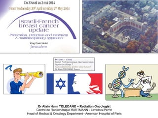
- 1. Dr Alain Haim TOLEDANO – Radiation Oncologist Centre de Radiothérapie HARTMANN - Levallois-Perret Head of Medical & Oncology Department– American Hospital of Paris
- 2. VOLTAIRE « L’art de la Médecine consiste à distraire le malade, pendant que la Nature le guérit » 18e siècle בזמן החולה של דעתו את להסיח מהותה הרפואה "אומנות ."אותו מרפא שהטבע
- 4. Therapeutic Choice : Why optimazing it ? Only one patient will have recurrence => This only one will receive maximal treatment Objective: please cut here Reality: StaJsJcally, each paJent has a recurrence risk. => Each paJent receive a maximal treatment, “probably“ efficient
- 5. 10% 90% 70% 30% 0% 50% 100% Local Disease Systemic Disease Ideal Classification (70% cured) Recommandations and consensus (10% cured) Better Select Prognostic Groups : For optimizing the choice of adjuvant treatment
- 6. Age Size N Grade Embols MulJfocality Metastases Pluridisciplinary Concertation Meeting The better way to chose treatment ? What kind of doctor you are ? האמפתי ,הסיכון טעם ,הסיכון קדומות,קבלת דעות .סבלנית הקשבה ,זמני ,פתוח ,דוגמטי In which kind of team do you exerce ? .מופרזת זהירות ,צודק שתמיד אחד ,לעצמו אדם כל ,מאוחד
- 7. Glivec* Before A.er 1 month Perfect Model : 1 abnormality, 1 medication : efficience Gastro Intestinal Stromal Tumor GIST The Belief
- 8. How to choose treatments ? Which pathological mechanisms?
- 9. GeneJc ConsJtuJonnal GeneJc SomaJc GENETIC The Hope In 100% cases : Cancer is a DNA disease Risk at < 70 years to have Breast Cancer Ovarian Cancer Muta7on gene BRAC1 57% (47-‐66%) 40% (35-‐46%) Muta7on gene BRAC2 49% (40-‐57%) 18% (13-‐23%)
- 10. “La taille moléculaire de l’ADN interdit, sans doute à tout jamais, de modifier le génome” ללא ,מאפשר אינו dnaה של המולקולרי הגודל הגנום את לשנות ,לצמיתות ספק Jacques Monod 1970 Jonathan ROTHBERG < 10 000 BASES DNA DURING A WHOLE LIFE 50 MILLIARDS PER HOUR Next GeneraJon Sequencing
- 11. . In each cell (cancer) exists 25 000 genes (3 Billions BP) . Which genes are determinant for tumoral aggressivity ? Sequencing human genom in 2003 0 500000000 1E+09 1,5E+09 2E+09 2,5E+09 3E+09 2003 2007 2008 2009 2010 2011 2012 2013 2014 2015 2016 2017 2018 3000000000 1500000 180000 45000 10000 4000 2000 1000 700 500 300 200 100 Coût du séquençage en dollars First Genom Sequencing : 13 years, 3 Billions $, 2000 researchers The Central Term " human genome " , it appeared only once, in all of the TORAH with a skip sequence ELS of (-1727) $
- 12. Mutation Frequencies in Common Cancers Dancey et al. The Gene'c Basis for Cancer Treatment Decisions. Cell 2012
- 13. 4918 The Journal of Clinical Investigation http://www.jci.org Volume 123 Number 11 November 2013 Tracking the clonal origin of lethal prostate cancer Michael C. Haffner,1 Timothy Mosbruger,1 David M. Esopi,1 Helen Fedor,2 Christopher M. Heaphy,2 David A. Walker,1 Nkosi Adejola,1 Meltem Gürel,1 Jessica Hicks,2 Alan K. Meeker,1,2,3 Marc K. Halushka,2 Jonathan W. Simons,4 William B. Isaacs,1,2,3 Angelo M. De Marzo,1,2,3 William G. Nelson,1,2,3 and Srinivasan Yegnasubramanian1 1Sidney Kimmel Comprehensive Cancer Center, 2Department of Pathology, and 3Brady Urological Institute, Johns Hopkins School of Medicine, Baltimore, Maryland, USA. 4Prostate Cancer Foundation, Santa Monica, California, USA. Recent controversies surrounding prostate cancer overtreatment emphasize the critical need to delineate the molecular features associated with progression to lethal metastatic disease. Here, we have used whole-genome sequencing and molecular pathological analyses to characterize the lethal cell clone in a patient who died of prostate cancer. We tracked the evolution of the lethal cell clone from the primary cancer to metastases through samples collected during disease progression and at the time of death. Surprisingly, these analyses revealed that the lethal clone arose from a small, relatively low-grade cancer focus in the primary tumor, and not from the bulk, higher-grade primary cancer or from a lymph node metastasis resected at prostatectomy. Despite being limited to one case, these findings highlight the potential importance of developing and implementing molecu- lar prognostic and predictive markers, such as alterations of tumor suppressor proteins PTEN or p53, to aug- ment current pathological evaluation and delineate clonal heterogeneity. Furthermore, this case illustrates the potential need in precision medicine to longitudinally sample metastatic lesions to capture the evolving constel- lation of alterations during progression. Similar comprehensive studies of additional prostate cancer cases are warranted to understand the extent to which these issues may challenge prostate cancer clinical management. Introduction Prostate cancer is the most frequently diagnosed malignancy and second leading cause of cancer-specific deaths in men in the United States (1). Clinically, prostate cancer is highly heterogeneous; manifestations vary from indolent localized tumors to widespread metastases. Given recent controversies surrounding overtreatment of prostate cancer, there is a critical need to understand the fea- tures of the primary tumor that are associated with progression to lethal disease (2, 3). Primary prostate cancers often harbor multiple morphologically and clonally distinct tumor foci (4–6). Despite the multifocal and multiclonal heterogeneity of primary prostate tumors, most dis- tant metastases from different anatomic sites in the same patient share the majority of genetic alterations, which suggests a mono- clonal origin of lethal metastatic cells (7, 8). Therefore, identify- ing the characteristics of the primary cancer lesion that ultimately can give rise to the lethal metastatic cell clone is of great interest. Studying the full spectrum of prostate cancer presentation and progression would require longitudinal, integrated analysis of the primary cancer and matched metachronous metastases sampled during disease progression and at death (9). Perhaps due to the protracted natural history of prostate cancer, such a study has not been conducted thus far. Here we present the case of a man with lethal metastatic pros- tate cancer for whom, through longitudinal sampling and com- prehensive genomic and pathological analysis, we identified the constellation of genomic alterations that characterized the lethal metastatic cell clone and traced its origin back to a specific lesion in the primary cancer. Results and Discussion The subject was diagnosed with prostate adenocarcinoma at age 47 years. His entire primary tumor and a single involved lymph node metastasis was initially removed by radical prostatectomy, but an elevated PSA level 5 years after surgery suggested systemic disease and prompted therapy with an investigational prostate cancer vaccine (GVAX; ref. 10), androgen ablation, systemic che- motherapy, and localized radiation (Supplemental Information and Supplemental Figure 1; supplemental material available online with this article; doi:10.1172/JCI70354DS1). Despite these interventions, 17 years after initial presentation, the patient suc- cumbed to overwhelming castrate-resistant prostate cancer at 64 years of age. After death, tissues from 7 metastatic sites were procured by rapid autopsy. To define the genetic features of the cell clone that gave rise to the lethal tumor burden, we performed whole-genome sequencing on 3 anatomically distinct autopsy metastases — M5 (liver), M38 (perigastric lymph node), and M40 (lung) — and germline DNA, with average sequencing coverage exceeding 50 (Supplemental Table 1 and ref. 11). We identified 85 coding mutations and 226 structural rearrangements (19 [8.4%] interchromosomal and 207 [91.6%] intrachromosomal) that were common to all 3 metastatic sites (Figure 1 and Supplemental Tables 2 and 3). None of the metastases harbored rearrangements involving TMPRSS2, ERG, or other ETS transcription factors. All 3 metastases also shared widespread copy number alterations (Supplemental Figure 2 and Supplemental Table 4). A >60-fold amplification of the AR locus was present in all distant hormone- refractory metastases (Supplemental Figure 3 and Supplemental Table 4). Although there was some genetic heterogeneity among Conflict of interest: Angelo M. De Marzo was employed at Predictive Biosciences Inc. during a portion of these studies. No funding or other support was provided by Predictive Biosciences Inc. for any of the work in this manuscript. Citation for this article: J Clin Invest. 2013;123(11):4918–4922. doi:10.1172/JCI70354. 4918 The Journal of Clinical Investigation http://www.jci.org Volume 123 Number 11 N protracted natural history of prostate cancer, such a study has not been conducted thus far. Here we present the case of a man with lethal metastatic pros- tate cancer for whom, through longitudinal sampling and com- prehensive genomic and pathological analysis, we identified the constellation of genomic alterations that characterized the lethal exceeding 50 (Supplement 85 coding mutations and 226 interchromosomal and 207 common to all 3 metastati Tables 2 and 3). None of the involving TMPRSS2, ERG, or 3 metastases also shared w (Supplemental Figure 2 and amplification of the AR locu refractory metastases (Supp Table 4). Although there wa Conflict of interest: Angelo M. De Marzo was employed at Predictive Biosciences Inc. during a portion of these studies. No funding or other support was provided by Predictive Biosciences Inc. for any of the work in this manuscript. Citation for this article: J Clin Invest. 2013;123(11):4918–4922. doi:10.1172/JCI70354. brief report mutations produced loss of PTEN staining and nuclear accumula- tionofp53,asexpected(Figure2,B–D,andSupplementalFigure5). Thus, from a molecular taxonomy perspective, this case belongs to a prostate cancer subtype characterized by the absence of ETS rear- rangement and the presence of SPOP mutations (12–14). Interestingly, in all of the autopsy metastases, we detected an 18-Mb inversion on chromosome X disrupting the ATRX gene, with associated loss of ATRX protein expression (Supplemental Figure 6). Loss-of-function alterations in ATRX are associated with massive intranuclear accumulation of telomeric DNA through alternative lengthening of telomeres (ALT) (15). Telomere-specific FISH showed telomeric aggregates, consistent with ALT, in all autopsy metastases (Figure 2E and Supplemental Figure 6). ATRX mutation may characterize a novel subgroup of metastatic pros- tate cancer; indeed, a recent study found ATRX alterations in a small subset of metastatic prostate cancers (14). To characterize the pathological landscape of the primary can- cer, we comprehensively examined sections sampling the entire radical prostatectomy specimen (composed of 36 blocks), obtained more than 17 years prior (Supplemental Figure 1). The spectrum of morphologies and grades included small, focal areas of Gleason pattern 3, large areas of Gleason pattern 4, and foci of intraductal and ductal adenocarcinoma (Supplemental Figure 7). To identify primary cancer lesions sharing characteristics of the autopsy metas- tases, we first evaluated PTEN immunohistochemical staining. The vast majority of the primary cancer showed strong PTEN staining. Interestingly, we identified only a single small (2.2 mm 1.3 mm) the one present in the autopsy metastases (Figure 3B and Supple- mental Figure 9). In contrast, this PTEN mutation was not present in DNA from 8 surrounding higher-grade lesions (P2–P9). Fur- thermore, the SPOP mutation was present in P1 as well as in P6 and P8, but not in any other sampled lesions from the primary cancer (Figure 3B and Supplemental Figure 9). Additionally, we detected the TP53 mutation in a subset of alleles from P1, but not from any other sampled region of the primary tumor (Figure 3B and Supplemental Figure 9), which suggests the emergence of a progressive subclone with TP53 mutation within P1. Together, these observations demonstrate a clonal relationship between P1 and the autopsy metastases and suggest that the lethal metastatic clone arose from P1 (a small, well-differentiated Gleason pattern 3 primary lesion), not from the prevalent Gleason pattern 4 cancer. This finding is particularly surprising since isolated Gleason pat- tern 3 lesions have shown no evidence of metastatic potential or progression to lethality (16, 17). Therefore, a Gleason pattern 3 lesion in close proximity to higher-grade lesions could have bio- logical properties different than those of isolated Gleason pattern 3 lesions. Furthermore, because P1 was the only part of the pri- mary cancer containing cells with index mutations in PTEN and TP53, which have previously been associated with aggressive dis- ease (18–20), comprehensive evaluation of PTEN and TP53 status could be useful for identifying lesions in the primary tumor that are more likely to progress. Overall, these data suggest that P1 ini- tially seeded a micrometastasis that escaped initial therapy and gave rise to all subsequent metastases, either directly or indirectly, Figure 2 Consensus genomic alterations and their phenotypic consequences in the autopsy metastases. (A) Anatomic distribution of study samples. Asterisks denote the 3 anatomically distinct autopsy metastases on which whole-genome sequencing was performed. (B–E) Molecular pheno- types of genomic alterations evaluated by immunohistochemistry (IHC) and telomere-specific FISH in representative metastasis M63. (B) AR amplification was associated with strong immunoreactivity for AR. (C) Mutations in TP53 (R248Q) resulted in nuclear accumulation of p53. (D) A frameshift deletion in the coding sequence of PTEN resulted in loss of PTEN immunostaining in neoplastic cells. Original magnification, 20. (E) The genomic inversion within the ATRX gene was associated with strong nuclear accumulation of telomeric sequence, consistent with ALT. Arrows indicate neoplastic cells. Scale bar: 10 m. brief rep which suggests that generation and selection of a cell clone harbor- ing these alterations was a later event, likely arising after androgen deprivation therapy (Supplemental Figure 3). Furthermore, a lung lesion that was biopsied 16 months prior to autopsy showed no evi- dence for ALT and ATRX alterations, despite having the PTEN, TP53, and SPOP mutations, amplification of AR, and high proliferation rates (Ki-67 index, >25%) similar to those of the autopsy metastases (Figure 3 and Supplemental Figures 9 and 10). This indicates that tions (Supplemental Figures 9 and 10), suggestive of an indep dent clonal/subclonal origin of this lesion. This finding prov proof-of-concept of the potential utility of repeated longitud evaluation of lesions during clinical management in order to ef tively target the evolving spectrum of molecular alterations du progression (21–24). These observations also suggest that m tiple tumor clones may arise, regress, and evolve during dis progression and treatment, similar to what has been observed Figure 3 Molecular and pathological findings in the primary tumor and their clonal relationship to the distant metastases.(A) PTEN staining in a cross-sectio the primary prostatectomy specimen (LA, left anterior; RA, right anterior; LP, left posterior; RP, right posterior).Individual tumor areas that were fur analyzed are indicated;green dotted outline denotes areas containing tumor glands.P1 was the only lesion in the primary tumor devoid of PTEN s ing in neoplastic cells.The adjacent P2 stained positive for PTEN, and in this regard was representative of the bulk tumor outside P1.Arrows indi tumor cells. Note that the surrounding normal stroma showed strong immunoreactivity for PTEN. Original magnification, 2 (top); 40 (bottom). S bar: 10 mm. (B) Summary of the analyzed consensus genomic alterations in the primary tumor and metastases.The presence and absence o consensus mutations are denoted by blue and gray, respectively.(C) Proposed model of disease progression in this index case, based on sequen and molecular pathological analyses. Phylogenetic relationships of distant metastases were calculated based on structural rearrangements. brief report Figure 3 Molecular and pathological findings in the primary tumor and their clonal relationship to the distant metastases.(A) PTEN staining in a cross-section of the primary prostatectomy specimen (LA, left anterior; RA, right anterior; LP, left posterior; RP, right posterior).Individual tumor areas that were further analyzed are indicated;green dotted outline denotes areas containing tumor glands.P1 was the only lesion in the primary tumor devoid of PTEN stain- ing in neoplastic cells.The adjacent P2 stained positive for PTEN, and in this regard was representative of the bulk tumor outside P1.Arrows indicate tumor cells. Note that the surrounding normal stroma showed strong immunoreactivity for PTEN. Original magnification, 2 (top); 40 (bottom). Scale bar: 10 mm. (B) Summary of the analyzed consensus genomic alterations in the primary tumor and metastases.The presence and absence of the consensus mutations are denoted by blue and gray, respectively.(C) Proposed model of disease progression in this index case, based on sequencing and molecular pathological analyses. Phylogenetic relationships of distant metastases were calculated based on structural rearrangements.
- 14. mRNA Genomics Proteomics Transcriptomics Epigenomics . Screening . TherapeuJc Is Genomic the Only True ? C o n T i n u u m
- 15. Is all the Genom Usefull ?
- 16. Sommes-‐nous programmés plutôt que façonnés par notre environnement ? ? סביבתנו ע"י מעוצבים מאשר מתוכנתים אנו האם
- 17. Les Rougon-Macquart : 20 romans Emile Zola, écrits entre 1871-1893 L’œuvre étudie : - L’influence du milieu sur l’Homme - Les tares héréditaires d’une Famille :הלימוד נושא םהאד על הסביבה השפעת המשפח של תורשתיים פגמים
- 18. Better Understanding = Better Efficience !
- 19. 0 200 400 600 800 1000 1200 * Manuscript submitted or published Analysis underway Sample acquisition phase Rare tumor project * Only accepting AA cases/500 target reached * * * * * * * * The Cancer Genome Atlas (TCGA) Network Members
- 20. GeneJc (transcriptomic) not genomic Clinical correlaJon Technical engeneering RT-‐PCR Markeqng Concept / Algorithm Intelligence of Genetic Tests
- 21. Tests and Biomarkers penetrate Medicine.. How will evoluate the need of Prediction, in a world governed by Statistics, with the Whole genome Sequencing ?
- 22. Screening, Molecular Profiling, Targeted Therapy…
- 23. « La médecine est un art et non une science exacte et rationnelle » 18e siècle 2013 מדויק מדע ולא אומנות הינה הרפואה יורציאונל
- 24. Baselga, Ann Oncol 2013 Clinical Medicine is of paramount importance for « Biological Medicine »
- 25. Charles Darwin, 1869 Ray Kurzweill, 2013 What will be the evolution of Civilization ? Transhumanism Genomic Has Changed Perspectives From Darwinism to Transhumanism
- 26. Redefine Doctors Ideal Characteristics
- 27. René-Théophile-Hyacinthe Laennec, (1781-1926) Médecin Français, Inventeur et metteur au point du diagnostic médical par auscultation (1819)- Inventeur du stéthoscope .הסטטוסקופ ממציא .האזנה ע״י הרפואי האבחון את ומיישם ממציא ,צרפתי רופא Alternatives to Clinical Medicine …
- 28. Analysis and clinical correlations of the « Big Data »
- 30. Test and Genomic Profile : What is the Future ? ואות לאפשר אלה לחזותו אין ,לעתיד בנוגע