Management of nmibc
•Download as PPTX, PDF•
5 likes•244 views

Management of non muscle invasibe bladder carcinoma

Recommended
Recommended
More Related Content
What's hot
What's hot (20)
Similar to Management of nmibc
Similar to Management of nmibc (20)
Recently uploaded
Recently uploaded (20)
Management of nmibc
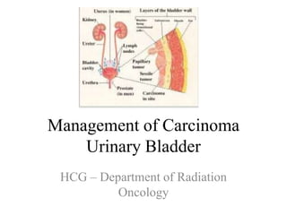
- 1. Management of Carcinoma Urinary Bladder HCG – Department of Radiation Oncology
- 3. ● Painless gross, episodic/ continuous hematuria 80% ● Bladder irritation/ UTI symptoms 30% Dysuria, Urgency , Increased frequency, Pyuria ● Storage symptoms 5% Strangury, Urinary retention ● Regional pelvic disease may cause flank pain 5% Nerve invasion, edema and ureteral obstruction ● Note: Only 10-20% of hematuria are diagnosed as bladder cancer
- 4. WORK UP
- 5. • Physical Examination (Digital rectal and Bimanual palpation before and after TURBT) – 1. Anterior vaginal wall in women and the prostate in men may reveal findings that suggest local extension of bladder cancer. 2. Extravesical involvement Sn 46% and Sp 82% 3. Size of tumor Sn of 93%, Sp 43% 4. Lesions near trigone and post bladder wall cannot be palpated A comparison between clinical and pathologic staging in patients with bladder cancer. Et al Mehrsai A
- 6. • CBC, Urine analysis, PSA, Biochemical profile , BUN , S.creatinine • Urine Cytology Sn 40-60% , Sp 98% “second morning” voided urine collected over three consecutive days 1. Sensitivity decreases in low grade as they shed less and are similar to normal cells 2. It should complement cystoscopy findings 3. Cannot replace Cystoscopy 4. Newer markers to identify proteins NMP,FDP , hyaluronic acid etc
- 7. • Cystoscopy with bladder mapping- • Tumor description (size and shape site, number and appearance and mucosal abnormalities) 1. Fluorescence scopy is more efficient (5-ALA) that accumulates in neoplastic cells 2. C/I in Acute infection and urethral stricture 3. Disadvantage - It cannot detect flat tumors efficiently • The pathological report should specify tumour location, tumour grade, depth of tumour invasion, presence of CIS, and whether the detrusor muscle is present in the specimen. Illustrative bladder map
- 8. If patient is diagnosed to have bladder tumor he needs to be investigated further for staging
- 9. • TURBT deep to detrusor muscle ( As absence is associated with higher chances of residue ) under anaesthesia (1) 1. Cold cup biopsy of suspicious area 2. When diffuse CIS is present, prostatic urtethra contains neoplastic cells in upto 25% 3. Sufficient resection to evaluate muscle invasion 4. Comments regarding residual, and muscle invasion (HPR) is important 1. Herr, H.W., et al. BJU Int, 2008. 102: 1242.
- 10. Carcinoma In Situ • Carcinoma in situ can present as a velvet- like, reddish area indistinguishable from inflammation, or it may not be visible at all. • In 128 men with T1G3 BC, the incidence of CIS in the prostatic urethra was 11.7% • The risk of prostatic urethra- or duct involvement is higher if the tumour is located on the trigone or bladder • Photodynamic diagnosis is performed using violet light after intravesical instillation of ALA • Without any treatment, approximately 54% of patients with CIS progress to muscle- invasive disease 1. Palou et al.
- 11. Imaging
- 12. • All imaging to be performed before TURBT , to avoid post surgical edema • CT urography detect papillary tumours in the urinary tract, which can be seen as filling defects or indicated by hydronephrosis • CT abdomen and pelvis to evaluate extravesical involvement and lymph nodal involvement
- 13. • MRI to distinguish superficial/invasive and intravesical/extravesical Sn -0.92 , Sp 0.80–90. Kobayashi S et al Eur Radiol. 2011;21:2178–2186 • PET CT in bladder cancer for pelvic node metastasis Lodde M,et al . BJU Int. 2010;106:658–663 • PET CT in bladder cancer for primary evaluation PET/CT and MRI in Bladder CancerKirsten Bouchelouche Lymph node Sensitivity Specificity PET CT 57% 33% CT 100% 100% Primary Sensitivity Specificity PET CT 85% 25% CT 77% 50%
- 14. Metastatic Work up • 25% of cases with tumours invading the muscular layer and in about 50% extending into the perivesical tissue present with metastatic disease (PET/CT and MRI in Bladder Cancer Kirsten Bouchelouche ) • Chest X ray to rule out lung mets- Sn 52% . EAU 2016 recommends CT to diagnose pulmonary metastases. • Bone scan for muscle invading disease, raised Alkaline Phosphatase , Bone pain • Renal Ultrasonography-can detect renal masses, hydronephrosis, and bladder intraluminal masses
- 15. Staging
- 16. AJCC Cancer Staging Handbook, Seventh Edition (2010)
- 17. Regional lymph nodes (N) Regional lymph nodes include both primary and secondary drainage regions. All other nodes above the aortic bifurcation are considered distant lymph nodes. NX Lymph nodes cannot be assessed N0 No lymph node metastasis N1 Single regional lymph node metastasis in the true pelvis (hypogastric, obturator, external iliac, or presacral lymph node) N2 Multiple regional lymph node metastasis in the true pelvis (hypogastric, obturator, external iliac, or presacral lymph node metastasis) N3 Lymph node metastasis to the common iliac lymph nodes Distant metastasis (M) MO No distant metastasis M1 Distant metastasis AJCC Cancer Staging Handbook, Seventh Edition (2010)
- 18. STAGE GROUPING 0a Ta N0 M0 0is Tis N0 M0 I T1 N0 M0 II T2a,b N0 M0 III T3a,b N0 M0 T4a N0 M0 IV T4b N0 M0 ANY T N1,2,3 M0 ANY T ANY N M1 AJCC Cancer Staging Handbook, Seventh Edition (2010)
- 19. Pathology
- 20. •Transitional Cell Carcinoma (TCC) 90% •Squamous cell Carcinoma- 5% more common in middle east Schistosomiasis Chronic catheterization •Adenocarcinoma – urachal 0.5%-2% •Small cell Carcinoma Rare
- 21. HISTOLOGIC GRADE • World Health Organization (WHO) has recommended changing bladder cancer grading to only two categories: 1) Low grade. Grade 1 is well differentiated and grade 2 is moderately well differentiated 2) High grade Grade 3 is poorly differentiated and grade 4 is undifferentiated. AJCC Cancer Staging Handbook, Seventh Edition (2010)
- 22. Non–Muscle-Invasive Disease (Superficial tumor) •Occur at the level of the bladder mucosa •Include carcinoma in situ (cis; Tis), •Papillary lesions (Ta), and •Invasion of(but not through) the lamina propria (T1). Muscle-Invasive Disease •T2, T3, and T4 tumors • That penetrate the muscularis propria • Are more aggressive and have a strong tendency to metastasize
- 23. Management of Non -Muscle Invasive Bladder Cancer
- 24. •SURGERY Transurethral resection Cystectomy •INTRAVESICAL THERAPY BCG Chemotherapy •RADIOTHERAPY
- 25. • Upto 80 % of them present with NMIBC • If residue consider for re TURBT and perform a second TURBT within 2-6 weeks after initial resection. Post TURBT alone Residue Complete Response Progression to MIBC Recurrence 53% 47% 15% 70%
- 26. • After CR risk stratify the patient as below • Millán-Rodríguez F 2000;164:680–4. [PubMed] Risk Groups Constitutes Recurrence at median of 40 months Progressio n at median of 40 months Mortality Low risk Grade 1 stage Ta disease and a single Grade 1 stage T1 tumor 37% 0% 0% Intermediate Risk Multiple grade 1 stage T1 tumors, grade 2 stage Ta disease and a single grade 2 stage T1 tumor 45% 1.8% 0.73% High Risk Multiple grade 2 stage T1 tumors, grade 3 stages Ta and T1 disease and any stage disease associated with CIS 54% 15% 9.5%
- 27. EORTC calculator • Adjuvant therapy post TURBT is decided by 2 factors 1. Probability of disease progression 2. Probability of recurrence And these factors depend on 1. Type 2. Stage 3. Grade 4. Multi centric disease 5. Size 6. Presence of CIS
- 31. Management 1. Smoking cessation 2. Adjuvant treatment • Intravesical Chemotherapy • Intravesical Immunotherapy 3.Radiation therapy 4. Surveillence
- 32. A single, immediate, post-operative intravesical instillation of chemotherapy • Indication – Low , Intermediate • Initiated within 24 hours after resection • MOA – Act by the destruction of circulating tumor cells resulting from TURB, and by an ablative effect (chemo-resection) on residual tumor cells at the resection site and on small overlooked tumors • C/I- Bladder perforation
- 33. Evidence • MMC, doxorubicin, and epirubicin showed benefit Sylvester RJ, Oosterlinck W, van der Meijden AP. J Urol. 2004;171:2186–90. quiz 2435. Arms Number of patients Recurrence rate Intravesical chemotherapy 750 48.4% Placebo/None 750 36.7%
- 34. Adjuvant instillation of intravesical chemotherapy • Indication- 1. Intermediate-risk disease 1-2 out of 4 risk factors (multiple tumors, size >3 cm, early recurrence <1 year, or frequent recurrences >1 per year) 2. No previous intravesical therapy • Contraindication 1. Bladder perforation • Treatment duration – Maximum of 1 year Kamat AM et al.. J Urol. 2014;192:305–15.
- 35. • Chemotherapeutic agents for intravesical therapy – Mitomycin-c 20-40mg weekly 6-8 wks, – Doxorubicin 50-60mg weekly 6-8 wks, • Sylvester RJ J Urol 2004;171(6 Pt 1):2186–2190.
- 36. METHOD OF ADMINISTRATION OF MITOMYCIN • 40mg of Mitomycin c in 20cc distill water can be instilled via a small tri lumen catheter. • Patients are recommended to limit fluid intake for 8-12 hours, and no fluid for 4 hours before treatment • The patient should lie prone for 15 minutes and then be allowed to move freely to bathe all parts of the bladder mucosa. The drug should to remain in the patient’s bladder for at least 1 hour (to a maximum of 2 hours). • Avoid direct skin contact during and after urinating as it may cause skin rash and irritation. • Following completion of the treatment, the Mitomycin should be drained from the patient’s bladder, by attaching a catheter drainage bag, allowing the Mitomycin to drain into the sealed bag. NCCN 2016
- 37. Intravesical Immunotherapy/BCG 1. Indication - high-risk tumors , Aggressive intermediate. Below is a trial comparing BCG and MMC. Benefit of Chemo immunotherap y vs Chemotherapy alone RR 95% CI P value Risk of recurrence 0.75 0.61-0.92 P = 0.006 Risk of progression 0.45 0.25-0.81 P = 0.007 Houghton BB. BJU Int. 2013;111:977–83
- 38. Duration of treatment – EORTC recommendation • In all patients either 1-year full-dose BCG treatment • (induction plus 3-weekly instillations at 3,6 and 12 months), or instillations of chemotherapy for a maximum of 1 year for intermediate risk • BCG is given at full dose, 3 years’ maintenance (three-weeky instillations 3, 6, 12, 18, 24, 30 and 36 months) for high risk
- 39. INTRAVESICAL BCG 1. Initiated 3-4 wks after resection 2.Contraindicated/With hold • bacteriuria, • Traumatic catheterisation • gross hematuria, • local symptoms, • systemic symptoms
- 40. BCG administration procedure • 120mg BCG in 50cc sterile normal saline can be instilled via a small catheter. • Patients are recommended to limit fluid intake for 8-12 hours, and to have no fluid intake for 4 hours before treatment. • For maximum effect the solution should be instilled when the bladder is completely empty and remain in direct bladder contact for 2 hours. • Avoid direct skin contact during and after urinating as it may cause skin rash and irritation. • Advised to sit while urinating and to empty the bladder completely. • Thorough cleansing of genital area and hands is advised. NCCN 2016
- 41. • Adverse effects from BCG are generally mild and and 60-80% of them present with these symptoms • Burning micturition • urgency. • frequency. • Fever • Skin rash. Reduction of Side Effects of Intravesical Therapy with Bacille Calmette-Guérin by Pentoxifylline?— An In Vitro Approach A. Böhle1,
- 42. Role of RT in High risk Superficial Bladder Cancer • No randomized studies • Dutch South Eastern Bladder Cancer Group had • Treatment appeared to be as effective as intravesical BCG / Mitomycin • Currently phase 2 evaluated T1 for TURBT followed by CTRT. Sample Size Stage Dose 121 T1G3 50/25#
- 43. Radical Cystectomy NMIBC • Indication 1. BCG Refractory tumors 2. The benefits and risks of immediate and delayed RC should be discussed with patients 3. In patients in whom RC is performed at the time of pathological NMIBC, the 5-year disease-free survival rate exceeds 80%(1) Stein, J.P., et al. J Clin Oncol, 2001. 19: 666.
- 44. Surveillence Risk group Surveillence Low Risk (grade 1 stage Ta disease and a single grade 1 stage T1 tumor) 3 months after resection . If Negative 9 month later , then annually Intermediate Risk(multiple grade 1 stage T1 tumors, grade 2 stage Ta disease and a single grade 2 stage T1 tumor) 3 monthly- for 2yrs 6 monthly for 5 yrs High Risk(multiple grade 2 stage T1 tumors, grade 3 stages Ta and T1 disease and any stage disease associated with CIS) 3 monthly- for 2yrs 6 monthly for 5 yrs
- 45. Muscle Invasive, Nonmetastatic Bladder Cancer
- 46. Muscle-Invasive Disease • Upto 30% of total Bladder Ca •T2, T3, and T4 tumors • That penetrate the muscularis propria • Are more aggressive and have a strong tendency to metastasize
- 47. Controversy • Radical cystectomy with pelvic lymphadenectomy is considered gold standard • No evidence comparing it with ChemoRT (bladder preservation) • New advancements in neoadjuvant and adjuvant chemotherapy, radiation therapy and bladder-preservation protocols should encourage bladder preservation • UK SPARE had poor recruitment
- 50. Neoadjuvant chemotherapy • Radical cystectomy provides 5-year survival in about 50% . • To improve these results ,NACT has been used since the 1980s • Most common regimens used for neoadjuvant chemotherapy is three 28-day cycles of MVAC as follows: methotrexate (30 mg/m2 on days 1, 15, and 22), vinblastine (3 mg/m2 on days 2, 15, and 22), doxorubicin (30 mg/m2 on day 2), and cisplatin (70 mg/m2 on day 2). • GC (gemcitabine/cisplatin) Stein JP 2006 Aug;24(3):296-304. David KA Urol 2007 Aug;178(2):451-4.
- 51. Advantages • Chemotherapy is delivered, when the burden of micrometastatic disease is expected to be low. • Tolerability of chemotherapy are expected to be better pre-cystectomy. • Favorable pathological status, by achieving pT0, pN0 and negative surgical margins. Disadvantages • Delayed cystectomy might compromise the outcome in patients not sensitive to. There are no trials indicating that delayed surgery, due to NAC, has a negative impact on survival. • Neoadjuvant chemotherapy does not seem to affect the outcome of surgical morbidity. In one randomized trial the same distribution of grade 3-4 postoperative complications was seen in both trial arms[1] Sternberg CN):1644-52[1]
- 52. European Association of Urology 2016 Recommendations • Neoadjuvant chemotherapy is recommended for T2-T4a, cN0M0 bladder cancer and should always be cisplatin-based combination therapy. • NACT is not recommended in patients who are ineligible for cisplatin-based combination chemotherapy.
- 53. Pre-operative radiotherapy in muscle- invasive bladder cancer Recommendations of European Association of Urology 1. Pre-operative radiotherapy is not recommended to improve survival 2. Pre-operative radiotherapy for operable MIBC can result in tumour down-staging after 4-6 weeks. Huncharek M Res 1998 May;18(3b):1931-4.
- 54. Cystectomy 1. Indications • MIBC T2-T4a, N0-Nx, M0 • high-risk and recurrent superficial tumours • BCG-resistant Tis, T1G3 • extensive papillary disease that cannot be controlled primary therapy • Salvage cystectomy if bladder preservation fails • In patients with inoperable locally advanced tumours (T4b), primary radical cystectomy is a palliative option.[1] 1. Nagele U World J Urol 2007 Aug;25(4):401-5.
- 55. • In men, standard RC includes removal of the bladder, prostate, seminal vesicles, distal ureters, and regional lymph nodes. Prostate-sparing cystectomy is an option in a subset of carefully selected patients with without involvement of the prostatic urethra and without prostate cancer. This procedure is oncologically safe [1] • In women, standard RC includes removal of the bladder, entire urethra and adjacent vagina, uterus, distal ureters, and regional lymph nodes [2]. • Orthotopic bladder cannot be offered for N2 disease[3] 1. Mertens, J Urol, 2014. 191: 1250. 2. Stenzl, A., et al. Series, 2005. 3: 138 3. Lebret, T., et al. Eur Urol, 2002. 42: 344.
- 56. European Association of Urology 2016 Recommendations • Offer sexual-preserving techniques to Male/ female patients motivated to preserve their sexual function since the majority will benefit. • Select patients based on: 1. Organ-confined disease; 2. Absence of tumor in bladder neck or urethra. 3. Do not offer pelvic organ-preserving radical cystectomy for Male /female patients as standard therapy for MIBC.
- 57. Lymph node dissection • The extent of LND has not been established to date. Standard lymphadenectomy in BC patients involves removal of nodal tissue cranially up to the common iliac bifurcation, with the ureter being the medial border, and including the internal iliac, presacral, obturator fossa and external iliac nodes [1] • No difference in outcome was reported between extended and super-extended LND in the two high-volume-centre studies identified [2] 1.Simone, G., et al. J Urol, 2013. 20: 390. 2. Liu JJ J Urol 2011 May;185
- 58. Multimodality bladder-preserving treatment • Combines TURBT, chemotherapy and radiation • Radiosensitising chemotherapy, cisplatin [1] or mitomycin C plus 5-fluorouracil can be used [2] • With MMT 5-year • CSS 50-82%[1,2] • OS 36-74%[3,4] 1. Milosevic, M., et al. 2007. 69: 80. 2.James, N.D., et al N Engl J Med. 2012. 366: 1477 3 Hoskin, P.J., et al. J Clin Oncol, 2010. 28: 4912. 4. Kaufman, D.S., et al. Urology, 2009. 73: 833.
- 59. Adjuvant chemotherapy Adjuvant cisplatin-based combination chemotherapy to patients with pT3/4 and/or pN+ disease if no neoadjuvant chemotherapy has been given[1,2,3] 1.Cohen, S.M., et al..Oncologist, 2006. 11: 630 2. Sylvester, R., et al. Ann Oncol, 2000. 11: 851. 3. David, K.A., et al.. J Urol, 2007. 178: 451.
- 60. Radiation techniques in Bladder Cancer • Definitive RT 1. Conventional Technique 2. 3D-CRT 3. IMRT • Palliative RT • Altered Fractionation • Brachytherapy
- 61. External Beam Irradiation • The standard protocol for radical EBRT as well as combined modality therapy. • It uses a four field iso-centric technique for both initial and boost field. • It consists of shaped anterior , posterior , right and left lateral fields • Induction and boost treatment fields will be discussed further
- 62. Simulation 1. Instruct patient to void urine 2. Insert Foley's catheter 3. Measure post void residual urine and replace with equal volume of bladder contrast + Additional 25mL contrast + 15mL of air. • Contrast defines the inner walls of bladder • Air aids visualization of anterior bladder on lateral simulation film • Contrast amount should not be less than post void residue 4. AP/PA and Lateral radiographs are taken or CT simulation is done
- 64. Induction Field 1. During first phase of treatment, bladder is treated along with 2 cm margin. 2. If using radiograph , contrast lines only inner wall hence another 5-10mm is added. 3. In men prostate is included and in women proximal 2 cm of urethra is included . 4. Limit the amount of small bowel irradiated.
- 65. Break to evaluate 1. After induction treatment or after a dose of 39- 42Gy, repeat cystoscopy is done . 2. If CR/Ta/Tis then they are advised to continue boost. 3. If the stage is more than T1 ,then advise cystectomy.
- 66. Boost Field 1. Tumor alone with 2 cm margin . 2. Tumor is delineated using information from bladder map during TURBT/Cystoscopy and CT/MRI 3. Another alternate is to treat the whole bladder and exclude the nodal volume 4. If tumor is in trigone or PL walls of bladder only lateral fields can be used for boost
- 67. Dose 1. Total dose of 65Gy(1.8-2Gy fractions, 5 days per week) together with chemo and max TURBT. 2. 40-45Gy is Induction dose 3. The tumor is then boost to full dose (15-20Gy) 4. In invasive cancer ,if there is complete response then local recurrence is limited to 15- 18% implying that dose is adequate in responders
- 68. Partial bladder treatment 1. Rationale – High dose (upto 80 Gy) can be given if 1/3 of bladder is spared . 2. Indications • <5cm in size • Unifocal disease • Without extensive Tis • No significant difference between arms Arms Dose/# 5 yr Local control Partial bladder irradiation 57.5/20 58% Whole bladder irradiation 52.5/20 59%
- 69. Treatment margins in Conformal radiotherapy 1. CTV to PTV margin , an isotropic 2 cm margin in all 3 dimensions. 2. As the greatest degree of bladder wall positional change occurred in cranial direction and least in antero-inferior direction , limited by pubic symphysis. 3. Anisotropic margin of 1.6cm anteriorly and posteriorly , 1.4cm laterally , 3cm superiorly , 1.4cm inferiorly has been recommended by Graham 4. Daily imaging 5. Fuducial based matching
- 70. 3D-CRT Patient position and immobilization • The patient should be planned and treated in the same position; supine with arms on their chest. Knee and ankle immobilization should be used to ensure patient positioning is reproducible. • The rectum should be empty of flatus and faeces. The use of daily micro- enemas may be considered. • Patients will be asked to empty their bladder 15 minutes prior to scan. • Whilst breathing normally, the patient should have a CT scan performed with 3–5-mm slice spacing. • Upper Extent -ischial tuberosities • Lower Extent -3 cm above the dome of the bladder or bottom of L5 (whichever is higher) • Reference radio-opaque tattoos should be made at the base of the abdomen and over each hip.
- 71. Volume Delineation • The GTV should integrate information from the staging CT or MRI as well as the diagnostic TURBT . MRI/CT fusion may be helpful, where available. • CTV constitutes the entire bladder • A standard approach is to define the PTV as the whole bladder identified by its non-involved outer bladder wall with a 1.5-cm margin plus extravesical extent of tumour with a 2-cm margin • All planning and treatment should be carried out with the bladder empty to minimize the risk of geographical miss and to keep the treated volumes as small aspossible. Patients with significant residual volumes post voiding should be considered for planning and treatment with a catheter in situ , although this is likely to increase urinary toxicity.
- 74. Dose constraints • Dose constraints (dose 2 Gy/fraction) used for organs at risk are as follows • Rectum: V66 < 30 % ,V60 < 50 % , V50 < 60 % , V40 < 70 % , V30 < 80 % . • Femoral heads: V50 < 50 % .[1] 1.Duchesne GM.International Journal of Radiation Oncology , Biology, Physics 2000 ; 47 : 379 – 88 .
- 75. Altered Fractionation • In T2-T4 tumors unsuited for cystectomy Numb er of patien ts Dose Survival at 5 yrs Local control Clinical complete response Hyperfractio nation 84 1 Gy three times a day to a dose of 84Gy 27% 12% 59% Conventiona l 84 2Gy everyday to a dose of 64Gy 18% 7% 36%
- 76. Palliative Radiotherapy • Palliative bladder irradiation is used in the treatment of bleeding from a primary tumor or a metastatic lesion to the bladder that cannot be controlled cystoscopically. • Symptomatic improvement can be achieved in 60-70% of patients • Schedules used: 30 Gy in 10 fractions 21 Gy in 3 fractions
- 77. Brachytherapy • Indications: - A solitary transitional cell carcinoma - Diameter less than 5 cm - Muscle invasion but with no extension through the bladder wall • Contraindications: tumor extending to perivesical fat and adjacent structures, multifocal, lymph node involvement.
- 78. • Initially preoperative EBRT of 3 x 3.5 Gy fractions for T1 tumors and 20 x 2 Gy for T2 tumors is delivered • Partial Cystectomy with routine iliac lymphadenectomy is performed. • Hollow Nylon tubes are placed intraoperatively for afterloading with Iridium sources
- 82. • Acute postoperative complications like thromboses, infections, delayed wound healing and fistula formation were seen in 19.5-30% of the cases. • Late complications: 25-39% were reported In the first year hematuria, stone formation, chronic cystitis were observed Symptomatic ulceration or fistula formation needing treatment or ureter stenosis with hydronephrosis is rare (1-6%) Chronic radiocystitis (0.6%)
- 83. Thank You!!
- 85. Risk group classification Risk of recurrence at 1 year Risk of progression at 1 year Adjuvant therapy Low Risk (grade 1 stage Ta disease and a single grade 1 stage T1 tumor) 15% 0.2% Not required Intermediate Risk(multiple grade 1 stage T1 tumors, grade 2 stage Ta disease and a single grade 2 stage T1 tumor) 38% 5% Required High Risk(multiple grade 2 stage T1 61% 17% Required
- 86. • Ta, G1 have 100 % recurrence rates at 5 yrs , but do not invade or metastasize – Hence TUR alone maybe sufficient Tumor Frequency of recurrence Ta(G2,G3) 20% No indication T1 (50% are G3) 50% Indication for Adjuvant therapy T1 with CIS 80% Indication for Adjuvant therapy Progression rate at 12months Overall survival at 5 yrs TURBT alone 63% TURBT 88%
- 87. Risk group classification Risk of recurrence at 1 year Risk of progression at 1 year Surveillence Low Risk (grade 1 stage Ta disease and a single grade 1 stage T1 tumor) 15% 0.2% 3 months after resection . If Negative 9 month later , then annually Intermediate Risk(multiple grade 1 stage T1 tumors, grade 2 stage Ta disease and a single grade 2 stage T1 tumor) 38% 5% 3 monthly- for 2yrs 6 monthly for 5 yrs High Risk(multiple grade 2 stage T1 61% 17% 3 monthly- for 2yrs 6 monthly for 5 yrs