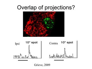
Substructure within the rat dLGN Claire Discenza Reinagel Lab Lab Meeting 9/23/09
Primary visual pathway
Substructure in the LGN: Monkey
Rat dLGN Monocular Binocular ipsilateral Optic Tract Adapted from Reese 1988
Contralateral  Ipsilateral Tracing and imaging methods 4 3 1 2
Retinal termini in dLGN
 
Zooming in on dLGN
 
How to define dLGN subdivisions? The distinct functional sublaminae are defined by anatomical, physiological and molecular criteria Anatomy: -- Anatomy of retinal projections --Overlapping/ Interleaving of ipsi and contra projections. -- Additional anatomic criteria Cellular organization Shapes, sizes and densities of cells (Immunostaining for neurons and glia)
First Analyses 1. Tracing of ipsilateral and contralateral regions in MATLAB, aligning these images for analyses. 2. Cytoarchitectonics to look for rat dLGN sub-structure in MATLAB and CellProfiler 3. Binocular region in rat dLGN? Looking for overlap
1. Tracing Geimsa dLGN
Aligning Geimsa tracings Aligned subcortical tracings from  one series of coronal sections containing dLGN
Unaligned dLGN outlines and fiduciary marks Aligned dLGN outlines and fiduciary marks
Aligned dLGN outlines
Aligned ipsilateral 'blob'
Examine the brain from all angles Sagittal (contra) Coronal (contra) ?? Coronal only
First Analyses 1. Tracing of ipsilateral and contralateral regions in MATLAB, aligning these images for analyses. 2. Cytoarchitectonics to look for rat dLGN sub-structure in MATLAB and CellProfiler 3. Binocular region in rat dLGN? Looking for overlap
Distinct dLGN regions? Looks like substructure Want to use blind algorithm
2. Cytoarchitecture with  CellProfiler and Matlab –  Choose the region of interest –  Mask the nissl image using the geimsa outlines –  Send the masked image through series of  CellProfiler analyses –  Look at output: Cell area, excentricity, density.
Aligning giemsa traces onto nissl sections for masking
1. Cytoarchitecture with  CellProfiler and Matlab
Cell Density Radius 10 pixels Radius 60 pixels Radius 100 pixels Radius 30 pixels
Larger, yellower dots mark larger cells Cell Area
Cell area in different ipsilateral 'blob' projections Area pixels Slice  Ipsi blob 1 Ipsi blob 2
First Analyses 1. Tracing of ipsilateral and contralateral regions in MATLAB, aligning these images for analyses. 2. Cytoarchitectonics to look for rat dLGN sub-structure in MATLAB and CellProfiler 3. Binocular region in rat dLGN? Looking for overlap
Retinal termini in dLGN
LGN binocular inputs -- CTB conjugated to Alexa 488 in one eye, 594 in the other eye.  -- visulize fine ipsi/contra projections in the dLGN 488 594


Overlap of projections? Grieve, 2009 Ipsi Contra
Torburg and Feller, 2003
Thanks to: Reinagel lab: Pam, Phil, Erik, & Carolyn. Kleinfeld lab for use of neurolucida and computer.  Karten Lab: Harvey & Agniezska. Burnham institute for photographs Brainmaps.org  Marin McDonald & Boulanger lab, Nick Wall & Callaway lab
THE END
Same Major Retinorecipients  Superior Colliculus (SC) Thalamus: (dLG), (IGL), (vLG) Accessory Optic System (AOS) Parabigeminal Nuc. (PBg) Pre-tectum (NOT),(OPT),(PPT) Suprachiasmatic Nucleus (SCN)


Tectum Detail
Surprises Inferior colliculus Projection to the Inf. Colliculus tectum
Eye Injections – Cholera Toxin  Contralateral Ipsilateral Inject cholera toxin B into vitreous humor. Wait 3 days. Remove brain, and slice. Incubate in a series of  antibodies against the subunit.  Stain by submitting antibodies to chemical reaction.
Kuss, Jäkel & Wichmann, 2005 (PsychoFun ) Phil Meier Acuity estimation 1.9  0.47 0.24 0.12 0.06 0 0.1 0.2 0.3 0.4 0.5 0.6 0.7 0.8 0.9 1 3 Days,  1057  Trials, Reversed Weibull Cycles/Degree p(Correct)
 
Rat dLGN Adapted from Reese and Jefferey, 1983 caudal rostral medial lateral 4 3 1 2 2 3 1 4 1 2 4 3 2 3 4 5 5 5 1 5 IPSI CONTRA 2 3 4 5 1 5 4 3 2 1
Brief Overview 1) The visual systems and the LGN 2) Why study the rat visual system? 3) Anatomy and the LGN 4) Methods: following the retinal ganglion cells to the rest of the brain. 5) Present and Future anatomical studies…
Why the Rat? In addition: Inexpensive, small, genetically/strain controlled, and visual animals. Similar to mouse. Knowledge of rodent visual systems can be followed up with genetic studies. Mouse coronal Rat coronal Brainmaps.org
 
How to define the Rat dLGN The distinct functional sublaminae are defined by anatomical, physiological and molecular criteria Anatomy first: -- Anatomy of retinal projections -- Additional anatomic criteria Cellular organization Shapes, sizes and densities of cells Immunostaining for    neurons (NeuN) glial cells (GFAP) GABAergic interneurons (GAD) ChAT (for tectal projections). -- Interleaving of ipsi and contra projections.
Substructure in rat dLGN Claire Discenza Reinagel Lab Lab Meeting 9/23/09

Discenza 9.23.09

  • 1.
    Substructure within therat dLGN Claire Discenza Reinagel Lab Lab Meeting 9/23/09
  • 2.
  • 3.
  • 4.
    Rat dLGN MonocularBinocular ipsilateral Optic Tract Adapted from Reese 1988
  • 5.
    Contralateral IpsilateralTracing and imaging methods 4 3 1 2
  • 6.
  • 7.
  • 8.
  • 9.
  • 10.
    How to definedLGN subdivisions? The distinct functional sublaminae are defined by anatomical, physiological and molecular criteria Anatomy: -- Anatomy of retinal projections --Overlapping/ Interleaving of ipsi and contra projections. -- Additional anatomic criteria Cellular organization Shapes, sizes and densities of cells (Immunostaining for neurons and glia)
  • 11.
    First Analyses 1.Tracing of ipsilateral and contralateral regions in MATLAB, aligning these images for analyses. 2. Cytoarchitectonics to look for rat dLGN sub-structure in MATLAB and CellProfiler 3. Binocular region in rat dLGN? Looking for overlap
  • 12.
    First Analyses 1.Tracing of ipsilateral and contralateral regions in MATLAB, aligning these images for analyses. 2. Cytoarchitectonics to look for rat dLGN sub-structure in MATLAB and CellProfiler 3. Binocular region in rat dLGN? Looking for overlap
  • 13.
  • 14.
    Aligning Geimsa tracingsAligned subcortical tracings from one series of coronal sections containing dLGN
  • 15.
    Unaligned dLGN outlinesand fiduciary marks Aligned dLGN outlines and fiduciary marks
  • 16.
  • 17.
  • 18.
    Examine the brainfrom all angles Sagittal (contra) Coronal (contra) ?? Coronal only
  • 19.
    First Analyses 1.Tracing of ipsilateral and contralateral regions in MATLAB, aligning these images for analyses. 2. Cytoarchitectonics to look for rat dLGN sub-structure in MATLAB and CellProfiler 3. Binocular region in rat dLGN? Looking for overlap
  • 20.
    Distinct dLGN regions?Looks like substructure Want to use blind algorithm
  • 21.
    2. Cytoarchitecture with CellProfiler and Matlab – Choose the region of interest – Mask the nissl image using the geimsa outlines – Send the masked image through series of CellProfiler analyses – Look at output: Cell area, excentricity, density.
  • 22.
    Aligning giemsa tracesonto nissl sections for masking
  • 23.
    1. Cytoarchitecture with CellProfiler and Matlab
  • 24.
    Cell Density Radius10 pixels Radius 60 pixels Radius 100 pixels Radius 30 pixels
  • 25.
    Larger, yellower dotsmark larger cells Cell Area
  • 26.
    Cell area indifferent ipsilateral 'blob' projections Area pixels Slice Ipsi blob 1 Ipsi blob 2
  • 27.
    First Analyses 1.Tracing of ipsilateral and contralateral regions in MATLAB, aligning these images for analyses. 2. Cytoarchitectonics to look for rat dLGN sub-structure in MATLAB and CellProfiler 3. Binocular region in rat dLGN? Looking for overlap
  • 28.
  • 29.
    LGN binocular inputs-- CTB conjugated to Alexa 488 in one eye, 594 in the other eye. -- visulize fine ipsi/contra projections in the dLGN 488 594
  • 30.
  • 31.
  • 32.
  • 33.
  • 34.
  • 35.
    Overlap of projections?Grieve, 2009 Ipsi Contra
  • 36.
  • 37.
    Thanks to: Reinagellab: Pam, Phil, Erik, & Carolyn. Kleinfeld lab for use of neurolucida and computer. Karten Lab: Harvey & Agniezska. Burnham institute for photographs Brainmaps.org Marin McDonald & Boulanger lab, Nick Wall & Callaway lab
  • 38.
  • 39.
    Same Major Retinorecipients Superior Colliculus (SC) Thalamus: (dLG), (IGL), (vLG) Accessory Optic System (AOS) Parabigeminal Nuc. (PBg) Pre-tectum (NOT),(OPT),(PPT) Suprachiasmatic Nucleus (SCN)
  • 40.
  • 41.
  • 42.
  • 43.
  • 44.
    Surprises Inferior colliculusProjection to the Inf. Colliculus tectum
  • 45.
    Eye Injections –Cholera Toxin Contralateral Ipsilateral Inject cholera toxin B into vitreous humor. Wait 3 days. Remove brain, and slice. Incubate in a series of antibodies against the subunit. Stain by submitting antibodies to chemical reaction.
  • 46.
    Kuss, Jäkel &Wichmann, 2005 (PsychoFun ) Phil Meier Acuity estimation 1.9 0.47 0.24 0.12 0.06 0 0.1 0.2 0.3 0.4 0.5 0.6 0.7 0.8 0.9 1 3 Days, 1057 Trials, Reversed Weibull Cycles/Degree p(Correct)
  • 47.
  • 48.
    Rat dLGN Adaptedfrom Reese and Jefferey, 1983 caudal rostral medial lateral 4 3 1 2 2 3 1 4 1 2 4 3 2 3 4 5 5 5 1 5 IPSI CONTRA 2 3 4 5 1 5 4 3 2 1
  • 49.
    Rat dLGN Adaptedfrom Reese and Jefferey, 1983 caudal rostral medial lateral 4 3 1 2 2 3 1 4 1 2 4 3 2 3 4 5 5 5 1 5 IPSI CONTRA 2 3 4 5 1 5 4 3 2 1
  • 50.
    Brief Overview 1)The visual systems and the LGN 2) Why study the rat visual system? 3) Anatomy and the LGN 4) Methods: following the retinal ganglion cells to the rest of the brain. 5) Present and Future anatomical studies…
  • 51.
    Why the Rat?In addition: Inexpensive, small, genetically/strain controlled, and visual animals. Similar to mouse. Knowledge of rodent visual systems can be followed up with genetic studies. Mouse coronal Rat coronal Brainmaps.org
  • 52.
  • 53.
    How to definethe Rat dLGN The distinct functional sublaminae are defined by anatomical, physiological and molecular criteria Anatomy first: -- Anatomy of retinal projections -- Additional anatomic criteria Cellular organization Shapes, sizes and densities of cells Immunostaining for neurons (NeuN) glial cells (GFAP) GABAergic interneurons (GAD) ChAT (for tectal projections). -- Interleaving of ipsi and contra projections.
  • 54.
    Substructure in ratdLGN Claire Discenza Reinagel Lab Lab Meeting 9/23/09