Cara's microbiology presentation
•Download as PPT, PDF•
2 likes•3,291 views

www.sustainabletechsys.com

Recommended
More Related Content
What's hot
What's hot (20)
Viewers also liked
Viewers also liked (20)
Similar to Cara's microbiology presentation
Similar to Cara's microbiology presentation (20)
More from Cara Mullen
More from Cara Mullen (14)
Recently uploaded
Recently uploaded (20)
Cara's microbiology presentation
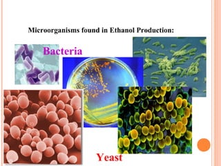
- 1. Microorganisms found in Ethanol Production: Bacteria Yeast
- 3. BACTERIA (SINGULAR: BACTERIUM) are Single-celled organisms which are Prokaryotes Yeast are a fungi which are Eukaryotes
- 4. THE MOST COMMON BACTERIAL SHAPES
- 6. Typical Eukaryotic Yeast Cell
- 7. Source of bacterial contamination Bacteria comes in On the corn, especially bad corn! On the trucks Bacteria are in the water Well water Cooling tower water Bacteria are in the air Higher in humid environments Summer time they thrive in moist environments Bacteria are on your person Skin, mouth
- 8. CERTAIN SPECIES OF BACTERIA AND WILD YEAST STRAINS LIVE FAVORABLY IN ETHANOL FERMENTATION CONDITIONS. THEY COMPETE WITH THE YEAST AND UTILIZE THE GLUCOSE. THIS LOWERS THE ETHANOL YIELDS AND INCREASES UNDESIRABLE ORGANIC ACIDS.
- 9. Stress factors for yeast • Temperature – 95˚F at start of fermentation good – Should lower temperature as alcohol concentration rises • Ethanol • CIP • Sulfite • Sugar • Acetic and/or lactic acid • Sodium • pH – Yeast perform well in acidic environments pH 3-4 – Acidic environment good for bacterial control
- 11. VINEGAR (ACETIC ACID) IS MADE FROM ETHANOL BY THE ACETIC ACID BACTERIUM, ACETOBACTER ACETI SAUERKRAUT IS MADE BY LACTIC ACID BACTERIA NATURALLY PRESENT ON CABBAGE PICKLES ARE MADE ESSENTIALLY BY THE SAME PROCESS FOR SAUERKRAUT WITH ORGANISMS: LEUCONOSTOC AND PEDIOCOCCUS
- 12. Comparison of relative efficiencies of different types of respiration
- 13. AEROBIC RESPIRATION: C6H1206 + 6O2 → 6H2O + 6CO2 + 2880 kJ or sugar + oxygen → water + carbon dioxide + heat
- 14. ANAEROBIC RESPIRATION WITH ETHANOL FORMATION (ALCOHOL FERMENTATION): C6H1206 → 2CH3CH2OH + 2CO2 + 210 kJ or sugar → ethanol + carbon dioxide + heat
- 15. ANAEROBIC RESPIRATION WITH LACTIC ACID FORMATION (FERMENTATION): C6H1206 → 2CH3CH(OH)COOH + 150 kJ or sugar → lactic acid + heat
- 16. Bacterial contamination Bacterial infections can cause large losses in profit Based on ~1% lactic acid growth at 13 wt% ethanol and $2 gal/ethanol For example a 50 MMGY ethanol plant infection causes loss of 1% loss = $1,000,000 per year 4% loss = $4,000,000 per year 1 organic acid molecule = 1 lost ethanol molecule C2H5 OH 1 lactic acid molecule = 1 lost ethanol molecule 2CH3CH(OH)COOH = 2CH3CH2 OH+ 2CO2 6C + 12H + 6O = 6C + 12H + 6O
- 17. Bacterial growth is difficult to control because they grow and live in similar environment as yeast do. Therefore, the bacteria compete with the yeast for nutrients and produce unwanted byproducts.
- 18. LACTIC ACID BACTERIA (LAB) Gram positive bacteria are Lactobacillus, Weisella, and Pediococcus species. Gram negative bacteria are Acetobacter and Gluconobacter species. Less common LAB contaminants: Luconostoc, Streptococcus, Aerococcus, Camobacterium, Enterococcus, Oenococcus, Teragenococcus, Vagococcus
- 19. LACTOBACILLUS
- 20. Lactic acid on Hplc • Lactic acid indicates bacterial contamination • Risk stuck fermentation • Primary source is a (LAB) lactic acid bacteria
- 21. Pediococcus • Gram positive cocci • Organized in pairs and Tetrads • All strains appear to have built-in resistance to high levels of penicillin and virginiamycin One hypothesis is that Pediococcus is more likely when corn has been stored on the ground
- 22. PEDIOCOCCUS
- 23. ACETOBACTER The bacteria are Gram negative and Gram variable no endospores catalase positive oxidase negative Is capable of metabolizing ethanol (Hoyer’s media) Durham tubes grow obligate aerobes incapable of fermentation.
- 24. ACETOBACTER
- 25. ACETIC ACID – Acetic acid “background” should be near detection limit of HPLC – Should strive to be below 0.05% – Primary source heterofermentative bacteria – Also aerobic acetic acid fermenters
- 26. GLUCONOBACTER Gram negative ovoid or rod shaped fermentation (acetic acid (acetaldhydes)/vinegar) non-motile or lophotrichous flagella Catalase positive obligately aerobic organisms optimal growth temperature is 25-30˚C, however, no growth occurs at 37˚C. They prefer pH of 5.5 6.0.
- 27. Gluconobacter
- 28. Weissella
- 29. WEISSELLA Lactobacillus “like” Gram positive short rod Some strains highly resistant to virginiamycin (acquired resistance??) All strains susceptible to 0.5ppm of penicillin
- 30. Leuconostoc
- 31. LEUCONOSTOC • Ovoid cocci often forming chains • Gram Positive • Facultatively anaerobic bacteria • Catalase-negative • LAB bacteria • Pickles and saurkraut
- 32. – pH can significantly decrease during an infection – Ethanol production will decrease with infections. The severity of the infection and the time the infection is present will dictate how much ethanol will be lost – Sugar usage will decrease meaning increased residual sugars present in the DDGS and Wet-cake causing a decrease in quality
- 33. Common contamination Sources • Heat exchangers • Yeast Prop • CO2 header • Fermentation – metal cracks • Dead legs • Leaking valves • Water/recycle • Air • Pipe work • Product storage
- 34. CIP (CLEANING IN PLACE) EVERYTHING NEEDS TO BE CLEANED – FERMENTERS – HEAT EXCHANGERS – MASH LINES – BEER/MASH INTERCHANGERS – YEAST PROPAGATION SYSTEM
- 35. Contamination sources for bacterial infections – Inadequate CIP – Dead legs • General cleanliness throughout plant especially in mash, yeast props, heat exchangers, and fermentation areas – Poor grain • Bacteria present in low numbers on good grain • Bacteria present in extremely high numbers on bad grain
- 36. NO PRACTICAL WAY TO CIP CO2 HEADER ENTIRE MASHING SYSTEM METHANATOR BACTERIA FLOAT OUT TO COOK WATER SYSTEM WATER TREATMENT SYSTEM
- 37. BACTERIALCIDAL OR BACTERIOSTATIC Antibiotics can reduce or kill bacteria Can be very specific Commercially available Can be expensive Creates resistance Antibiotic companies often can help lab to help id resistant strains Alternatives: Hop Acids substitute for antibiotics Very expensive Chemical washes Steam, Bleach, Hydrogen peroxide, Caustic, Chlorine dioxide, Iodophor, Ammonium biflouride Lower in cost but not selective Destroys yeast cells
- 38. GRAM STAIN 1. Primary stain: crystal violet stains all cells purple 2. Mordant: Gram’s iodine crystallizes purple stain in cells 3. Decolorizer: 95% ethanol dissolves lipid layers in cell walls, allows crystallized purple stain to wash out 4. Counterstain: safranin enters vacant cells turning them red
- 39. GRAM STAIN
- 41. GRAM STAIN PROCEDURE Innoculate organism onto the slide by placing a drop of DI water on the slide using sterile loop. Place slide on warmer low until dry. Do NOT over heat. Cover slide with Crystal Violet for 1 minute. Rinse with DI water. Cover slide with Iodine for 1 minute. Rinse with DI water. Drizzle alcohol over slide at a slant for 10 seconds. Rinse with DI water. Cover slide with Safranin for 1 minute. Rinse with DI water. Dry slide on warmer or sitting at a slant to air dry.
- 42. SERIAL DILUTION AND STANDARD PLATE COUNTS Standard plate count: One method of measuring bacterial growth Agar plate: A petri dish containing a nutrient medium solidified with agar Serial dilutions are used to dilute the original bacterial culture before you transfer known volume of culture onto agar plate
- 44. CALCULATION OF THE NUMBER OF BACTERIA PER MILLILITER OF CULTURE USING SERIAL DILUTION Pour plate: made by first adding 1.0ml of diluted culture to 9ml of agar Spread plate: made by adding 0.1ml of diluted culture to surface of solid medium
- 46. ANOTHER WAY TO MEASURE BACTERIAL GROWTH Petroff-Hausser counting chamber Bacterial suspension is introduced onto chamber with a calibrated pipette Microorganisms are counted in specific calibrated areas Number per unit volume is calculated using an appropriate formula
- 48. MOST PROBABLE NUMBER (MPN) Method to estimate number of cells Used when samples contain too few organisms to give reliable measures of population size by standard plate count Series of progressively greater dilutions Typical MPN test consists of five tubes of each of three volumes (e.g. 10, 1, and 0.1ml)
- 49. ANY QUESTIONS??