SlideShare a Scribd company logo
1 of 45
CT Coronary Angiography :
Indication & Interpretation
Dr Awadhesh Sharma
Indications of CTA in
current scenario
Stable chest pain
1. No Known CAD:
 Appropriate as the first line test in stable typical or atypical chest pain, or other symptoms
which are thought to represent a possible anginal equivalent (e.g. dyspnoea on exertion,
jaw pain).
 After a nonconclusive functional test, in order to obtain more precision regarding diagnosis
and prognosis, if such information will influence subsequent patient management.
 May be appropriate in some asymptomatic high-risk individuals, such as those with a
higher likelihood of non-calcified plaque.
 Rarely appropriate in very low-risk symptomatic patients – those under age 40 with non-
cardiac symptoms – or those with low- to intermediate-risk asymptomatic patients.
Indications..
2. Known CAD:
 It is appropriate to perform CTA as a first line test for evaluating patients with known CAD
who present with stable typical or atypical chest pain, or other symptoms which are
thought to represent a possible anginal equivalent (e.g. dyspnea on exertion, jaw pain).
3. Functional imaging:
 It may be appropriate to perform CT derived FFR and CT myocardial perfusion Imaging to
evaluate the functional significance of intermediate stenoses on CTA (30-70% diameter
stenosis).
 Add FFRCT and stress-CTP to CTA to increase specificity, positive predictive value, and
diagnostic accuracy.
 CTP can be a valuable alternative when CT-FFR is technically difficult.
Indications..
4. Stable Coronary Artery Disease: CCTA Post-Revascularization
 In intra-coronary stent ≥ 3.0 mm, implementing measures to improve stent imaging
accuracy, such as heart-rate control, iterative, sharp kernel, and mono-energetic
reconstruction.
 CCTA is appropriate to evaluate patients with prior CABG, particularly for graft patency,
and to visualize grafts and other structures prior to cardiac surgery re-do.
 Protocols to optimize stent imaging should be developed and followed. It may also be
appropriate to perform coronary CTA in symptomatic patients with stents <3.0 mm,
especially those known to have thin stent struts (<100 mm) in proximal, non-bifurcation
locations.
Indications..
5. Stable Coronary Artery Disease: CCTA in Other Conditions
Asymptomatic high risk subjects:
 may be appropriate in selected asymptomatic high risk individuals, especially in those who
have a higher likelihood of having a large amount of non-calcified plaque.
 Asymptomatic low or intermediate risk : rarely appropriate.
 Coronary artery bypass grafts : It is appropriate to perform CTA for evaluation of
patients with prior CABG, particularly if graft patency is the primary objective.
Other Indications:
 CTA is appropriate for coronary artery evaluation before non-coronary cardiac
surgery as an equivalent alternative to invasive angiography in patients with low-
to-intermediate probability of CAD and younger patients with primarily non-
degenerative valvular conditions.
 CTA is appropriate to exclude coronary artery disease in patients with suspected
non-ischemic cardiomyopathy.
 Late enhancement CT imaging may be appropriate to pinpoint infiltrative heart
disease or scar in some patients who have non-ischemic or ischemic
cardiomyopathy who cannot undergo cardiac MRI.
 CTA is appropriate for the evaluation of coronary anomalies.
 Limited delay image CTA (60 seconds-to-90 seconds) is appropriate alternative to TEE
to exclude LA/LAA thrombus, as well as in patients where TEE-associated risks
outweigh the benefits.
 Late enhancement CT imaging may be appropriate to evaluate myocardial viability in
some patients who cannot undergo cardiac MRI if it has the potential to impact
diagnosis and treatment.
Coronary anomalies  (A) Three-dimensional multi-detector row
computed tomography reconstruction of a
right-sided single coronary artery with a pre-
pulmonary course of the left main stem in a
42-year-old man. The left main coronary
artery (black arrows) is originating from the
proximal part of the right coronary artery
(black arrowheads; left panel) than following
a pre-pulmonary course to the anterior
interventricular groove, where the left main
coronary artery splits in the left anterior
descending coronary angiography (LAD), an
intermediate branch (RIM), and the
circumflex coronary artery (RCX, right panel).
Ao, ascending aorta; PA, pulmonary artery.
 (B) Transaxial multi-detector row computed
tomography image of a right-sided single
coronary artery with an interarterial path of
the left main stem in a 64-year-old man. The
left main coronary artery (white arrowheads)
originates from the proximal part of the right
coronary artery (black arrow) than following
an interarterial path between the ascending
aorta and the pulmonary trunk. The white
arrows indicate the mid part of the circumflex
coronary artery. Ao, ascending aorta; LA, left
atrium; LV, left ventricle.
Implementation In Clinical Practice
1. Medical versus Invasive Treatment
 A central aim of evaluation for CAD is to identify patients who need appropriate
revascularization to improve prognosis or symptoms not responding to medical
therapy, as well as those that can be managed with medical therapy alone.
 Stenosis severity still remains the primary arbiter of therapeutic decisions, but more
and more data now suggest that anatomy coupled with a physiologic correlate is a
better or even possibly, a necessary way for optimal decision-making.
2. Role of CTA for guiding further non-invasive evaluation:
 CTA facilitates decision making by dividing patients into multiple informative categories.
 Those with a negative CTA or demonstration of non-obstructive CAD would generally
exclude flow limiting CAD with high certainty and avoid downstream testing.
 CTA seems to allow for more appropriate use of statins and anti-platelet therapies better
than when using non CTA methods for CAD diagnosis.
 CTA and FFRCT may allow for even more uniform down-stream interventions and
narrow the differences between revascularization rates between men and women unlike
what happens after usual stress testing imaging.
How to Interpret CTA
Anterior projection:
Left Main or left coronary artery
(LCA)
Left anterior descending (LAD)
diagonal branches (D1, D2)
septal branches
Circumflex (Cx)
Marginal branches (M1,M2)
Right coronary artery
Acute marginal branch (AM)
AV node branch
Posterior descending artery
(PDA)
RAO projection:
Lateral Projection.
CAD-RADS:
 Cad-Rads is the Coronary Artery Disease-Reporting and Data System.
 It was published in 2016 by the Society of Cardiovascular Computed Tomography
(SCCT), the American College of Radiology (ACR) and the North American Society
for Cardiovascular Imaging (NASCI) and it has been endorsed by the American
College of Cardiology (ACC) .
 Cad-Rads is developed to standardize reporting of coronary CTA, to improve
communication and to guide therapy.
Post CABG ..
Vulnerable plaque…
Vulnerable plaque features include:
Low-attenuation plaque
Positive remodeling
Spotty calcification
Napkin-ring sign
If two or more of these features are present modifier “V” should
be added to the CAD-RADS category.
Plaque Characteristics..
 There are three plaque types on coronary CTA:
 Calcified- Atherosclerotic plaque in which the entire plaque appears as calcium density
(>130 HU on non-enhanced CT).
 Partially calcified- Atherosclerotic lesion with 2 components of which one is
calcification.
 Non-calcified- Plaque with no calcium content
Low-attenuation plaque- Lesions associated with plaque rupture frequently have a large lipid
rich core. Lipid on CT appears as low attenuation. Plaques with <30 HU on CTA were found to
be present significantly more often in patients with acute coronary syndrome
Positive remodelling- is defined as a compensatory outward enlargement of the vessel wall at
the site of the atherosclerotic lesion with preservation of the coronary lumen.
Higher lipid content and abundance of macrophages.
Can present with an acute coronary syndrome without any prior cardiac history.
Spotty calcification- usually defined as calcifications < 3 mm .
Small (< 1 mm), intermediate (1-3 mm) and large (> 3 mm) calcifications.
Small spotty calcifications on CTA are associated with high-risk plaques.
Napkin-ring sign- high-risk plaque feature on CTA.
It is defined as a central low-attenuation area adjacent to the coronary lumen and a higher “ring-
like” attenuation tissue surrounding this central area..
On histology the area of low-attenuation corresponds to the large necrotic core, while the “ring-
like” outer area correlates with fibrous plaque tissue.
The Napkin-ring sign is strongly associated with major adverse cardiovascular events.
Fig. 2
(A1) Presence of positive
remodeling (yellow arrows) and
low attenuation plaques (LAP,
red arrow) are the most
important determinants of
plaque vulnerability.
(A2) Stable plaques lack both
these features.
(A3) Major adverse cardiac
events by the presence of 1 or
both features in a follow up of --
- patients for 2 years, and 300
patients for up to 10 years.
(A4) Patients with HRP had 45
and 10 folds higher likelihood
of adverse outcomes,
respectively.
Presence of significant
stenosis over and above
HRP features (A5) and
interval progression in
plaque magnitude (A6)
increased the likelihood of
adverse events further.
Greater number of adverse
plaque characteristics
were associated with
greater of adverse
outcomes (A7) and the
HRP characteristics were
associated with abnormal
fractional flow reserve
regardless of luminal
stenosis (A8).
Imaging of coronary atherosclerotic plaque by
multidetector row computed tomography. The contrast-
enhanced multi-detector row computed tomography
data set shows a noncalcified plaque in the proximal
right coronary artery with substantial positive
remodelling and only a mild associated reduction of the
coronary lumen.
Case 1-
 (a) Sixty-one year old man with
vague chest pain, not typical for
angina with normal treadmill. 3D-
VR images clearly shows segment
of significant stenosis in large
diagonal branch.
 (b) The segment of stenosis, which
was missed on initial reading of
axial images as it is difficult to
visualise well, until 3D-VR images
were reviewed.
 (c) Segment of stenosis also clearly
visible on a reconstructed 3D-MIP
reconstructed to simulate LAO
cranial angio view.
 (d) Lesion seen on corresponding
angiogram view. Segment of
stenosis was subsequently treated
by PCI. Note the stenosis appears
more severe on the 3D images,
due to parameters used to create
the images
 (a) 58 year old man.16 MSCT scan.
In this CPR image, a short segment
of significant stenosis in the distal
RCA, is clearly visible and there is
associated non calcified plaque
seen surrounding the contrast
filled lumen (arrow).
 (b) The fibrous plaque in the lumen
of the RCA is also well visualised
on the axial image surrounding the
contrast filled narrowed lumen
(arrow).
 (c) At coronary angiography, the
segment of stenosis is seen and
corresponds to the CTCA findings
(a) 53 year old man with CTCA showing soft and
calcified plaque in left main extending to
bifurcation and proximal LAD. Could be easily
misread as associated with significant
stenosis by inexperienced reader.
(b) Conventional angiography recommended
because of segment of significant stenosis in
mid LAD (not shown here). Coronary
angiogram shows slight narrowing and
tapering of lumen of distal left main segment
but no significant segment of stenosis is
identified (arrow).
(c) Cross sectional image at orthogonal plane of
contrast filled normal lumen of left main
proximal to lesion in Fig. a (arrow).
(d) Cross sectional image of lumen of distal left
main at level of lesion in Fig. a showing soft
and calcified plaques (arrowhead) and
contrast filled lumen (arrow) with narrowing
of less than 20% of its diameter.
 A potential pitfall in the
assessment of coronary artery
stenosis is to mistake a motion
artefact for a noncalcified
plaque.
 This might particularly occur in
coronary CTA datasets of
reduced image quality. One
should always check a second
reconstruction time-point for the
presence of any noncalcified
plaque.
 If the plaque is seen only on one
of the reconstruction time
points, a motion artefact has to
be expected mimicking the
finding.
Curved-planar reformations of the right coronary artery
(RCA) at two different reconstruction time-points: At 75%
of the R-R interval a noncalcified plaque in the distal RCA
is suspected. Reviewing this area at 45% of the R-R
interval shows no evidence of plaque, proving the “lesion”
in diastole to be a motion artefact
Motion Artifact
summary
 In general, CTA has the advantage of visualizing the stenosis and the atheromatous
plaque as opposed to making an educated guess about its presence, as with
physiologic testing.
 CTA has excellent sensitivity for identifying flow limiting disease and has very high
negative predictive value, making it the strongest test to rule out flow limiting CAD,
especially in patients with low to intermediate risk.
This Photo by Unknown Author is licensed under CC BY-SA

More Related Content

Similar to CT angiography.pptx

coronary angiography, LV angiogram and coronary anomalies
coronary angiography, LV angiogram and coronary anomaliescoronary angiography, LV angiogram and coronary anomalies
coronary angiography, LV angiogram and coronary anomaliesSalman Ahmed
 
Imaging of aortic pathologies
Imaging of aortic pathologies Imaging of aortic pathologies
Imaging of aortic pathologies Pankaj Kaira
 
TRANS-CRANIAL DOPPLER
TRANS-CRANIAL DOPPLER  TRANS-CRANIAL DOPPLER
TRANS-CRANIAL DOPPLER RiyadhWaheed
 
Aortic disasters ahmed
Aortic disasters ahmedAortic disasters ahmed
Aortic disasters ahmedEM OMSB
 
Cardiac CT.pptx
Cardiac CT.pptxCardiac CT.pptx
Cardiac CT.pptxAbinVl1
 
Carotid artery disease
Carotid artery diseaseCarotid artery disease
Carotid artery diseaseBlerim Ademi
 
Thoraco abdominal aortic aneurysm(TAAA) management
Thoraco abdominal aortic aneurysm(TAAA) managementThoraco abdominal aortic aneurysm(TAAA) management
Thoraco abdominal aortic aneurysm(TAAA) managementDhaval Bhimani
 
magnetic resonance angiography
magnetic resonance angiographymagnetic resonance angiography
magnetic resonance angiographyqavi786
 
CT Imaging for Acute Aortic Syndrome
CT Imaging for Acute Aortic SyndromeCT Imaging for Acute Aortic Syndrome
CT Imaging for Acute Aortic SyndromeSun Yai-Cheng
 
Magnetic resonance angiography
Magnetic resonance angiographyMagnetic resonance angiography
Magnetic resonance angiographyRahman Ud Din
 
peripheral vascular disease
peripheral vascular diseaseperipheral vascular disease
peripheral vascular diseaseLei Zhu
 
coronaryangiogram-140520060959-phpapp01.pdf
coronaryangiogram-140520060959-phpapp01.pdfcoronaryangiogram-140520060959-phpapp01.pdf
coronaryangiogram-140520060959-phpapp01.pdfjiregnaetichadako
 
LMCA : Evolution of management GABG to PCI
LMCA : Evolution of management  GABG to PCILMCA : Evolution of management  GABG to PCI
LMCA : Evolution of management GABG to PCINAJEEB ULLAH SOFI
 
Role of MDCT tin coronary artery part 3 (manifestation of coronary artery dis...
Role of MDCT tin coronary artery part 3 (manifestation of coronary artery dis...Role of MDCT tin coronary artery part 3 (manifestation of coronary artery dis...
Role of MDCT tin coronary artery part 3 (manifestation of coronary artery dis...AHMED ESAWY
 
extracranial and intracranial cerebral collateral circulation .pptx
extracranial and intracranial cerebral collateral circulation .pptxextracranial and intracranial cerebral collateral circulation .pptx
extracranial and intracranial cerebral collateral circulation .pptxDr.Ahmed M Khalaf
 

Similar to CT angiography.pptx (20)

coronary angiography, LV angiogram and coronary anomalies
coronary angiography, LV angiogram and coronary anomaliescoronary angiography, LV angiogram and coronary anomalies
coronary angiography, LV angiogram and coronary anomalies
 
Imaging of aortic pathologies
Imaging of aortic pathologies Imaging of aortic pathologies
Imaging of aortic pathologies
 
TRANS-CRANIAL DOPPLER
TRANS-CRANIAL DOPPLER  TRANS-CRANIAL DOPPLER
TRANS-CRANIAL DOPPLER
 
Aortic disasters ahmed
Aortic disasters ahmedAortic disasters ahmed
Aortic disasters ahmed
 
Cardiac CT.pptx
Cardiac CT.pptxCardiac CT.pptx
Cardiac CT.pptx
 
Coronary ectasia
Coronary ectasia Coronary ectasia
Coronary ectasia
 
Carotid artery disease
Carotid artery diseaseCarotid artery disease
Carotid artery disease
 
Thoraco abdominal aortic aneurysm(TAAA) management
Thoraco abdominal aortic aneurysm(TAAA) managementThoraco abdominal aortic aneurysm(TAAA) management
Thoraco abdominal aortic aneurysm(TAAA) management
 
Acute brain attack 911
Acute brain attack  911Acute brain attack  911
Acute brain attack 911
 
magnetic resonance angiography
magnetic resonance angiographymagnetic resonance angiography
magnetic resonance angiography
 
CT Imaging for Acute Aortic Syndrome
CT Imaging for Acute Aortic SyndromeCT Imaging for Acute Aortic Syndrome
CT Imaging for Acute Aortic Syndrome
 
Magnetic resonance angiography
Magnetic resonance angiographyMagnetic resonance angiography
Magnetic resonance angiography
 
peripheral vascular disease
peripheral vascular diseaseperipheral vascular disease
peripheral vascular disease
 
Basics of coronary angiography
Basics of coronary angiographyBasics of coronary angiography
Basics of coronary angiography
 
Coronary angiogram
Coronary angiogramCoronary angiogram
Coronary angiogram
 
coronaryangiogram-140520060959-phpapp01.pdf
coronaryangiogram-140520060959-phpapp01.pdfcoronaryangiogram-140520060959-phpapp01.pdf
coronaryangiogram-140520060959-phpapp01.pdf
 
Cxr congenital
Cxr  congenitalCxr  congenital
Cxr congenital
 
LMCA : Evolution of management GABG to PCI
LMCA : Evolution of management  GABG to PCILMCA : Evolution of management  GABG to PCI
LMCA : Evolution of management GABG to PCI
 
Role of MDCT tin coronary artery part 3 (manifestation of coronary artery dis...
Role of MDCT tin coronary artery part 3 (manifestation of coronary artery dis...Role of MDCT tin coronary artery part 3 (manifestation of coronary artery dis...
Role of MDCT tin coronary artery part 3 (manifestation of coronary artery dis...
 
extracranial and intracranial cerebral collateral circulation .pptx
extracranial and intracranial cerebral collateral circulation .pptxextracranial and intracranial cerebral collateral circulation .pptx
extracranial and intracranial cerebral collateral circulation .pptx
 

More from LPS Institute of Cardiology Kanpur UP India

More from LPS Institute of Cardiology Kanpur UP India (20)

Cardiac Murmur & Dynamic Auscultation.pptx
Cardiac Murmur & Dynamic Auscultation.pptxCardiac Murmur & Dynamic Auscultation.pptx
Cardiac Murmur & Dynamic Auscultation.pptx
 
Pragmatic Use of Rosuvastatin for CVD Prevention
Pragmatic Use of Rosuvastatin for CVD PreventionPragmatic Use of Rosuvastatin for CVD Prevention
Pragmatic Use of Rosuvastatin for CVD Prevention
 
HEART FAILURE TREATMENT RECENT ADVANCES 2024
HEART FAILURE TREATMENT RECENT ADVANCES 2024HEART FAILURE TREATMENT RECENT ADVANCES 2024
HEART FAILURE TREATMENT RECENT ADVANCES 2024
 
Primary care management in Acute Coronary Syndrome
Primary care management in Acute Coronary SyndromePrimary care management in Acute Coronary Syndrome
Primary care management in Acute Coronary Syndrome
 
Heart sounds,murmurs & Dynamic auscultation.pptx
Heart sounds,murmurs & Dynamic auscultation.pptxHeart sounds,murmurs & Dynamic auscultation.pptx
Heart sounds,murmurs & Dynamic auscultation.pptx
 
Cardiac Sounds, Murmurs & Dynamic Auscultation.pptx
Cardiac Sounds, Murmurs & Dynamic Auscultation.pptxCardiac Sounds, Murmurs & Dynamic Auscultation.pptx
Cardiac Sounds, Murmurs & Dynamic Auscultation.pptx
 
acute rheumatic fever .pptx
acute rheumatic fever .pptxacute rheumatic fever .pptx
acute rheumatic fever .pptx
 
ffr.pptx
ffr.pptxffr.pptx
ffr.pptx
 
Mechanical Circulatory Support.pptx
Mechanical Circulatory Support.pptxMechanical Circulatory Support.pptx
Mechanical Circulatory Support.pptx
 
PACEMAKER BASIC AND TIMING CYCLE .pptx
PACEMAKER BASIC AND TIMING CYCLE .pptxPACEMAKER BASIC AND TIMING CYCLE .pptx
PACEMAKER BASIC AND TIMING CYCLE .pptx
 
Cardiac Murmur & Dynamic Auscultation.pptx
Cardiac Murmur & Dynamic Auscultation.pptxCardiac Murmur & Dynamic Auscultation.pptx
Cardiac Murmur & Dynamic Auscultation.pptx
 
Vitamin D and heart disease
Vitamin D and heart diseaseVitamin D and heart disease
Vitamin D and heart disease
 
CALCIFIED CORONARY ARTERY LESIONS
CALCIFIED CORONARY ARTERY LESIONSCALCIFIED CORONARY ARTERY LESIONS
CALCIFIED CORONARY ARTERY LESIONS
 
Azelnidipine.pptx
Azelnidipine.pptxAzelnidipine.pptx
Azelnidipine.pptx
 
DAPT & Statin Fixed dose combination.pptx
DAPT & Statin Fixed dose combination.pptxDAPT & Statin Fixed dose combination.pptx
DAPT & Statin Fixed dose combination.pptx
 
TRANSESOPHAGEAL ECHOCARDIOGRAPHY.pptx
TRANSESOPHAGEAL ECHOCARDIOGRAPHY.pptxTRANSESOPHAGEAL ECHOCARDIOGRAPHY.pptx
TRANSESOPHAGEAL ECHOCARDIOGRAPHY.pptx
 
Advances in treatment of Pulmonary Arterial Hypertension
Advances in treatment of Pulmonary Arterial HypertensionAdvances in treatment of Pulmonary Arterial Hypertension
Advances in treatment of Pulmonary Arterial Hypertension
 
pulmonary hypertension.pptx
pulmonary hypertension.pptxpulmonary hypertension.pptx
pulmonary hypertension.pptx
 
CALCIUM CHANNEL BLOCKERS AND CARDIOVASCULAR SAFETY.pptx
CALCIUM CHANNEL BLOCKERS AND CARDIOVASCULAR SAFETY.pptxCALCIUM CHANNEL BLOCKERS AND CARDIOVASCULAR SAFETY.pptx
CALCIUM CHANNEL BLOCKERS AND CARDIOVASCULAR SAFETY.pptx
 
Dapagliflozin
DapagliflozinDapagliflozin
Dapagliflozin
 

Recently uploaded

Grade 9 Q4-MELC1-Active and Passive Voice.pptx
Grade 9 Q4-MELC1-Active and Passive Voice.pptxGrade 9 Q4-MELC1-Active and Passive Voice.pptx
Grade 9 Q4-MELC1-Active and Passive Voice.pptxChelloAnnAsuncion2
 
Introduction to ArtificiaI Intelligence in Higher Education
Introduction to ArtificiaI Intelligence in Higher EducationIntroduction to ArtificiaI Intelligence in Higher Education
Introduction to ArtificiaI Intelligence in Higher Educationpboyjonauth
 
Hierarchy of management that covers different levels of management
Hierarchy of management that covers different levels of managementHierarchy of management that covers different levels of management
Hierarchy of management that covers different levels of managementmkooblal
 
Introduction to AI in Higher Education_draft.pptx
Introduction to AI in Higher Education_draft.pptxIntroduction to AI in Higher Education_draft.pptx
Introduction to AI in Higher Education_draft.pptxpboyjonauth
 
Quarter 4 Peace-education.pptx Catch Up Friday
Quarter 4 Peace-education.pptx Catch Up FridayQuarter 4 Peace-education.pptx Catch Up Friday
Quarter 4 Peace-education.pptx Catch Up FridayMakMakNepo
 
AMERICAN LANGUAGE HUB_Level2_Student'sBook_Answerkey.pdf
AMERICAN LANGUAGE HUB_Level2_Student'sBook_Answerkey.pdfAMERICAN LANGUAGE HUB_Level2_Student'sBook_Answerkey.pdf
AMERICAN LANGUAGE HUB_Level2_Student'sBook_Answerkey.pdfphamnguyenenglishnb
 
Atmosphere science 7 quarter 4 .........
Atmosphere science 7 quarter 4 .........Atmosphere science 7 quarter 4 .........
Atmosphere science 7 quarter 4 .........LeaCamillePacle
 
Field Attribute Index Feature in Odoo 17
Field Attribute Index Feature in Odoo 17Field Attribute Index Feature in Odoo 17
Field Attribute Index Feature in Odoo 17Celine George
 
MULTIDISCIPLINRY NATURE OF THE ENVIRONMENTAL STUDIES.pptx
MULTIDISCIPLINRY NATURE OF THE ENVIRONMENTAL STUDIES.pptxMULTIDISCIPLINRY NATURE OF THE ENVIRONMENTAL STUDIES.pptx
MULTIDISCIPLINRY NATURE OF THE ENVIRONMENTAL STUDIES.pptxAnupkumar Sharma
 
Full Stack Web Development Course for Beginners
Full Stack Web Development Course  for BeginnersFull Stack Web Development Course  for Beginners
Full Stack Web Development Course for BeginnersSabitha Banu
 
Procuring digital preservation CAN be quick and painless with our new dynamic...
Procuring digital preservation CAN be quick and painless with our new dynamic...Procuring digital preservation CAN be quick and painless with our new dynamic...
Procuring digital preservation CAN be quick and painless with our new dynamic...Jisc
 
How to do quick user assign in kanban in Odoo 17 ERP
How to do quick user assign in kanban in Odoo 17 ERPHow to do quick user assign in kanban in Odoo 17 ERP
How to do quick user assign in kanban in Odoo 17 ERPCeline George
 
Roles & Responsibilities in Pharmacovigilance
Roles & Responsibilities in PharmacovigilanceRoles & Responsibilities in Pharmacovigilance
Roles & Responsibilities in PharmacovigilanceSamikshaHamane
 
Proudly South Africa powerpoint Thorisha.pptx
Proudly South Africa powerpoint Thorisha.pptxProudly South Africa powerpoint Thorisha.pptx
Proudly South Africa powerpoint Thorisha.pptxthorishapillay1
 
How to Configure Email Server in Odoo 17
How to Configure Email Server in Odoo 17How to Configure Email Server in Odoo 17
How to Configure Email Server in Odoo 17Celine George
 

Recently uploaded (20)

Grade 9 Q4-MELC1-Active and Passive Voice.pptx
Grade 9 Q4-MELC1-Active and Passive Voice.pptxGrade 9 Q4-MELC1-Active and Passive Voice.pptx
Grade 9 Q4-MELC1-Active and Passive Voice.pptx
 
Introduction to ArtificiaI Intelligence in Higher Education
Introduction to ArtificiaI Intelligence in Higher EducationIntroduction to ArtificiaI Intelligence in Higher Education
Introduction to ArtificiaI Intelligence in Higher Education
 
Rapple "Scholarly Communications and the Sustainable Development Goals"
Rapple "Scholarly Communications and the Sustainable Development Goals"Rapple "Scholarly Communications and the Sustainable Development Goals"
Rapple "Scholarly Communications and the Sustainable Development Goals"
 
Hierarchy of management that covers different levels of management
Hierarchy of management that covers different levels of managementHierarchy of management that covers different levels of management
Hierarchy of management that covers different levels of management
 
Introduction to AI in Higher Education_draft.pptx
Introduction to AI in Higher Education_draft.pptxIntroduction to AI in Higher Education_draft.pptx
Introduction to AI in Higher Education_draft.pptx
 
9953330565 Low Rate Call Girls In Rohini Delhi NCR
9953330565 Low Rate Call Girls In Rohini  Delhi NCR9953330565 Low Rate Call Girls In Rohini  Delhi NCR
9953330565 Low Rate Call Girls In Rohini Delhi NCR
 
Quarter 4 Peace-education.pptx Catch Up Friday
Quarter 4 Peace-education.pptx Catch Up FridayQuarter 4 Peace-education.pptx Catch Up Friday
Quarter 4 Peace-education.pptx Catch Up Friday
 
Model Call Girl in Bikash Puri Delhi reach out to us at 🔝9953056974🔝
Model Call Girl in Bikash Puri  Delhi reach out to us at 🔝9953056974🔝Model Call Girl in Bikash Puri  Delhi reach out to us at 🔝9953056974🔝
Model Call Girl in Bikash Puri Delhi reach out to us at 🔝9953056974🔝
 
Raw materials used in Herbal Cosmetics.pptx
Raw materials used in Herbal Cosmetics.pptxRaw materials used in Herbal Cosmetics.pptx
Raw materials used in Herbal Cosmetics.pptx
 
AMERICAN LANGUAGE HUB_Level2_Student'sBook_Answerkey.pdf
AMERICAN LANGUAGE HUB_Level2_Student'sBook_Answerkey.pdfAMERICAN LANGUAGE HUB_Level2_Student'sBook_Answerkey.pdf
AMERICAN LANGUAGE HUB_Level2_Student'sBook_Answerkey.pdf
 
Atmosphere science 7 quarter 4 .........
Atmosphere science 7 quarter 4 .........Atmosphere science 7 quarter 4 .........
Atmosphere science 7 quarter 4 .........
 
Field Attribute Index Feature in Odoo 17
Field Attribute Index Feature in Odoo 17Field Attribute Index Feature in Odoo 17
Field Attribute Index Feature in Odoo 17
 
MULTIDISCIPLINRY NATURE OF THE ENVIRONMENTAL STUDIES.pptx
MULTIDISCIPLINRY NATURE OF THE ENVIRONMENTAL STUDIES.pptxMULTIDISCIPLINRY NATURE OF THE ENVIRONMENTAL STUDIES.pptx
MULTIDISCIPLINRY NATURE OF THE ENVIRONMENTAL STUDIES.pptx
 
Full Stack Web Development Course for Beginners
Full Stack Web Development Course  for BeginnersFull Stack Web Development Course  for Beginners
Full Stack Web Development Course for Beginners
 
Procuring digital preservation CAN be quick and painless with our new dynamic...
Procuring digital preservation CAN be quick and painless with our new dynamic...Procuring digital preservation CAN be quick and painless with our new dynamic...
Procuring digital preservation CAN be quick and painless with our new dynamic...
 
How to do quick user assign in kanban in Odoo 17 ERP
How to do quick user assign in kanban in Odoo 17 ERPHow to do quick user assign in kanban in Odoo 17 ERP
How to do quick user assign in kanban in Odoo 17 ERP
 
Roles & Responsibilities in Pharmacovigilance
Roles & Responsibilities in PharmacovigilanceRoles & Responsibilities in Pharmacovigilance
Roles & Responsibilities in Pharmacovigilance
 
Proudly South Africa powerpoint Thorisha.pptx
Proudly South Africa powerpoint Thorisha.pptxProudly South Africa powerpoint Thorisha.pptx
Proudly South Africa powerpoint Thorisha.pptx
 
TataKelola dan KamSiber Kecerdasan Buatan v022.pdf
TataKelola dan KamSiber Kecerdasan Buatan v022.pdfTataKelola dan KamSiber Kecerdasan Buatan v022.pdf
TataKelola dan KamSiber Kecerdasan Buatan v022.pdf
 
How to Configure Email Server in Odoo 17
How to Configure Email Server in Odoo 17How to Configure Email Server in Odoo 17
How to Configure Email Server in Odoo 17
 

CT angiography.pptx

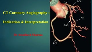
  • 1. CT Coronary Angiography : Indication & Interpretation Dr Awadhesh Sharma
  • 2. Indications of CTA in current scenario
  • 3.
  • 4.
  • 5. Stable chest pain 1. No Known CAD:  Appropriate as the first line test in stable typical or atypical chest pain, or other symptoms which are thought to represent a possible anginal equivalent (e.g. dyspnoea on exertion, jaw pain).  After a nonconclusive functional test, in order to obtain more precision regarding diagnosis and prognosis, if such information will influence subsequent patient management.  May be appropriate in some asymptomatic high-risk individuals, such as those with a higher likelihood of non-calcified plaque.  Rarely appropriate in very low-risk symptomatic patients – those under age 40 with non- cardiac symptoms – or those with low- to intermediate-risk asymptomatic patients.
  • 6. Indications.. 2. Known CAD:  It is appropriate to perform CTA as a first line test for evaluating patients with known CAD who present with stable typical or atypical chest pain, or other symptoms which are thought to represent a possible anginal equivalent (e.g. dyspnea on exertion, jaw pain). 3. Functional imaging:  It may be appropriate to perform CT derived FFR and CT myocardial perfusion Imaging to evaluate the functional significance of intermediate stenoses on CTA (30-70% diameter stenosis).  Add FFRCT and stress-CTP to CTA to increase specificity, positive predictive value, and diagnostic accuracy.  CTP can be a valuable alternative when CT-FFR is technically difficult.
  • 7. Indications.. 4. Stable Coronary Artery Disease: CCTA Post-Revascularization  In intra-coronary stent ≥ 3.0 mm, implementing measures to improve stent imaging accuracy, such as heart-rate control, iterative, sharp kernel, and mono-energetic reconstruction.  CCTA is appropriate to evaluate patients with prior CABG, particularly for graft patency, and to visualize grafts and other structures prior to cardiac surgery re-do.  Protocols to optimize stent imaging should be developed and followed. It may also be appropriate to perform coronary CTA in symptomatic patients with stents <3.0 mm, especially those known to have thin stent struts (<100 mm) in proximal, non-bifurcation locations.
  • 8. Indications.. 5. Stable Coronary Artery Disease: CCTA in Other Conditions Asymptomatic high risk subjects:  may be appropriate in selected asymptomatic high risk individuals, especially in those who have a higher likelihood of having a large amount of non-calcified plaque.  Asymptomatic low or intermediate risk : rarely appropriate.  Coronary artery bypass grafts : It is appropriate to perform CTA for evaluation of patients with prior CABG, particularly if graft patency is the primary objective.
  • 9. Other Indications:  CTA is appropriate for coronary artery evaluation before non-coronary cardiac surgery as an equivalent alternative to invasive angiography in patients with low- to-intermediate probability of CAD and younger patients with primarily non- degenerative valvular conditions.  CTA is appropriate to exclude coronary artery disease in patients with suspected non-ischemic cardiomyopathy.  Late enhancement CT imaging may be appropriate to pinpoint infiltrative heart disease or scar in some patients who have non-ischemic or ischemic cardiomyopathy who cannot undergo cardiac MRI.
  • 10.  CTA is appropriate for the evaluation of coronary anomalies.  Limited delay image CTA (60 seconds-to-90 seconds) is appropriate alternative to TEE to exclude LA/LAA thrombus, as well as in patients where TEE-associated risks outweigh the benefits.  Late enhancement CT imaging may be appropriate to evaluate myocardial viability in some patients who cannot undergo cardiac MRI if it has the potential to impact diagnosis and treatment.
  • 11. Coronary anomalies  (A) Three-dimensional multi-detector row computed tomography reconstruction of a right-sided single coronary artery with a pre- pulmonary course of the left main stem in a 42-year-old man. The left main coronary artery (black arrows) is originating from the proximal part of the right coronary artery (black arrowheads; left panel) than following a pre-pulmonary course to the anterior interventricular groove, where the left main coronary artery splits in the left anterior descending coronary angiography (LAD), an intermediate branch (RIM), and the circumflex coronary artery (RCX, right panel). Ao, ascending aorta; PA, pulmonary artery.  (B) Transaxial multi-detector row computed tomography image of a right-sided single coronary artery with an interarterial path of the left main stem in a 64-year-old man. The left main coronary artery (white arrowheads) originates from the proximal part of the right coronary artery (black arrow) than following an interarterial path between the ascending aorta and the pulmonary trunk. The white arrows indicate the mid part of the circumflex coronary artery. Ao, ascending aorta; LA, left atrium; LV, left ventricle.
  • 13. 1. Medical versus Invasive Treatment  A central aim of evaluation for CAD is to identify patients who need appropriate revascularization to improve prognosis or symptoms not responding to medical therapy, as well as those that can be managed with medical therapy alone.  Stenosis severity still remains the primary arbiter of therapeutic decisions, but more and more data now suggest that anatomy coupled with a physiologic correlate is a better or even possibly, a necessary way for optimal decision-making.
  • 14. 2. Role of CTA for guiding further non-invasive evaluation:  CTA facilitates decision making by dividing patients into multiple informative categories.  Those with a negative CTA or demonstration of non-obstructive CAD would generally exclude flow limiting CAD with high certainty and avoid downstream testing.  CTA seems to allow for more appropriate use of statins and anti-platelet therapies better than when using non CTA methods for CAD diagnosis.  CTA and FFRCT may allow for even more uniform down-stream interventions and narrow the differences between revascularization rates between men and women unlike what happens after usual stress testing imaging.
  • 16.
  • 17. Anterior projection: Left Main or left coronary artery (LCA) Left anterior descending (LAD) diagonal branches (D1, D2) septal branches Circumflex (Cx) Marginal branches (M1,M2) Right coronary artery Acute marginal branch (AM) AV node branch Posterior descending artery (PDA)
  • 18.
  • 21.
  • 22.
  • 23.
  • 24.
  • 25. CAD-RADS:  Cad-Rads is the Coronary Artery Disease-Reporting and Data System.  It was published in 2016 by the Society of Cardiovascular Computed Tomography (SCCT), the American College of Radiology (ACR) and the North American Society for Cardiovascular Imaging (NASCI) and it has been endorsed by the American College of Cardiology (ACC) .  Cad-Rads is developed to standardize reporting of coronary CTA, to improve communication and to guide therapy.
  • 26.
  • 27.
  • 29. Vulnerable plaque… Vulnerable plaque features include: Low-attenuation plaque Positive remodeling Spotty calcification Napkin-ring sign If two or more of these features are present modifier “V” should be added to the CAD-RADS category.
  • 30. Plaque Characteristics..  There are three plaque types on coronary CTA:  Calcified- Atherosclerotic plaque in which the entire plaque appears as calcium density (>130 HU on non-enhanced CT).  Partially calcified- Atherosclerotic lesion with 2 components of which one is calcification.  Non-calcified- Plaque with no calcium content
  • 31. Low-attenuation plaque- Lesions associated with plaque rupture frequently have a large lipid rich core. Lipid on CT appears as low attenuation. Plaques with <30 HU on CTA were found to be present significantly more often in patients with acute coronary syndrome
  • 32. Positive remodelling- is defined as a compensatory outward enlargement of the vessel wall at the site of the atherosclerotic lesion with preservation of the coronary lumen. Higher lipid content and abundance of macrophages. Can present with an acute coronary syndrome without any prior cardiac history.
  • 33. Spotty calcification- usually defined as calcifications < 3 mm . Small (< 1 mm), intermediate (1-3 mm) and large (> 3 mm) calcifications. Small spotty calcifications on CTA are associated with high-risk plaques.
  • 34. Napkin-ring sign- high-risk plaque feature on CTA. It is defined as a central low-attenuation area adjacent to the coronary lumen and a higher “ring- like” attenuation tissue surrounding this central area.. On histology the area of low-attenuation corresponds to the large necrotic core, while the “ring- like” outer area correlates with fibrous plaque tissue. The Napkin-ring sign is strongly associated with major adverse cardiovascular events.
  • 35. Fig. 2 (A1) Presence of positive remodeling (yellow arrows) and low attenuation plaques (LAP, red arrow) are the most important determinants of plaque vulnerability. (A2) Stable plaques lack both these features. (A3) Major adverse cardiac events by the presence of 1 or both features in a follow up of -- - patients for 2 years, and 300 patients for up to 10 years. (A4) Patients with HRP had 45 and 10 folds higher likelihood of adverse outcomes, respectively.
  • 36. Presence of significant stenosis over and above HRP features (A5) and interval progression in plaque magnitude (A6) increased the likelihood of adverse events further. Greater number of adverse plaque characteristics were associated with greater of adverse outcomes (A7) and the HRP characteristics were associated with abnormal fractional flow reserve regardless of luminal stenosis (A8).
  • 37. Imaging of coronary atherosclerotic plaque by multidetector row computed tomography. The contrast- enhanced multi-detector row computed tomography data set shows a noncalcified plaque in the proximal right coronary artery with substantial positive remodelling and only a mild associated reduction of the coronary lumen.
  • 39.
  • 40.  (a) Sixty-one year old man with vague chest pain, not typical for angina with normal treadmill. 3D- VR images clearly shows segment of significant stenosis in large diagonal branch.  (b) The segment of stenosis, which was missed on initial reading of axial images as it is difficult to visualise well, until 3D-VR images were reviewed.  (c) Segment of stenosis also clearly visible on a reconstructed 3D-MIP reconstructed to simulate LAO cranial angio view.  (d) Lesion seen on corresponding angiogram view. Segment of stenosis was subsequently treated by PCI. Note the stenosis appears more severe on the 3D images, due to parameters used to create the images
  • 41.  (a) 58 year old man.16 MSCT scan. In this CPR image, a short segment of significant stenosis in the distal RCA, is clearly visible and there is associated non calcified plaque seen surrounding the contrast filled lumen (arrow).  (b) The fibrous plaque in the lumen of the RCA is also well visualised on the axial image surrounding the contrast filled narrowed lumen (arrow).  (c) At coronary angiography, the segment of stenosis is seen and corresponds to the CTCA findings
  • 42. (a) 53 year old man with CTCA showing soft and calcified plaque in left main extending to bifurcation and proximal LAD. Could be easily misread as associated with significant stenosis by inexperienced reader. (b) Conventional angiography recommended because of segment of significant stenosis in mid LAD (not shown here). Coronary angiogram shows slight narrowing and tapering of lumen of distal left main segment but no significant segment of stenosis is identified (arrow). (c) Cross sectional image at orthogonal plane of contrast filled normal lumen of left main proximal to lesion in Fig. a (arrow). (d) Cross sectional image of lumen of distal left main at level of lesion in Fig. a showing soft and calcified plaques (arrowhead) and contrast filled lumen (arrow) with narrowing of less than 20% of its diameter.
  • 43.  A potential pitfall in the assessment of coronary artery stenosis is to mistake a motion artefact for a noncalcified plaque.  This might particularly occur in coronary CTA datasets of reduced image quality. One should always check a second reconstruction time-point for the presence of any noncalcified plaque.  If the plaque is seen only on one of the reconstruction time points, a motion artefact has to be expected mimicking the finding. Curved-planar reformations of the right coronary artery (RCA) at two different reconstruction time-points: At 75% of the R-R interval a noncalcified plaque in the distal RCA is suspected. Reviewing this area at 45% of the R-R interval shows no evidence of plaque, proving the “lesion” in diastole to be a motion artefact Motion Artifact
  • 44. summary  In general, CTA has the advantage of visualizing the stenosis and the atheromatous plaque as opposed to making an educated guess about its presence, as with physiologic testing.  CTA has excellent sensitivity for identifying flow limiting disease and has very high negative predictive value, making it the strongest test to rule out flow limiting CAD, especially in patients with low to intermediate risk.
  • 45. This Photo by Unknown Author is licensed under CC BY-SA

Editor's Notes

  1. Central Illustration Role of CTA in chronic CAD. Also please see Table 1.
  2. CT angiography for detection of high-risk plaques. (A1) Presence of positive remodeling (yellow arrows) and low attenuation plaques (LAP, red arrow) are the most important determinants of plaque vulnerability. (A2) Stable plaques lack both these features. Major adverse cardiac events by the presence of 1 or both features in a follow up of --- patients for 2 years (A3), and 300 patients for up to 10 years. (A4) Patients with HRP had 45 and 10 folds higher likelihood of adverse outcomes, respectively. Presence of significant stenosis over and above HRP features (A5) and interval progression in plaque magnitude (A6) increased the likelihood of adverse events further. Greater number of adverse plaque characteristics were associated with greater of adverse outcomes (A7) and the HRP characteristics were associated with abnormal fractional flow reserve regardless of luminal stenosis (A8). (B) Potential indicators of inflammation by CTA as a complementary feature for identification of plaque vulnerability. It can be detected either by simultaneous PET imaging with F-18 FDG (that targets macrophage infiltration) (A1 & A2), or by fat attenuation index of perivascular fat (that represents lower prevalence of adipocytes consequent to greater cytokines in neointima) (A3 & A4). Modified from Motoyama et al. JACC 2007, Motoyama et al. JACC 2009, Lee et al. JACC 2019 Ahmadi et al. JACC-Imaging 2018, Rogers et al. JACC-Imaging 2010, Antoniades et al. Lancet 2018.
  3. CT angiography for detection of high-risk plaques. (A1) Presence of positive remodeling (yellow arrows) and low attenuation plaques (LAP, red arrow) are the most important determinants of plaque vulnerability. (A2) Stable plaques lack both these features. Major adverse cardiac events by the presence of 1 or both features in a follow up of --- patients for 2 years (A3), and 300 patients for up to 10 years. (A4) Patients with HRP had 45 and 10 folds higher likelihood of adverse outcomes, respectively. Presence of significant stenosis over and above HRP features (A5) and interval progression in plaque magnitude (A6) increased the likelihood of adverse events further. Greater number of adverse plaque characteristics were associated with greater of adverse outcomes (A7) and the HRP characteristics were associated with abnormal fractional flow reserve regardless of luminal stenosis (A8). (B) Potential indicators of inflammation by CTA as a complementary feature for identification of plaque vulnerability. It can be detected either by simultaneous PET imaging with F-18 FDG (that targets macrophage infiltration) (A1 & A2), or by fat attenuation index of perivascular fat (that represents lower prevalence of adipocytes consequent to greater cytokines in neointima) (A3 & A4). Modified from Motoyama et al. JACC 2007, Motoyama et al. JACC 2009, Lee et al. JACC 2019 Ahmadi et al. JACC-Imaging 2018, Rogers et al. JACC-Imaging 2010, Antoniades et al. Lancet 2018.
  4. On average, the CT attenuation within ‘fibrous’ plaques is higher than within ‘lipid-rich’ plaques (mean attenuation values of 91–116 vs. 47–71 HU)
  5. One should always compare ‘‘lumen to lumen’’, the contrast filled lumen with the lesion to the normal lumen proximal or distal to the lesion, rather than compare ‘‘wall to wall’’ as this will lead to overestimation of the stenosis, especially if there is positive remodelling