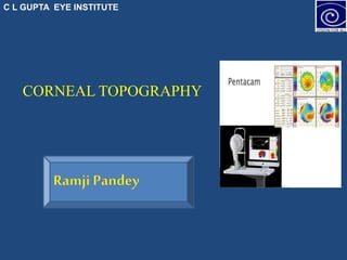
corneal topography by ramji pandey ...C L GUPTA eye institute
•Download as PPTX, PDF•
3 likes•111 views

myppt for pentacam

Recommended
More Related Content
What's hot
What's hot (20)
Similar to corneal topography by ramji pandey ...C L GUPTA eye institute
Similar to corneal topography by ramji pandey ...C L GUPTA eye institute (20)
More from C L GUPTA Eye Institute
More from C L GUPTA Eye Institute (12)
Recently uploaded
Recently uploaded (20)
corneal topography by ramji pandey ...C L GUPTA eye institute
- 1. C L GUPTA EYE INSTITUTE CORNEAL TOPOGRAPHY
- 2. C L GUPTA EYE INSTITUTE THE SCHEIMPFLUG PRINCIPLE • The Scheimpflug principle, It is a geometric rule that describes the orientation of the plane of focus of an optical system (such as a camera) when the lens plane is not parallel to the image plane 4/15/2021 2
- 3. C L GUPTA EYE INSTITUTE THE DEVICE •The light source - UV-free blue LED’s (wavelength=475 nm). 2 cameras 1 st -Located in the center for the purposes of detection of the size and orientation of the pupil and to control fixation. • 2 nd - Mounted on rotating wheel to capture images from the anterior segment. This rotating process supplies pictures in three dimensions and also allows the center of the cornea to be measured precisely. 4/15/2021 3
- 4. C L GUPTA EYE INSTITUTE
- 5. C L GUPTA EYE INSTITUTE Assessment of Corneal Surface
- 6. C L GUPTA EYE INSTITUTE Corneal topography Measurements with a keratometer is insufficient and limited to the central 3 mm. Its provides us with a detailed description of various curvature and shape characteristics of the cornea. Provides helpful information for the illustration of astigmatism, detection of corneal pathologies and perfection of contact lens fitting. 4/15/2021 6 Anderson D, Kojima R. Topography: A clinical pearl. Optom Manag 2007 Feb;42(2):35
- 7. C L GUPTA EYE INSTITUTE CLINICALAPPLICATION 1. Pachymetry 2. Scheimpflug Imaging in LASIK 3. Topography and contact lenses. 4. Topography in RK. 5. Post keratoplasty astigmatism. 6. Keraoconus screening 7. Corneal Pathologies 8. Anterior Chamber implantation of phakic IOLs 9) Glaucoma Screening • a) Effect of pilocarpine on anterior chamber depth and anterior chamber volume in eyes with narrow angle and open angles 4/15/2021 7
- 8. C L GUPTA EYE INSTITUTE PENTACAM • The OCULUS Pentacam/Pentacam HR is a rotating Scheimpflug camera. • The rotational measuring procedure generates Scheimpflug images in three dimensions The Pentacam calculates a 3-dimensional model of the anterior eye segment from as many as 25.000 (HR: 138.000) true elevation points. • It takes a maximum of 2 seconds to generate a complete image of the anterior eye segment.
- 9. C L GUPTA EYE INSTITUTE Review of Pentacam main Page • This page should be displayed with the four main Refractive maps – Anterior sagittal curvature map, – Anterior and posterior elevation maps and – Thickness map • Belin/Ambrosis Enhanced ectasia maps • Topography maps for Keratoconus indices
- 10. C L GUPTA EYE INSTITUTE 4/15/2021 10
- 11. C L GUPTA EYE INSTITUTE ANTERIOR SAGITTAL MAP Steep areas hot colours (red and orange), while flat areas cold colours (green and blue). Normally, the inferior (I) point has a higher value than the superior (S) one, and the I-S difference should be < 1.5 D. The superior point may rarely have a higher value than the inferior one ,the S-I difference should be < 2.5D 4/15/2021 11
- 12. C L GUPTA EYE INSTITUTE CON 4/15/2021 12
- 13. C L GUPTA EYE INSTITUTE 4/15/2021 13
- 14. C L GUPTA EYE INSTITUTE 4/15/2021 14
- 15. C L GUPTA EYE INSTITUTE Classification of various patterns on axial map of placido based topography. Top A, round; B, oval; C, superior steepening; D, inferior steepening; E, irregular; F, symmetric bow tie; G, symmetric bow tie with skewed radial axes; H, asymmetric bow tie with inferior steepening (AB/IS); I, asymmetric bow tie with superior steepening; J, asymmetric bow tie with skewed radial axes (AB/SRAX
- 16. C L GUPTA EYE INSTITUTE • K1 or Rf: Horizontal curvature power of the cornea in the central 3 mm circle expressed in diopters • K2 or Rs: Vertical curvature power of the cornea in the central 3 mm expressed in diopters. • Km: Mean curvature power of the cornea in the central 3 mm expressed in diopters. • Rh: Horizontal curvature radius of the central 3 mm expressed in mm. • Rv: Vertical curvature radius of the central 3 mm expressed in mm. • Rm: Mean curvature radius of the central 3 mm expressed in mm. • Qs: “Quality specification” should be displayed “OK”
- 17. C L GUPTA EYE INSTITUTE Important figures: • when taking the decision—that any anterior K readings should not be more than 47D on the front sagittal curvature map. Recently, with the availability of thin flap technology, the power 49D became acceptable. More than 49D is risky regardless of the patient’s refractive error. • Corneal astigmatism on either surface should not be higher than 6D; otherwise it is a risk factor. • Against the rule astigmatism is considered suspicious.
- 18. C L GUPTA EYE INSTITUTE ELEVATION MAPS 4/15/2021 18
- 19. C L GUPTA EYE INSTITUTE Elevation Map • The computer considers all points above the reference surface as elevations, being displayed as positive values, and considers all points below the reference surface as depressions, being displayed as negative values (in microns) • The elevation maps are more accurate than curvature maps in evaluating both surfaces of the cornea. They are less affected by tear film disturbance and use of contact lenses
- 20. C L GUPTA EYE INSTITUTE • The elevation values on the front surface map should not exceed +12 μ. • Values between +13 μ and +15 μ are suspected, • and any value > +15 μ is considered a risk factor. • The elevation values on the back surface map should not exceed +17 μ. • Values between +18 μ and +20 μ are suspected, and • Any value >+20 μ is considered a risk factor.
- 21. C L GUPTA EYE INSTITUTE Important considerations • Be careful when any value of the central 4 mm of the elevation maps is more than +15 μ for the anterior surface, and more than +20 μ for the posterior surface. • Be careful when the “back-front” difference is more than +5 μ at the same point. • Be careful when there is an isolated island on either surface
- 22. C L GUPTA EYE INSTITUTE CORNEAL THICKNESS MAP
- 23. C L GUPTA EYE INSTITUTE • Pachy apex: Corneal thickness at the apex. The computer considers the apex as the origin of the coordinates, X for the horizontal and Y for the vertical Therefore, zero is displayed in both squares of pachy apex coordinates. • Pupil center: Corneal thickness in the pupil center. The Xand Y-coordinates show the position of the pupil center from the apex. The two coordinates differ according to pupil medriasis or miosis, because the pupil center is often shifted superotemporally when dilated. • Thinnest location: Thinnest point in the cornea. It is the most important in the decision procedure
- 24. C L GUPTA EYE INSTITUTE • Cornea volume: studies are carried out to realize the relaSome tionship between cornea volume and ectatic changes. • Chamber volume: Volume less than 100 mm3 should alert us to check the patient for angle closure glaucoma. • AC depth (Int.): Central anterior chamber depth, measured from the inner surface of corneal endothelium to the iris. • It is important for phakic IOLs. It should not be less than 2.8 mm to keep the corneal endothelium intact. • KPD: The average value of keratometric power deviation. The normal value is less than +0.75. When the value is more than +1.5, it is abnormal indicating an abnormal cornea .
- 25. C L GUPTA EYE INSTITUTE How to start reading Pentacam Refractive Map
- 26. C L GUPTA EYE INSTITUTE Based on severity of curvature • Mild - <45D in both meridian • Moderate 45 – 52D in both meridian • Severe - >52D in both meridian • Advanced - >62D in both meridian 4/15/2021 26
- 27. C L GUPTA EYE INSTITUTE • 4/15/2021 27 THANK YOU