GROWTH & DEVELOPMENT OF MANDIBLE
•
4 likes•529 views

email antarleenasengupta@gmail.com for full seminar

Recommended
More Related Content
What's hot
What's hot (20)
Similar to GROWTH & DEVELOPMENT OF MANDIBLE
Similar to GROWTH & DEVELOPMENT OF MANDIBLE (20)
More from Dr Antarleena Sengupta
More from Dr Antarleena Sengupta (16)
Recently uploaded
Recently uploaded (20)
GROWTH & DEVELOPMENT OF MANDIBLE
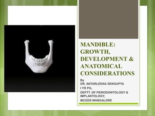
- 1. MANDIBLE: GROWTH, DEVELOPMENT & ANATOMICAL CONSIDERATIONS By DR. ANTARLEENA SENGUPTA I YR PG, DEPTT. OF PERIODONTOLOGY & IMPLANTOLOGY, MCODS MANGALORE
- 2. CONTENTS INTRODUCTION ANATOMY BODY OF MANDIBLE OUTER SURFACE INNER SURFACE RAMUS LATERAL SURFACE MEDIAL SURFACE CORONOID PROCESS CONDYLAR PROCESS ATTACHMENTS & RELATIONS MUSCLE ATTACHMENTS FORAMINA BLOOD SUPPLY NERVE SUPPLY LYMPHATICS STRUCTURES RELATED TO MANDIBLE 2
- 3. contd. GROWTH OF MANDIBLE PRENATAL EMBRYOLOGY MECKEL’S CARTILAGE ENDOCHONDRAL BONE FORMATION CONDYLAR PROCESS CORONOID PROCESS MENTAL REGION POST-NATAL EMBRYOLOGY RAMUS BODY ANGLE LINGUAL TUBEROSITY ALVEOLAR PROCESS CHIN CONDYLAR PROCESS CORONOID PROCESS THEORIES OF GROWTH ENLOW’S EXPANDING V PRINCIPLE ENLOW’S COUNTERPART PRINCIPLE AGE CHANGES IN MANDIBLE INFANTS & CHILDREN ADULTS OLD AGE ANATOMIC CONSIDERATIONS DEVELOPMENTAL ANOMALIES SURGICAL CONSIDERATIONS ANATOMIC SPACES CONCLUSION REFERENCES 3
- 5. ANTERIOR VIEW LATERAL VIEW 5
- 8. 8 MENTAL PROTUBERANCE MENTAL TUBERCLES ANTERIOR VIEW
- 9. BODY Each half of the body has outer and inner surfaces, and upper and lower borders. The outer surface presents the following features: 1. The symphysis menti 2. The mental protuberance 3. The mental foramen 4. The oblique line 9
- 10. OUTER SURFACE Anterior border LATERAL VIEW 10
- 12. INNER SURFACE has the following features: Mylohyoid Line Submandibular fossa Sublingual fossa Superior and Inferior genial tubercles Mylohyoid groove 12
- 14. UPPER BORDER The upper or alveolar border bears sockets for the teeth. 14
- 15. LOWER BORDER 15 The lower border of the mandible is also called the base. Near the midline the base shows an oval depression called the digastric fossa.
- 16. RAMUS 16 ANTERIOR VIEW LATERAL VIEW • LATERAL SURFACE of ramus is flat and bears a number of oblique ridges. U L P A
- 17. MEDIAL SURFACE of ramus has the following: Mandibular foramen Lingula Mylohyoid groove Mandibular notch Angle of mandible 17 Lingula Submandibular fossaMylohyoid groove Mandibular notch
- 18. Coronoid process Condyloid process Head Neck Pterygoid fovea 18 Superior view Superior view Pterygoid fovea Neck
- 19. RADIOGRAPHIC VIEW OF MANDIBLE 19
- 20. ATTACHMENTS & RELATIONS OF MANDIBLE MUSCLE ATTACHMENTS 20 LATERAL VIEW
- 21. 21 MEDIAL VIEW
- 22. FORAMINA & RELATIONS OF MANDIBLE 22 Mandibular foramen Mental foramen
- 23. BLOOD SUPPLY Central blood supply Peripheral blood supply 23 via the INFERIOR ALVEOLAR ARTERY except the coronoid process, which is supplied by temporalis muscle vessels. via the PERIOSTEAL VESSELS, which run parallel to cortical surface of bone, giving off NUTRIENT VESSELS those penetrate cortical bone and anastomose with the branches of inferior alveolar artery.
- 24. NERVE SUPPLY Derived from mandibular branch (V3) of trigeminal nerve. 1. Long Buccal Nerve – supplies mucosa opposite the posterior-most mandibular molars(6,7,8) on their buccal aspect. 2. Inferior Alveolar Nerve – supplies all lower jaw teeth, lower lip, buccal mucosa from incisors to premolar & the skin over the chin. 3. Lingual Nerve – sensory supply to anterior 2/3rd of tongue, the mucosa on the lingual aspect of lower teeth & floor of the mouth. 24
- 26. LYMPHATICS 26 Submandibular lymph nodes Submental lymph nodes Jugulo-Omohyoid group of deep cervical lymph nodes Jugulo-Digastric group of deep cervical lymph nodes Mandible + lower teeth Small wedge in symphysis Lower incisors Extremely posterior
- 27. STRUCTURES RELATED TO MANDIBLE SALIVARY GLANDS: Parotid Submandibular sublingual LYMPH NODES: Parotid Submandibular Submental ARTERIES: Maxillary Superficial temporal Masseteric Inferior alveolar Mylohyoid Mental Facial 27
- 28. NERVES: Lingual Auriculotemporal Masseteric Inferior alveolar Mylohyoid Mental MoM: insertions of Temporalis Masseter Medial pterygoid Lateral pterygoid LIGAMENTS: Lateral ligament of TMJ Stylomandibular Sphenomandibular and Pterygomandibular raphe 28
- 29. PRENATAL DEVELOPMENT OF MANDIBLE MECKEL’S CARTILAGE Derived from 1st branchial arch 41st-45th day IU Extends from cartilaginous otic capsule to midline(symphysis) Provides template for guiding growth of mandible MANDIBULAR DIVISION, TRIGEMINAL NERVE(V3)→ first structure to develop 2 ossification centres: 1 for each half; arises 6th wk. IU 29
- 30. Ossifying membrane located lateral to the Meckel’s Cartilage Spreads below & around IAN and incisive branch and upwards to form a trough to accommodate developing tooth buds Dorsally and ventrally spreads to form body and ramus of mandible Ossification continues→ Meckel’s cartilage surrounded by bone→ invaded by bone→ ossification stops at lingula 30 Continued growth into middle ear : develops auditory ossicles To sphenoid bone to form a remnant of Meckel’s cartilage Sphenomandibular ligament
- 31. ENDOCHONDRAL BONE FORMATION THE CONDYLAR PROCESS: 5th wk. IU 10th- 14th wk. IU THE CORONOID PROCESS: 10th – 14th wk. IU Accessory cartilage gets incorporated into expanding ramus; disappears before birth. 31 Area of mesenchymal condensation seen above ventral part of the developing mandible Develops into a cone-shaped cartilage → replaced by mid-fetal life; upper end persists into adulthood Secondary cartilage of coronoid process grows in response to developing temporalis muscle.
- 32. MENTAL REGION: 2 small cartilages appear on either side of symphysis 7th wk. IU 1st yr. postnatal 32 Formation of mental ossicles Incorporated in intramembranous ossification Complete ossification
- 33. POSTNATAL DEVELOPMENT OF MANDIBLE Divided into development of following functional parts: Ramus Corpus or Body of mandible Angle of the mandible Lingual tuberosity Alveolar process Chin Condyle Coronoid process 33 THREE FORMS OF GROWTH can be seen in the mandible: o Vertical o Transverse o rotational
- 34. VERTICAL GROWTH of the mandible is quite pronounced. The mandible has to keep pace with the descent of the maxilla and must also maintain the interocclusal vertical direction. 34
- 35. TRANSVERSE GROWTH of the mandible is achieved principally by the divergence of the condyles as they grow posteriorly(Enlow’s V principle) Buccal bone deposition on the body and ramus 35
- 36. In ROTATIONAL GROWTH, the matrix surrounding the mandible acts to moderate the shape changes of the bone rotating with it. 36
- 37. THEORIES OF GROWTH ENLOW’S EXPANDING ‘V’ PRINCIPLE ENLOW’S COUNTERPART PRINCIPLE 37 States that the growth of any given facial/cranial part relates specifically to other structural and geometric counterparts in the face and cranium. The growth, movement & enlargement of these bones occur towards the wide ends of the ‘V’ as a result of differential deposition & selective resorption.
- 38. AGE CHANGES IN MANDIBLE 38
- 40. AGNATHIA Characterized by hypoplasia or absence of mandible. More commonly, only a portion of jaw is missing. Partial absence of mandible is more common. Entire mandible on one side may be missing or more frequently, only the condyle or the entire ramus. Bilateral agenesis of condyles and ramus have also been reported. 40
- 41. MICROGNATHIA Means small jaw, either the maxilla or mandible may be involved. True micrognathia is classified as: Congenital Acquired 41
- 42. CONGENITAL MICROGNATHIA Etiology: I. Idiopathic II. Assoc’d. with congenital heart disease III. Pierre-Robin Syndrome Follows a hereditary pattern. Agenesis of condyles results in true micrognathia. Such cases may be due to posterior positioning of the mandible with regard to the skull or to a steep mandibular angle resulting in apparent retrusion of mandible. 42
- 43. ACQUIRED MICROGNATHIA Postnatal origin. Usually results from a disturbance in the area of TMJ. Since the normal growth of the mandible depend on normally developing condyles as well as muscles, condylar ankylosis may result in deficient mandible. Clinically it is characterized by severe retrusion of the chin, a steep mandibular angle, and a deficient chin button 43
- 44. MACROGNATHIA Macrognathia refers to the condition of abnormally large jaws. An increase in both the jaws is frequently proportional to generalized increase in entire skeleton. Often associated with other conditions like: Paget’s disease of bone Acromegaly Leontiasis ossea 44
- 45. Etiology: unknown, although cases may follow hereditary patterns. In many instances the prognathism is due to disparity in the size of maxilla to mandible. The angle between the ramus and the body influence the relation of mandible to maxilla. Thus prognathic patients tend to have long rami which form a steep angle with the body of the mandible. 45
- 46. FACIAL HEMIHYPERTROPHY One of the rare developmental disorder. Asymmetric over growth of one or more body parts. Represents hyperplasia rather than hypertrophy. It is of 3 types, namely: Simple hyperplasia Complex hyperplasia Hemifacial hyperplasia F:M > 2:1, often affecting on right side. 46
- 47. Asymmetry starts at birth. Enlargement is more accentuated at the age of 6 and continues ‘til the overall growth ceases. Enlargement of mandible and teeth on the affected side. The bone is wider and thicker. Premature shedding of the deciduous teeth. Roots of teeth are sometimes proportionately enlarged but maybe short. Permanent teeth on the affected side is often enlarged, most frequently involving cuspid, premolars, and 1st molar. Permanent teeth on affected side develops more rapidly and erupt before their counterpart on the uninvolved side. Macroglossia 47
- 48. 48
- 49. PAGET’S DISEASE Characterized by excessive growth and abnormal remodeling of bone. Results in bones which are weak, enlarged and extensively vascularized. Etiology: unknown, there may be evidence of genetic link. Possible etiologic factors: viral infections Inflammatory cause Autoimmune connective tissue 49 o Recognized most commonly after the age of 50 years. o Its prevalence increases with age. o Male:female> 1:1 o Jaws are involved more commonly. o The most common complaint is bone pain. o This pain is perceived as dull aching pain deep below the soft tissues. o It may persist or exacerbate during the night. o The involved bone becomes warm to the touch due to increased vascularity
- 50. CHERUBISM Autosomal dominant The gene is mapped to chromosome 4p16. Facial appearance is similar to plump-cheeked angels, hence the name cherubism. First described in the year 1953 by Jones. Jaw lesions are usually painless and symmetric. Lesions which are firm and non- tender to palpate involve molar to coronoid regions, often associated with cervical lymphadenopathy. This contributes to the characteristic full-faced appearance. 50
- 51. CHERUBISM- RADIOGRAPHIC APPEARANCE Bilateral multilocular radiolucencies in the posterior mandible. These lesions tend to show varying degree of remission and involution after puberty. There maybe displacement, rotation of the teeth. Premature exfoliation, delayed eruption. 51
- 52. EXOSTOSES Normal anatomic variation. Hinders removal of plaque by patient. May have to be removed to improve the prognosis of neighbouring teeth. Most common in lingual area of canine and premolars, above mylohyoid muscle. Also found on buccal/labial surfaces of mandibular teeth. 52
- 53. ANATOMIC SPACES Several anatomic spaces or compartments are found close to the operative field of periodontal & implant surgery sites. Contain loose connective tissue– easily distended by hemorrhage, inflammatory fluid, and infection. Surgical invasion of these areas may result in dangerous hemorrhage (intraoperative) or infections (postoperative) & should be carefully avoided. 53
- 54. ANATOMIC SPACES 54 A. SUBMENTAL SPACE: between mylohyoid muscle superiorly and platysma inferiorly. Infection results in swelling of the region; more dangerous as it proceeds posteriorly. B. MASTICATOR SPACE: contains masseter, pterygoid (lat. and med.), tendon of insertion of temporalis, ramus and posterior mandible. Infection leads to swelling of face, severe trismus and pain. C. SUBLINGUAL SPACE: below the oral mucosa in anterior part of floor of mouth. Infection of this space raises floor of mouth, displacing the tongue, resulting in pain & difficulty swallowing. D. SUBMANDIBULAR SPACE: external to sublingual space below mylohyoid and hyoglossus muscle. Contains the submandibular gland and connected with sublingual space. Infections lead to obliterated submandibular line+ pain in swallowing.
- 55. SURGICAL CONSIDERATIONS Surgical trauma (pressure, manipulation and postsurgical swelling) to the mental nerve produces paresthesia of lip– recovers slowly. Partial/complete cutting of the nerve can result in permanent paresthesia/dysesthesia. 55
- 56. SURGICAL CONSIDERATIONS In partially/completely edentulous patients, disappearance of alveolar portion brings mandibular canal and mental foramen closer to superior border. In such patients, during evaluation for placement of implants, the distance between canal and superior surface of the bone as well as location of mental foramen must be carefully determined to avoid surgical injury to the nerve. 56
- 57. SURGICAL CONSIDERATIONS The lingual nerve lies close to the surface of the oral mucosa in the third molar area and goes deeper as it travels forward. It can be damaged during anesthetic injections (and during extraction procedures). It can be injured when a periodontal partial- thickness flap is raised in third molar region or when releasing incisions are made in the area. 57
- 58. SURGICAL CONSIDERATIONS The alveolar process, which provides the supporting bone to the teeth, has a narrower distal curvature than the body of mandible, creating a flat surface in the posterior area between the teeth and the anterior border of the ramus. This results in the formation of external oblique ridge, which runs downward and forward to region of second/first molar, creating a shelflike bony area. Resective osseous therapy may be difficult in this area because of the amount of bone that must be removed distally toward ramus to achieve resection of a periodontal osseous defect on the distal aspect of mandibular 2nd/3rd molar. 58
- 59. SURGICAL CONSIDERATIONS Distal flap procedures in distal to the last molar can be performed effectively only if there exists sufficient space. 59
- 60. CONCLUSION Familiarity with the location and appearance of the mental nerve reduces likelihood of injury to the nerve. Determining the amount of available bone is critical for placement of implants. 60
- 61. REFERENCES 1. B D Chaurasia’s Human Anatomy vol. 3 2. Human Embryology, Inderbir Singh 3. Clinical Periodontology by Carranza, 10th edition, vol 2 4. Shafer’s Textbook of Oral Pathology 5. Contemporary Orthodontics by W R Profitt 6. Handbook of Local Anesthesia, S F Malamed 7. Image references: Wikimedia Commons Gray’s anatomy, 41st edition 61
- 62. 62