
Recommended
More Related Content
What's hot
What's hot (20)
Similar to human cell anatomy and function
Similar to human cell anatomy and function (20)
Recently uploaded
Recently uploaded (20)
human cell anatomy and function
- 2. CELL INTRODUCTION „ •All the living things are composed of cells. • A single cell is the smallest unit that has all the characteristics of life. •Cell is defined as the structural and functional unit of the living body.
- 3. General Characteristics of Cell Each cell in the body: 1. Needs nutrition and oxygen 2. Produces its own energy necessary for its growth, repair and other activities 3. Eliminates carbon dioxide and other metabolic wastes, 4. Maintains the medium, i.e. the environment for its survival
- 4. 5. Shows immediate response to the entry of invaders like bacteria or toxic substances into the body 6. Reproduces by division. There are some exceptions like neuron, which do not reproduce
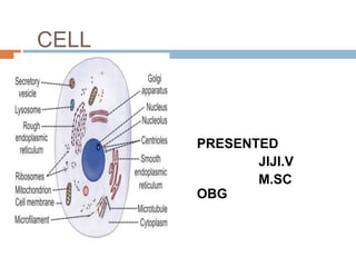
- 5. STRUCTURE OF THE CELL Each cell is formed by a cell body and a membrane covering the cell body called the cell membrane. Cellbody has two parts, namely nucleus and cytoplasm surrounding the nucleus . Thus, the structureof the cell is studied under three headings: 1. Cell membrane 2. Cytoplasm 3. Nucleus.
- 6. Cell membrane is a protective sheath, enveloping the cell body. It is also known as plasma membrane or plasmalemma. This membrane separates the fluid outside the cell called extracellular fluid (ECF) and the fluid inside the cell called intracellular fluid (ICF). The cellmembrane is a semipermeable membrane. So, there is free exchange of certain substances between ECF and ICF. CELL MEMBRANE
- 7. Thickness of the cell membrane varies From 75 to111Å
- 8. COMPOSITION OF CELL MEMBRANE Cell membrane is composed of three types of substances: 1. Proteins (55%) 2. Lipids (40%) 3. Carbohydrates (5%).
- 9. 1.The materials dissolved in lipid layer also move to all areas of the cell membrane. Major lipids are: 1. Phospholipids 2. Cholesterol. 2.Lipid layer of the cell membrane is a semipermeablemembrane and allows only the fat-soluble substances to pass through it. Thus, the fat-soluble substances like oxygen, carbon dioxide and alcohol can pass through this lipid layer. 3.The water-soluble substances such as glucose, urea and electrolytes cannot pass through this.
- 10. Protein layers of the cell membrane are electron- dense layers. These layers cover the two surfaces of the central lipid layer. Protein layers give protection to the central lipid layer. The protein substances present in these layers are mostly glycoproteins. Protein molecules are classified into two categories: 1. Integral proteins or transmembrane proteins. 2. Peripheral proteins or peripheral membraneproteins
- 11. Examples of integral protein: i. Cell adhesion proteins ii. Cell junction proteins iii. Some carrier (transport) proteins iv. Channel proteins v. Some hormone receptors vi. Antigens vii. Some enzymes Examples of peripheral proteins: i. Proteins of cytoskeleton ii. Some carrier (transport) proteins iii. Some enzymesPeripheral proteins or The proteins which are partially embedded in the outer and inner surfaces of the cell membrane and do not penetrate the cell membrane. Peripheral proteins Integral or transmembrane proteins are the proteins that pass through entire thickness of cell membrane from one side to the other side. These proteins are tightly bound with the cell membrane.
- 12. Some of the carbohydrate molecules present in cell membrane are attached to proteins and form glycoproteins (proteoglycans). Some carbohydrate molecules are attached to lipids and form glycolipids. Carbohydrate molecules form a thin and loose covering over the entire surface of the cell membrane called glycocalyx.
- 13. 1. Carbohydrate molecules are negatively charged and do not permit the negatively charged substances to move in and out of the cell 2. Glycocalyx from the neighboring cells helps in the tight fixation of cells with one another
- 14. FUNCTIONS OF CELL MEMBRANE 1. Protective function: Cell membrane protects the cytoplasm and the organelles present in the cytoplasm 2. Selective permeability: Cell membrane acts as a semipermeable membrane, which allows only some substances to pass through it and acts as a barrier for other substances 3. Absorptive function: Nutrients are absorbed into the cell through the cell membrane
- 15. 4. Excretory function: Metabolites and other waste products from the cell are excreted out through thecell membrane 5. Exchange of gases: Oxygen enters the cell from theblood and carbon dioxide leaves the cell and enters the blood through the cell membrane 6. Maintenance of shape and size of the cell: Cell membraneis responsible for the maintenance of shapeand size of the cell
- 16. Cytoplasm of the cell is the jelly like material formed by80% of water. It contains a clear liquid portion called cytosol and various particles of different shape and size. These particles are proteins, carbohydrates, lipidsor electrolytes in nature. Cytoplasm also contains manyorganelles with distinct structure and function. Cytoplasm is made up of two zones: 1. Ectoplasm: Peripheral part of cytoplasm, situatedjust beneath the cell membrane 2. Endoplasm: Inner part of cytoplasm, interposed between the ectoplasm and the nucleus.
- 17. Organelles with limiting membrane 1. Endoplasmic reticulum 2. Golgi apparatus 3. Lysosome 4. Peroxisome 5. Centrosome and centrioles 6. Secretory vesicles 7. Mitochondria 8. Nucleus Cytoplasmic organelles are the cellular structures embedded in the cytoplasm. Organelles are consideredas small organs of the cell. Some organelles are bound 1, limiting membrane and 2.not have limitingmembrane Organelles without limiting membrane 1. Ribosomes 2. Cytoskeleton
- 18. „a.ENDOPLASMIC RETICULUM •Endoplasmic reticulum is a network of tubular and microsomal vesicular structures which are interconnected with one another. •It is covered by a limiting membrane which is formed by proteins and bilayered lipids. •The lumenof endoplasmic reticulum contains a fluid medium called endoplasmic matrix. •The endoplasmic reticulum forms the link between nucleus and cell membrane by connecting the cell membrane with the nuclear membrane.
- 20. Types of Endoplasmic Reticulum Endoplasmic reticulum is of two types: 1.Rough endoplasmic reticulum 2.Smooth endoplasmic reticulum. Both the types are interconnected and continuous with one another. 1.Rough Endoplasmic Reticulum It is the endoplasmic reticulum with rough, bumpy or bead-like appearance. Rough appearance is due to the attachment of granular ribosomes to its outer surface. Hence, it is also called the granular endoplasmic reticulum.
- 21. FunctionsofRoughEndoplasmicReticulum • Synthesis of proteins •Degradation of worn-out organelles 1.Rough endoplamic
- 22. It is the endoplasmic reticulum with smooth appearance. It is also called a granular reticulum. It is formed by many interconnected tubules. So, it is also called tubular endoplasmic reticulum. Functions of Smooth Endoplasmic Reticulum 1. Synthesis of non-protein proteins such as cholesterol and steroid. 2.Storage and metabolism of calcium 3.Catabolism and detoxification
- 23. • Golgi apparatus or Golgi body or Golgi complex is a membrane-bound organelle, involved in the processing of proteins. • It is present in all the cells except red blood cells. It is named after the discoverer Camillo Golgi. • Usually, each cell has one Golgi apparatus. Some of the cells may have more than one Golgi apparatus. • Each Golgi apparatus consists of 5 to 8 flattened membranous sacs called the
- 24. •Golgi apparatus is situated near the nucleus. It hastwo ends or faces, namely cis face and trans face. •The cis face is positioned near the endoplasmic reticulum.Reticular vesicles from endoplasmic reticulum enter the Golgi apparatus through cis face. •The trans faceis situated near the cell membrane. The processed substances make their exit from Golgi apparatus through trans face .
- 25. Functions of Golgi Apparatus Major functions of Golgi apparatus are processing, packing, labeling and delivery of proteins and other 1. Processing of materials Here, the glycoproteins and lipids are modified and processed. 2. Packaging - Packed in the form of secretory granules, secretory vesicles and lysosomes, which are transported either out of the cell or to another part of the cell. Because of this, Golgi apparatus is called the ‘post office of the cell’. 3. Labeling - the processed and packed materials and labels them (such as phosphate group), depending upon the chemical content for delivery. The Golgi apparatus is called ‘shipping department of the cell’.
- 26. •Lysosomes are the membrane-bound vesicular organelles found throughout the cytoplasm. • The lysosomes are formed by Golgi apparatus. •Among the organelles of the cytoplasm, the lysosomes have the thickest covering membrane. •The membrane is formed by a bilayered lipid material. •It has many small granules which contain hydrolytic enzymes
- 27. Lysosomes are of two types: 1. Primary lysosome, which is pinched off from Golgi apparatus. It is inactive in spite of having hydrolytic Enzymes 2. Secondary lysosome, which is the active lyso some. It is formed by the fusion of a primary lysosome
- 28. PEROXISOMES Peroxisomes or microbodies are the membrane limitedvesicles like the lysosomes Peroxisomes contain some oxidative enzymes such as catalase, urate oxidase and Damino acid oxidase. Functions of Peroxisomes •Breakdown the fatty acids by means of a process called betaoxidation: •Large number of peroxisomes are present in the cells of liver, which is the major organ for detoxification •Form the major site of oxygen utilization in the cells • Accelerate gluconeogenesis from fats • Degrade purine to uric acid • Participate in the formation of myelin • Play a role in the formation of bile acids. „
- 30. Centrosome is the membrane-bound cellular organelle situated almost in the center of cell, close to nucleus. It consists of two cylindrical structures called centrioles which are made up of proteins. Centrioles are responsiblefor the movement of chromosomes during cell division
- 31. SECRETORY VESICLES Secretory vesicles are the organelles with limiting membrane and contain the secretory substances. Secretory vesicles are present throughout the cytoplasm. When necessary, these vesicles are ruptured and secretory substances are released into the cytoplasm. „
- 32. MITOCHONDRION Mitochondrion (plural = mitochondria) is a membrane bound Cytoplasmic organelle concerned with production of energy. It is a rod-shaped or oval-shaped structure with a diameter of 0.5 to 1 μ. It is covered by a bilayered membrane .
- 33. The outer membrane is smooth and encloses the contents of mitochondrion. This membrane contains various enzymes such as acetyl-CoA synthetase and glycerolphosphate acetyltransferase. The inner membrane is folded in the form of shelf-like inward projections called cristae and it covers the inner matrix space. Cristae contain many enzymes and other protein molecules which are involved in respiration and synthesis of adenosine triphosphate (ATP). Because of these functions is collectively known as respiratory chain or electron transport system. MITOCHONDRION
- 34. Mitochondrion moves freely in the cytoplasm of the cell. It is capable of reproducing itself. Mitochondrion contains its own deoxyribonucleic acid (DNA), which is responsible for many enzymatic actions. Functions of Mitochondrion 1. Production of energy Mitochondrion is called the ‘power house’ or ‘power plant’ of the cell because it produces the energy required for cellular functions. 2. Synthesis of ATP The components of respiratory chain in mitochondrion are responsible for the synthesis of ATP by utilizing the energy by oxidative phosphorylation 3.Other functions Other functions of mitochondria include storage of calcium and detoxification of ammonia in liver
- 35. Mitochondrion moves freely in the cytoplasm of the cell. It is capable of reproducing itself. Mitochondrion contains its own deoxyribonucleic acid (DNA), which is responsible for many enzymatic actions. Functions of Mitochondrion 1. Production of energy Mitochondrion is called the ‘power house’ or ‘power plant’ of the cell because it produces the energy required for cellular functions. 2. Synthesis of ATP The components of respiratory chain in mitochondrion are responsible for the synthesis of ATP by utilizing the energy by oxidative phosphorylation 3.Other functions Other functions of mitochondria include storage of calcium and detoxification of ammonia in liver
- 36. •Cytoskeleton is the cellular organelle present throughout •the cytoplasm. •It determines the shape of the cell and gives support to the cell. •It is a complex network of structures with varying sizes. In addition to determining the shape of the cell. • •it is also essential for the cellular movements and the response of the cell to external stimuli. Cytoskeleton consists of three major protein components: 1. Microtubule 2. Intermediate filaments 3. Microfilaments.
- 38. RIBOSOMES 1. Ribosomes are the organelles without limiting membrane. 2. These organelles are granular and small dot-like structures with a diameter of 15 nm. 3. Ribosomes are made up of 35% of proteins and 65% of ribonucleic acid (RNA). 4. RNA present in ribosomes is called ribosomal RNA (rRNA).
- 40. Types of Ribosomes i. Ribosomes that are attached to rough endoplasmic reticulum ii. Free ribosomes that are distributed in the cytoplasm. Functions of Ribosomes 1. Ribosomes are called ‘protein factories’ because oftheir role in the synthesis of proteins. Messenger RNA (mRNA) carries the genetic code for protein synthesis from nucleus to the ribosomes. 2. Ribosomes attached to rough endoplasmic reticulum are involved in the synthesis of proteins such as the enzymatic proteins, hormonal proteins, lysosomal proteins and the proteins of the cell membrane. 3. Free ribosomes are responsible for the synthesis of proteins in
- 41. Nucleus is the most prominent and the largest cellular organelle. It has a diameter of 10 μ to 22 μ and occupies about 10% of total volume of the cell •Nucleus is present in all the cells in the body except the red blood cells. •The cells with nucleus are called eukaryotes and those without nucleus are known as prokaryotes. •Presence of nucleus is necessary for cell division. •Most of the cells have only one nucleus (uninucleated •cells). •Few types of cells like skeletal muscle cells have •many nuclei (multinucleated cells). „
- 42. Nucleus is located in the center of the cell. It is mostly spherical in shape. However, the shape and situation of nucleus vary in some cells. STRUCTURE OF NUCLEUS Nucleus is covered by a membrane called nuclear mem - brane and contains many components. Major compo nents of nucleus are • Nucleoplasm, • Chromatin and •Nucleolus.
- 43. Nuclear Membrane Nuclear membrane is double layered and porous in nature. This allows the nucleoplasm to communicate with the cytoplasm. The outer layer of nuclear membrane is continuous with the membrane of endoplasmic reticulum. The space between the two layers of nuclear membrane is continuous with the lumen of endoplasmic reticulum. Pores of the nuclear membrane are guarded (lined) by protein molecules. Diameter of the pores is about 80 to 100 nm. However, it is decreased to about 7 to 9 nm because of the attachment of protein moleculeswith the periphery of the pores. Exchange of materials between nucleoplasm and cytoplasm
- 45. 1. Nucleoplasm is a highly viscous fluid that forms the ground substance of the nucleus. It is similar to cytoplasm present outside the nucleus. 2. Nucleoplasm surrounds chromatin and nucleolus. 3. It contains dense fibrillar network of proteins called the nuclear matrix and many substances such as nucleotides and enzymes. 4. The nuclear matrix forms the structural framework for organizing chromatin. 5. The soluble liquid part of nucleoplasm is known as nuclear hyaloplasm
- 48. .DNA is a double helix which wraps around central core of eight histone molecules to form the fundamental packing unit of chromatin called nucleosome. Nucleosomes are packed together tightly with the help of a histone molecule to form a chromatin fiber. .Just before cell division, the chromatin condenses to form chromosome. Chromatin is a thread-like material made up of large molecules of DNA.
- 49. Chromosomes Chromosome is the rod-shaped nuclear structure that carries a complete blueprint of all the hereditary characteristics of that species . A chromosome is formed from a single DNA molecule coiled around histone molecules. Each DNA contains many genes. The chromosomes are not visible in the nucleus under microscope. Only during cell division, the chromosomes are visible under microscope. This is because DNA becomes more tightly packed just before cell division. All the dividing cells of the body except reproductive cells contain 23 pairs of chromosomes
- 50. The cells with 23 pairs of chromosomes are called diploid cells. The reproductive cells called gametes or sex cells contain only 23 single chromosomes. These cells are called haploid cells.
- 51. CHROMOSOME
- 52. •Nucleolus is a small, round granular structure of the nucleus. •Each nucleus contains one or more nucleoli. •The nucleolus contains RNA and some proteins, which are similar to those found in ribosomes. •The RNA is synthesized by five different pairs of chromosomes and stored in the nucleolus. Later, it is condensed to form the subunits of ribosomes. •All the subunits formed in the nucleolus are transported to cytoplasm through the pores of nuclear membrane. •In the cytoplasm, these subunits fuse to form ribosomes, which play an essential role in the formation of proteins.
- 53. •Nucleolus is a small, round granular structure of the nucleus. •Each nucleus contains one or more nucleoli. •The nucleolus contains RNA and some proteins, which are similar to those found in ribosomes. •The RNA is synthesized by five different pairs of chromosomes and stored in the nucleolus. Later, it is condensed to form the subunits of ribosomes. •All the subunits formed in the nucleolus are transported to cytoplasm through the pores of nuclear membrane. •In the cytoplasm, these subunits fuse to form ribosomes, which play an essential role in the formation of proteins.
- 54. 1. Control of all the cell activities that include metabolism, protein synthesis, growth and reproduction (cell division) 2. Synthesis of RNA 3. Formation of subunits of ribosomes. 4. Sending genetic instruction to the cytoplasm for protein synthesis through messenger RNA (mRNA)
- 55. 5. Control of the cell division through genes 6. Storage of hereditary information (in genes) and transformation of this information from one generation of the species to the next.
- 56. DNA is present in the nucleus (chromosome) and mitochondria of the cell. The DNA present in the nucleus is responsible for the formation of RNA. RNA regulates the synthesis of proteins by ribosomes. DNA in mitochondria is called non- chromosomal DNA
- 58. DNA Among the three components of DNA structure, sugar is the one which forms the backbone of the DNA molecule. It is also called deoxyribose. The nitrogenous bases of the opposite strands form hydrogen bonds, forming a ladder-like structure.
- 59. DNA
- 60. Each chain of DNA molecule consists of many nucleotides. Each nucleotide is formed by: 1. Deoxyribose – sugar 2. Phosphate 3. One of the following organic (nitrogenous) bases: Purines – Adenine (A) – Guanine (G) Pyrimidines – Thymine (T) – Cytosine (C) •The adenine of one strand binds specifically with thymine of opposite strand. •Similarly, the cytosine of one strand binds with guanine of the other strand. •DNA forms the component of chromosomes, which carries the hereditary information. The hereditary information that is encoded in DNA is called genome.
- 61. DNA The two strands of DNA run in opposite directions. These strands are held together by the hydrogen bond that is present between the two complementary bases. The strands are helically twisted, where each strand forms a right-handed coil and ten nucleotides make up a single turn. The pitch of each helix is 3.4 nm. Hence, the distance between two consecutive base pairs (i.e., hydrogen-bonded bases of the opposite strands) is 0.34 nm.
- 62. DNA DNA
- 63. Gene is a portion of DNA molecule that contains the message or code for the synthesis of a specific proteinfrom amino acids. It is like a book that contains the information necessary for protein synthesis. Gene is considered as the basic hereditary unit of the cell. In the nucleotide of DNA, three of the successive base pairs are together called a triplet or a codon. „ 30000 genes
- 65. Each codon codes or forms code word (information) for one amino acid. There are 20 amino acids and there is separate code for each amino acid. For example, the triplet CCA is the code for glycine and GGC is the code for proline. Thus, each gene forms the code word for a particular protein to be synthesized in ribosome (outside the nucleus) from amino acids.
- 66. GENETIC DISORDERS „ 1. A genetic disorder is a disorder that occurs because of the abnormalities in an individual’s genetic material (genome). 2. Genetic disorders are either hereditary disorders or due to defect in genes. Causes of Gene Disorders Genetic disorders occur due to two causes: 1. Genetic variation: Presence of a different form of gene 2. Genetic mutation: Generally, mutation means an alteration or a change in nature, form, or quality. Genetic mutation refers to change of the DNA sequence within a gene or chromosome of an organism, which results in the creation of a new character.
- 67. RIBONUCL EIC ACID 1. Ribonucleic acid (RNA) is a nucleic acid that contains a long chain of nucleotide units. It is similar to DNA but contains ribose instead of deoxyribose. 2. Various functions coded in the genes are carried out in the cytoplasm of the cell by RNA. RNA is formed from DNA.
- 68. RNA rna STRUCTURE OF RNA Each RNA molecule consists of a single strand of polynucleotide unlike the double stranded DNA. Eachnucleotide in RNA is formed by: 1. Ribose – sugar. 2. Phosphate. 3. One of the following organic bases: Purines – Adenine (A) – Guanine (G) Pyrimidines – Uracil (U)– Cytosine (C). Uracil replaces the thymine of DNA and it has similar structure of thymine. „
- 70. RNA TYPES OF RNA •RNA is of three types. • Each type of RNA plays a specific role in protein synthesis 1. Messenger RNA (mRNA) Messenger RNA carries the genetic code of the aminoacid sequence for synthesis of protein from the DNA to the cytoplasm. 2. Transfer RNA (tRNA) Transfer RNA is responsible for decoding the genetic message present in mRNA. 3. Ribosomal RNA (rRNA) Ribosomal RNA is present within the ribosome and forms a part of the structure of ribosome. It is responsible for the assembly of protein from amino acids in the ribosome
- 71. Functions of RNA • Facilitate the translation of DNA into proteins • Functions as an adapter molecule in protein synthesis • Serves as a messenger between the DNA and the ribosomes. • They are the carrier of genetic information in all living cells • Promotes the ribosomes to choose the right amino acid which is required in building up of new proteins in the body.