Micrography Image Analysis with Cell Broadband Engine Project
•
0 likes•38 views

Research project and development of a computer-aided diagnosis system prototype that provided anatomic pathologists with a valid support in the diagnosis of the carcinoma of the cervix. Based on advanced machine learning and high performance computing algorithms on large images taken in the Papanicolaou (Pap) test.

Recommended
Recommended
More Related Content
What's hot
What's hot (18)
Similar to Micrography Image Analysis with Cell Broadband Engine Project
Similar to Micrography Image Analysis with Cell Broadband Engine Project (20)
Recently uploaded
Recently uploaded (20)
Micrography Image Analysis with Cell Broadband Engine Project
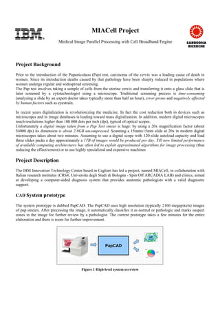
- 1. MIACell Project Medical Image Parallel Processing with Cell Broadband Engine Project Background Prior to the introduction of the Papanicolaou (Pap) test, carcinoma of the cervix was a leading cause of death in women. Since its introduction deaths caused by that pathology have been sharply reduced in populations where women undergo regular and widespread screening. The Pap test involves taking a sample of cells from the uterine cervix and transferring it onto a glass slide that is later screened by a cytotechnologist using a microscope. Traditional screening process is time-consuming (analyzing a slide by an expert doctor takes typically more than half an hour), error-prone and negatively affected by human factors such as eyestrain. In recent years digitalization is revolutionizing the medicine. In fact the cost reduction both in devices such as microscopes and in image databases is leading toward mass digitalization. In addition, modern digital microscopes reach resolutions higher than 100.000 dots per inch (dpi), typical of optical scopes. Unfortunately a digital image taken from a Pap Test smear is huge: by using a 20x magnification factor (about 54000 dpi) its dimension is about 2.8GB uncompressed. Scanning a 15mmx15mm slide at 20x in modern digital microscopes takes about two minutes. Assuming to use a digital scope with 120-slide autoload capacity and load three slides packs a day approximately a 1TB of images would be produced per day. Till now limited performance of available computing architectures has often led to exploit approximated algorithms for image processing (thus reducing the effectiveness) or to use highly specialized and expensive machines Project Description The IBM Innovation Technology Center based in Cagliari has led a project, named MIACell, in collaboration with Italian research institutes (CRS4, Università degli Studi di Bologna - Spin Off ARCADIA LAB) and clinics, aimed at developing a computer-aided diagnosis system that provides anatomic pathologists with a valid diagnostic support. CAD System prototype The system prototype is dubbed PapCAD. The PapCAD uses high resolution (typically 2160 megapixels) images of pap smears. After processing the image, it automatically classifies it as normal or pathologic and marks suspect zones in the image for further review by a pathologist. The current prototype takes a few minutes for the entire elaboration and there is room for further improvement. PapCADPapCAD Figure 1 High-level system overview
- 2. Algorithms and Technology With advanced machine learning and pattern recognition algorithms, the system self-learns analyzing and comparing several images. The more it is trained the more its internal model is accurate. The problem is computationally demanding since many huge images have to be analyzed. The PapCAD approaches this task by adopting a hybrid solution, based on both traditional SMPs and IBM Cell Broadband Engine accelerators for some highly computing power demanding tasks. What’s more, in order to reduce development and developer learning times, the software cross-platform development process was inspired by agile programming principles and state-of-the-art parallelizing and auto-vectorizing compilers were leveraged to achieve rapidly good system performance while keeping the code portable and at a high level. Pathologists Benefits In conclusion, the PapCAD is not meant to replace the pathologists but to help them to improve their diagnosis accuracy and therefore improve treatment of patients. Some benefits derive from digitalizing the analysis process. Doctors, even remotely, can navigate the image like a map and search for pathologic cells using a PC. The images can be archived into a database, which has great value for the entire scientific community. The PapCAD provides the doctor automatically with a list of likely pathologic zones and mark them in the image for review (see Figure 2). Using the PapCAD a low experienced pathologist can reach the accuracy of a more expert colleague and an expert pathologist can save a lot of time and improve his productivity. The reach of this technology is not limited to pap tests, but it can be exploited for different biologic images. For example, the same technology has been successfully deployed for digital mammography. Contacts Andrea Corona IBM Cagliari Innovation Technical Center - Italy andrea.corona@it.ibm.com Robert Alexander IBM Systems & Technology Group - Italy rja@it.ibm.com Figure 2 Pap smear image after PapCAD processing