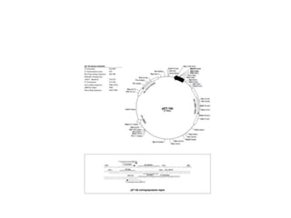
1. Rational Computational Design of
Inhibitors Preventing the Selective
Binding of Glutathione-
Conjugated Substrates to Human
Aldose Reductase
Lu Z. A.1, Lee J. D.2, Tomlinson S.2, Watowich S.2
1CPRIT Summer Undergraduate Research Program, University of Texas Medical Branch, Galveston TX
2University of Texas Medical Branch, Galveston TX
2. Background
• Human Aldose Reductase (hALR2) is a member of the
aldo-keto reductase enzyme family
• Reduces toxic aldehydes, glucose, and glutathione-
conjugated aldehydes
• Uses NADPH as a cofactor
3.
4. • Glutathione-conjugated aldehydes have stronger affinity
for Aldose reductase than toxic aldehydes
– Aldose reductase has a specific binding site for GSH
• The reduction of glutathione conjugated aldehydes has
been shown to increase inflammatory responses, tumor
growth, and lead to metastasis of certain cancers
(Srivastava, 2011)
5.
6. Our Concern
• Inhibition of the glutathione conjugated aldehyde
pathway may also inhibit the degradation of toxic lipid
aldehydes
• The quest for a selective inhibitor that will block the
glutathione conjugated pathway without compromising
the reduction of toxic lipid aldehydes
7. Goals
• Obtain and characterize aldose reductase
• Develop an assay to test aldose reductase activity for
glutathione-conjugated substrates
• Identify inhibitors using computational modeling and
simulations
• Test inhibitors that selectively target
glutathione-tagged substrates
8. We believe that we can discover inhibitors which will
specifically exclude glutathione-tagged aldehydes, allowing
for the continued reduction (and elimination) of other toxic
aldehydes
Motivating Hypothesis
9. MATERIALS and METHODS
• Plasmid with Aldose Reductase gene in pET
vector sent to us from Podjarny, Alberto from
France
• Methods for transformation and induction
based off of paper from Keneth h. Gabbay
(1991)
10. Transformation
• Human aldose reductase, inserted into a pET-15b
plasmid (Podjarny group)
• Contains a His-tag sequence
• Standard transformation protocol was used – adapted
from the Gabbay group (1991)
– BL21(DE3) E. coli cells
• Antibiotic resistance for selection
– carbenicillin
J. Biol. Chem. 1991, 266, 24031-24037.
11.
12. Protein Over-Expression
• Small-scale induction of colonies with isopropyl β-D-1-
thiogalactopyranoside (IPTG)
– IPTG induction inactivates the lac repressor,
activating transcription of the protein of interest
• Protein Bands emerged around ~38 kDa
• Medium-scale induction of two “lucky” colonies
– Goal: to make “bucket-loads” of desired protein
14. Purification
• Two colonies were tested for culture growth and protein
over-expression
• Cells were lysed, desired protein was purified using a Ni-
sepharose column
• hALR eluted using increasing imidazole concentrations
• Protein concentration ~ 300 nM
• Purity of elutions verified by SDS-PAGE
• Identity checked by mass spectroscopy
15. ladder soluble insoluble flow wash 25 mM 50 mM 100 mM250 mM beads
113kD
92kD
52.9kD
35.4kD
29kD
21.5kD
Protein with methionine: 38016.7 Da
Protein without methionine: 37867.5 Da
Methionine 149.21 Da
hALR2 ~38kD
16.
17. Preliminary Activity Analysis
(NADPH Assay)
• Protein dialyzed in buffer containing 1 mM DTT
(dithiothreitol)
• Protein concentrations determined by UV-Vis
spectroscopy at 280 nm
– Molar extinction coefficients predicted by the ExPASy
ProtParam online tool based on the protein sequence
• Reaction Conditions - 6.6% w/v (NH4)2SO4, 33 mM
NaH2PO4, pH 6.6, 0.11 mM NADPH,, 4.7 mM DL-
glyceraldehyde, 0.59 ug of enzyme, 1% DMSO (Podjarny,
2005)
• Measuring the decrease in NADPH, which absorbs at
340 nm
18. 0
0.1
0.2
0.3
0.4
0.5
0.6
0.7
0 10 20 30
A340
Time (minutes)
Activity 1 day after induction
NADPH-1
Enz4-1
Enz11-1
0
0.1
0.2
0.3
0.4
0.5
0.6
0.7
0 5 10 15 20 25 30
A340
Time (min)
Activity 10 days after induction
NADPH
Enz4
Enz11
19. Further Steps
• Acquire kinetic parameters (analyze reaction data
using Michaelis-Menten formalism and Dynafit
program)
• Acquire kinetic parameters for HNE and GS-HNE
20. Applications
• Novel drug candidates to inhibit cancerous tumor
growths
• Chemical interventions for inflammatory diseases
• Reduce toxicity of inhibitory drugs
21. Acknowledgments
• Dr. Stan Watowich, Mr. Jonathan Lee, Dr. Suzanne Tomlinson, Ms. Andrea
Garces, Dr. Usha Viswanathan
• UTMB GSBS
• Gulf Coast Consortium
This research was funded by the CPRIT Summer Undergraduate Program in
Computational Cancer Biology, training grant award RP 101489 from the Cancer
Prevention & Research Institute of Texas (CPRIT).
22. References
• Barski, O. A.; Gabbay, K. H.; Grimshaw, C. E.; Bohren, K. M. Mechanism of human aldehyde reductase: Characterization of the active site
pocket. Biochemistry 1995, 34, 11264-11275.
• Srivastava S. K., Yadav U. C., Reddy A. B., Saxena A., Tammali R., Shoeb M., Ansari N. H., Bhatnagar A., Petrash M. J., Srivastava S.,
Ramana K. V. (2011). Aldose reductase inhibition suppresses oxidative stress-induced inflammatory disorders. Chem. Biol. Interact. 191,
330–338. doi: 10.1016/j.cbi.2011.02.023.
• Van Zandt C., Jones M. L., Gunn D. E., Geraci L., Jones H., Sawicki D.R., Sredy J., Jacot J.L., DiCioccio A. T., Petrova T., Mitschler A.,
and, Podjarny A.D. Discovery of 3-[(4,5,7-Trifluorobenzothiazol-2-yl)methyl]indole-N-acetic Acid (Lidorestat) and Congeners as Highly
Potent and Selective Inhibitors of Aldose Reductase for Treatment of Chronic Diabetic Complications. Journal of Medicinal
Chemistry 2005 48 (9), 3141-3152.
• Wu J. T., Wu L. H., and Knight J. H.. Stability of NADPH: Effect of Various Factors on the Kinetics of Degradation. J. Med. Chem. 2005,
48, 3141-3152.