Separate DNA by size with agarose gel electrophoresis
•
14 likes•2,848 views

Agarose gel electrophoresis is used to separate DNA fragments by size. DNA has a negative charge and will migrate toward the positive electrode in an electrical field. Shorter DNA fragments move faster through an agarose gel than longer fragments. Samples are run alongside a DNA ladder to determine the size of unknown DNA fragments. After electrophoresis, gels are stained with ethidium bromide and visualized under UV light to see DNA band patterns. This allows analysis of results such as identifying the presence of specific genes.

Recommended
More Related Content
What's hot
What's hot (20)
Similar to Separate DNA by size with agarose gel electrophoresis
Similar to Separate DNA by size with agarose gel electrophoresis (20)
More from YOUSIF H M SHARIF
Recently uploaded
Recently uploaded (20)
Separate DNA by size with agarose gel electrophoresis
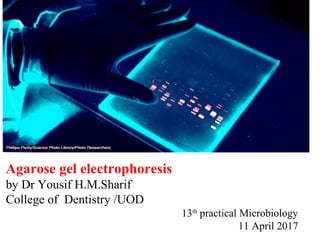
- 1. Agarose gel electrophoresis by Dr Yousif H.M.Sharif College of Dentistry /UOD 13th practical Microbiology 11 April 2017
- 2. Why electrophoresis? • To separate DNA fragments from each other • To determine the sizes of DNA fragments • To determine the presence or amount of DNA • To analyze restriction digestion products
- 3. Background on Agarose Gel Electrophoresis • Agarose gel electrophoresis is a molecular method to separate DNA or RNA molecules by size. • This is achieved by moving negatively charged nucleic acid molecules through an agarose matrix with an electric field (electrophoresis). • Shorter molecules move faster and migrate faster than longer ones .
- 4. • DNA is negatively charged. +- Power DNA • When placed in an electrical field, DNA will migrate toward the positive pole (anode). H O2 • An agarose gel is used to slow the movement of DNA and separate by size.
- 5. +- Power DNA How fast will the DNA migrate? strength of the electrical field, buffer, density of agarose gel… Size of the DNA! *Small DNA move faster than large DNA …gel electrophoresis separates DNA according to size small large
- 6. Material required for agarose gel electrophoresis Electrophoresis chamber Agarose gel Gel casting tray Running Buffer Staining agent (dye) A comb DNA ladder Sample to be separate
- 7. Casting tray Gel combs Power supply Gel tank Cover Electrical leads Electrophoresis Equipment
- 8. Agarose What is Agarose ? Agarose is a linear polymer extracted from seaweed. D-galactose 3,6-anhydro L-galactose
- 9. Which running buffer Should I use ? • TBE (Tris-borate-EDTA) 1X if you look for small DNA fragment ,Nice band • TAE (Tris-acetate EDTA) 1X as running buffer –Large DNA fragment ,less prone to overheat
- 10. %Percent agarose •Most agarose gels are made with between 0.7% (good separation or resolution of large 5–10kb DNA fragments) and 2% (good resolution for small 0.2–1kb fragments) agarose dissolved in electrophoresis buffer. •1 % Running Buffers The most common being: Tris/Acetate/EDTA (TAE) •Tris-acid solutions are effective buffers for slightly basic conditions, which keep DNA deprotonated and soluble in water. •the role of the EDTA is to protect the nucleic acids against enzymatic degradation
- 11. Preparing a 2% W/V Agarose Gel • 2% W/V agarose = 2 grams agarose dissolved in 100 milliliters (mL) of Buffer • For our purposes each gel tray requires 25 mLs of gel • To prepare 25 ml of a 2% agarose gel 2 grams = X grams 100mls 25 mls X = 0.5 grams agarose • Weight 0.5 grams agarose and dissolve in 25 mls buffer
- 12. Procedure of making agarose gel • An agarose gel is prepared by combining agarose powder and a buffer solution.
- 13. Agarose Buffer Flask for boiling 1.Make a 1% agarose solution in 20ml TAE. ( 0.2g agarose in 20ml TAE OR TBE ) 2. Carefully bring the solution just to the boil to dissolve the agarose. 3. Let the solution cool down to about 60 °C at room temperature. Stir or swirl the solution while cooling. 4.After complete dissolve and let it cool around 40C, we simply add 2microliter of safe- Dye then pour in to gel tray STEPS
- 14. Agarose Buffer Solution Combine the agarose powder and buffer solution. Use a flask that is several times larger than the volume of buffer.
- 15. Gel casting tray & combs
- 16. Seal the edges of the casting tray and put in the combs. Place the casting tray on a level surface. None of the gel combs should be touching the surface of the casting tray. Preparing the Casting Tray
- 17. Agarose is insoluble at room temperature (left). The agarose solution is boiled until clear (right). Gently swirl the solution periodically when heating to allow all the grains of agarose to dissolve. ***Be careful when boiling - the agarose solution may become superheated and may boil violently if it has been heated too long in a microwave oven. Melting the Agarose
- 18. Allow the agarose solution to cool slightly (~60ºC) and then carefully pour the melted agarose solution into the casting tray. Avoid air bubbles. Pouring the gel
- 19. Each of the gel combs should be submerged in the melted agarose solution.
- 20. When cooled, the agarose polymerizes, forming a flexible gel. It should appear lighter in color when completely cooled (30-45 minutes). Carefully remove the combs and tape.
- 21. Place the gel in the electrophoresis chamber.
- 22. buffer Add enough electrophoresis buffer to cover the gel to a depth of at least 1 mm. Make sure each well is filled with buffer. Cathode (negative) Anode (positive) wells DNA
- 23. Loading DYE: • Bromophenol Blue (for color) • Glycerol (for weight) Sample Preparation Mix the samples of DNA with Loading dye. This allows the samples to be seen when loading onto the gel, and increases the density of the samples, causing them to sink into the gel wells.
- 24. Loading the Gel Carefully place the pipette tip over a well and gently expel the sample. The sample should sink into the well. Be careful not to puncture the gel with the pipette tip.
- 25. Place the cover on the electrophoresis chamber, connecting the electrical leads. Connect the electrical leads to the power supply. Be sure the leads are attached correctly - DNA migrates toward the anode (red). When the power is turned on, bubbles should form on the electrodes in the electrophoresis chamber. Running the Gel
- 26. wells Bromophenol Blue Cathode (-) Anode (+) Gel After the current is applied, make sure the Gel is running in the correct direction. Bromophenol blue will run in the same direction as the DNA. DNA (-)
- 27. 100 200 300 1,650 1,000 500 850 650 400 12,000 bp 5,000 2,000 DNA Ladder Standard DNA ladder (DNAs of known sizes) on the gel makes it easy to determine the sizes of unknown DNAs. - + DNA migration bromophenol blue Note: bromophenol blue migrates at approximately the same rate as a 300 bp DNA molecule
- 29. Visualising the results • The most common dye used to make DNA or RNA bands visible for agarose gel electrophoresis is ethidium bromide, usually abbreviated as EtBr. It fluoresces under UV light when intercalated into DNA (or RNA). By running DNA through an EtBr-treated gel and visualizing it with UV light. EtBr is a known mutagen,But, safer alternatives are available. Methylen Blue ,Bio-safe DNA stain etc.
- 30. Figure shows 1%Agarose gel electrophoresis of the PCR-amplified mecA methicillin resistance gene. Lanes: 1: 50-bp ladder; 2: Negative control (S. aureus ATCC 8325-4); 3: Positive control (S. aureus strain COL); 4-7: S. aureus isolates showing 162 bpmecAamplicon Result analysis and interpretation of Gel electrophoresis BP DNA ladder _VE +VE S4 S5 S6 S7 Control