
Recommended
More Related Content
What's hot
What's hot (20)
Similar to Vipin ich
Similar to Vipin ich (20)
Recently uploaded
Recently uploaded (20)
Vipin ich
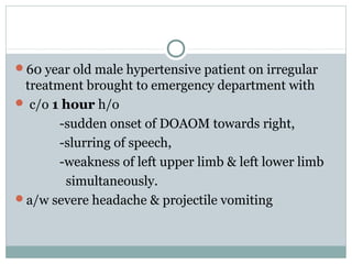
- 1. 60 year old male hypertensive patient on irregular treatment brought to emergency department with c/o 1 hour h/o -sudden onset of DOAOM towards right, -slurring of speech, -weakness of left upper limb & left lower limb simultaneously. a/w severe headache & projectile vomiting
- 2. O/e BP – 200/100mmHg, PR – 52/minute, RR – 24/minute, TEMP - normal Neuro – L side UMN facial palsy, non fluent aphasia, power grade 0/5 UL & LL left side, plantar extensor with brisk DTR left side. no neck rigidity Other systems - WNL
- 3. What will do next? ASSESS Airway, breathing, circulation Check RBS = NORMAL URGENT NCCT BRAIN
- 6. HEMORRHAGIC STROKE -DR VIPIN VENGAYIL DNB RESIDENT
- 7. Etiology of ICH (overall) Hypertension (m/c) Vascular malformations Cerebral amyloid angiopathy Intracranial tumors Bleeding diathesis, anticoagulation, fibrinolysis Granulomatous angiitis & vasculitides Sympathomimetic agents (amphetamine, cocaine) Hemorrhage into cerebral infarct Trauma
- 8. Clinical features Features of RAISED ICT Headache, vomiting, decreased LOC Correlated with hematoma size and prognosis Progressive over time Seizures in lobar ICH Focal neurological deficits depending on the location of ICH
- 9. Hypertension and ICH Most important risk factor (>70% of 1ry ICH) Bifurcation of small penetrating arteries (50–700 μm diameter) Atherosclerosis Lipid deposition, layering of platelet and fibrin aggregates, breakage of elastic lamina, atrophy and fragmentation of smooth muscle, dissections, and granular or vesicular cellular degeneration Charcot and Bouchard aneurysm Fibrinoid necrosis of the subendothelium focal dilatations rupture of microaneurysm
- 11. N Engl J Med 2001;344(19):1450–1460
- 12. N Engl J Med 2001;344(19):1450–1460 Lobar hemorrhage 25% • Penetrating cortical branches of ACA, MCA, & PCA • Peripheral location ∴ lower frequency of coma • Lower mortality • Better functional outcome
- 13. N Engl J Med 2001;344(19):1450–1460 Basal ganglia 35-40% • Ascending lenticulostriate branches of MCA • Wide spectrum of severity extending to coma and decerebrate rigidity • Ventricular extension carries very poor prognosis
- 15. N Engl J Med 2001;344(19):1450–1460 Thalamus 10-15% • Ascending thalamogeniculate branches of PCA • Abrupt hydrocephalus from aqueductal obstruction from intraventricular clot • Responds to ventriculostomy
- 18. N Engl J Med 2001;344(19):1450–1460 Pons 5% • Paramedian branches of the basilar artery • Bilateral carries very poor prognosis (coma, quadriplegia, decerebrate posturing, horizontal ophthalmoplegia, pinpoint reactive pupils)
- 20. N Engl J Med 2001;344(19):1450–1460 Cerebellum 5-10% • Penetrating branches of the PICA, AICA, SCA • Abrupt onset vertigo, h/a, n/v, inability to walk in absence of weakness • Ipsilateral ataxia, horizontal gaze palsy, peripheral facial palsy • Unpredictable deterioration to coma
- 23. Lobar hemorrhage
- 25. Vascular malformations Aneurysms, AVM, cavernous angiomas Younger, female patients, familial history Imaging may show concurrent SAH Dx by MRI and cerebral angiography Usually supratentorial, lobar ICH Cavernous angioma: on MRI (T2) central nidus of irregular bright signal mixed with mottled hypointensity, surrounded by peripheral hypointense ring
- 36. Management principles A-B-C: Airway support Decreased level of consciousness Bulbar muscle dysfunction Blood pressure control Anticoagulation reversal Intracranial pressure control Monitoring “Neurological and cardiovascular deterioration greatest in the 24hours following symptom onset”
- 45. Elevated icp Hypertonic saline & mannitol Elevate head end of bed Intubation & acute hyperventilation Csf drainage if necessary
- 46. ICP monitoring consider if Reduced LOC (GCS <8) Clinical evidence of cerebral herniation Significant IVH Hydrocephalus waves A (plateau)- pathologic B (respiratory) C (Cardiac) waveforms
- 47. Blood pressure & ICH BP is elevated on admission even in patients who have no history of hypertension MAP > 120mmHg in over 2/3 of patients Precipitant of the hemorrhage? Reflection of chronic hypertension? Attempt to maintain CPP? Sympathetic activation 2ry to pain & anxiety? Tends to return to baseline 7-10 days post ICH
- 48. Acute management of BP How fast should BP be lowered? Rapidly lowering MAP by ≈ 15% does not lower CBF Reductions of 20% can affect CBF Current guidelines suggest a reduction of ≤ 20% in the first 24 hrs Which agents should be used? Short and rapidly acting IV antihypertensive Labetalol, hydralazine, esmolol, nicardipine Sodium nitroprusside and nitroglycerin should avoid d/t vasodilation and increase ICP
- 51. General Monitoring and Nursing Care In a Canadian study of 49 hospitals that included ICH patients, a higher proportion of registered nurses at the hospital and better nurse- physician communication were independently associated with lower 30-day mortality even after adjustment for disease severity, comorbidities & hospital characteristics
- 54. Other principles Glucose should be monitored. Both hyperglycemia and hypoglycemia should be avoided (150 - 200mg%) Clinical seizures or electrographic seizures should be treated with anti seizure drugs Prophylactic antiseizure medication is not recommended
- 55. Ivh 45% pt, a/w poor outcomes Although the insertion of a VC should theoretically aid in drainage of blood and CSF from the ventricles, VC use alone may be ineffective because of difficulty maintaining catheter patency & slow removal of intraventricular blood. Thus, there has been recent interest in the use of thrombolytic agents as adjuncts to VC use in the setting of IVH IVH: Recommendations 1. Although intraventricular administration of rtPA in IVH appears to have a fairly low complication rate, the efficacy and safety of this treatment are uncertain (Class IIb; Level of Evidence B). (Revised from the previous recommendation) 2. The efficacy of endoscopic treatment of IVH is uncertain
- 56. Hematoma expansion Hematoma enlargement >70% have hematoma enlargement w/in 3 hrs of symptom onset; 1/3 clinically significant Most occur within 3 hrs, can be up to 12 hrs Independent predictor of worse outcome & ↑ mortality
- 57. Hematoma expansion Journal of the Neurological Sciences 261 (2007) 99–107
- 60. Recombinant Factor VIIa Factor VIIa has locally action at sites of tissue injury and vascular-wall disruption by binding tissue factor & generating thrombin and activating platelets Recombinant FVIIa directly activates fX on the surface of activated plts resulting in acceleration of coagulation Factor Seven for Acute Hemorrhagic Stroke (FAST) trial, N Engl J Med 2008;358:2127-37 841 patients, within 4 hours of onset of stroke Placebo vs. 20 μg/kg vs. 80 μg/kg of rFVIIa 1ry end point: 90-day functional outcome or death
- 61. Recombinant Factor VIIa Significant reduction in growth of hematoma volume in the 80 μg/kg group No significant difference in functional outcome and mortality Venous thromboembolic events were similar in all three groups (limits its usage) Arterial thromboembolic events were significantly more frequent in the 80 μg/kg group
- 62. ABC of hematoma size Broderick, JP et al. Stroke 1993;24:987-993 1.26 million subjects from Greater Cincinnati
- 63. CT-A “Spot Sign” Focal area of enhancement within the hematoma on CTA have been shown to be: Independent predictor of hematoma expansion Associated with longer median hospital stay Independent of time to presentation Sensitivity 91%, specificity 89%, NPV 96%
- 65. CT-A “Spot Sign” Recent proposal of a “Spot Sign” definition (Can J Neurol Sci 2009;36:456-461) Serpiginous and/or spot-like appearance Within the margin of the parenchymal hematoma without connection to an outside vessel >1.5mm diameter in maximal axial dimention >Double the HU density compared to background hematoma (>150 HU) Multiple or single in number Comparison to unenhanced CT for mimickers Calcifications (tumour, choroid, infectious, etc)
- 66. Anticoagulation associated ICH Goal of treatment: fully reverse INR to normal range High dose Vitamin K 10-20 mg IV for 3days Effect takes 12-24hrs Helps achieving sustained reversal of INR Fresh frozen plasma 15-20ml/kg Volume overload, insufficient factor IX ABO compatibility, thawing, infusion time (30hrs) Prothrombin Complex Concentrate(PCC,Octaplex) Combination of II, VII, IX, X, variable protein C and S Dosage dependant on initial INR Smaller volume, correct INR as fast as 30 min
- 67. Anticoagulation associated ICH ICH associated with IV heparin Rapidly normalize activated partial thromboplastin time Protamine sulfate 1 mg per 100 U heparin, adjusted for time since last heparin dose 30-60 min: 0.5 to 0.75 mg per 100U heparin 60-120 min: 0.375 to 0.5 mg per 100 U heparin >120min: 0.25 to 0.375 mg per 100 U heparin Slow IV injection (<5 mg/min, max dose 50 mg) Beware of systemic hypotension
- 68. AAICH – restarting anticoagulation 1% recurrent ICH in initial 3 mths post ICH Risk estimated to double with anticoagulation Stroke. 2007;38:200 1-2023
- 69. Miscellaneous Venous thromboembolism prophylaxis Intermittent pneumatic compression Heparin SQ prophylaxis (3-5 d if no bleeding) IVC filter (proximal venous thrombosis) Hyperglycemia Associated with poor outcome and ↑ mortality Marker of outcome or contributor? Hyperpyrexia Associated with poor outcome and neuro deterioration Septic workup, treat with antipyretics or cooling devices Often central in origin
- 71. 4. Supratentorial hematoma evacuation in deteriorating patients might be considered as a life-saving measure (New recommendation) 5. DC with or without hematoma evacuation might reduce mortality for patients with supratentorial ICH who are in a coma, have large hematomas with significant midline shift, or have elevated ICP refractory to medical management (New recommendation) 6. The effectiveness of minimally invasive clot evacuation with stereotactic or endoscopic aspiration with or without thrombolytic usage is uncertain (Revised from the previous guideline)
- 73. Decompressive craniectomy Surgical removal of cranial bone flap to relieve intracranial pressure Useful in large ischemic CVA with profound edema Role in traumatic brain injury still needs to be established
- 75. Minimally Invasive Neurosurgery for Hemorrhage (newer) Evacuation Minimally invasive surgery generally refers to the concept of creating minimal trauma to normal appearing tissue during the process of removing the hematoma This stands in distinction from the open craniotomy in which a large bone flap is created; the brain is exposed, retracted, and manipulated to inspect the site of bleeding and suction blood from multiple areas.
- 76. 1.endoscopic evacuation of the hematoma In endoscopic evacuation, a small burr hole is created, and an endoscope measuring from 5 to 8 mm diameter is inserted through normal brain tissue into the hematoma. Suction and irrigation are applied to remove the hematoma. The brain is then visualized via the endoscope to determine the site of bleeding and to determine the amount of ICH evacuated.
- 77. 2. stereotactic aspiration and thrombolysis This technique involves using image guidance to place a catheter into the main body of the hematoma and aspirate blood. A catheter is left in the body of the hematoma, and during the course of several days, repeated small doses of thrombolytics (recombinant tissue-type plasminogen activator) are instilled via the catheter into the brain to clear the blood slowly during the course of 72 to 96 hours.
- 79. Conclusions ICH has an increasing incidence, but continues to have a very poor prognosis Hypertension is a major risk factor Acute BP reduction of 15-20% is safe Anticoagulation should be reversed ASAP Intraventricular extension & midline shift a/w poor prognosis Early detection/ imaging & treatment DECREASES morbidity & mortality but for a limited extent Physiotherapy & rehabilitation play definite role.
- 81. THANK YOU..
Editor's Notes
- Posterior compartment
- FFP has all the coagulation factors Cryoppt has factor VIII, fibrinogen, fibronectin, factor XIII, and von Willebrand factor (vWF)
- Nonvalvular AF no CVA 5% per year risk of cerebral embolism 12% if previous stroke 4% in prosthetic valvee
- Acutely used in hemiparetic/plegic patients How tight of a control is still controversial