Electrocardiogram interpretation
•Download as PPTX, PDF•
38 likes•6,563 views

Cardiac conduction system 12 lead system rules for normal ECG. Determination of heart rate

Recommended
More Related Content
What's hot
What's hot (20)
Similar to Electrocardiogram interpretation
Similar to Electrocardiogram interpretation (20)
More from Dr. Vitthalrao Vikhe Patil Foundation's College of Physiotherapy, Ahmednagar
More from Dr. Vitthalrao Vikhe Patil Foundation's College of Physiotherapy, Ahmednagar (20)
Recently uploaded
Recently uploaded (20)
Electrocardiogram interpretation
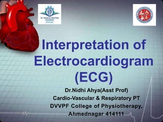
- 1. Interpretation of Electrocardiogram (ECG) Dr.Nidhi Ahya(Asst Prof) Cardio-Vascular & Respiratory PT DVVPF College of Physiotherapy, Ahmednagar 414111
- 2. Objectives • Cardiac conduction system • 12 lead system • rules for normal ECG. • Determination of heart rate.
- 11. Lead Systems- 12 lead ECG Bipolar Limb Leads -3 Precordial Chest Leads-6 Augmented Leads-3
- 13. Anatomic Groups
- 14. Determine regularity Look at the R-R distances Regular -are they equidistant apart? Occasionally irregular? Regularly irregular? Irregularly irregular? R R
- 15. The 10 rules for a normal ECG
- 16. Rule 1 PR interval Millivolts Milliseconds 0 200 400 600 -0.5 0 0.5 1.0 P R T Q S PR interval should be 120 to 200 milliseconds (0.12- 0.2 sec) or 3 to 5 little squares
- 17. Rule 2 Millivolts Milliseconds 0 200 400 600 -0.5 0 0.5 1.0 QRS P R T Q S The width of the QRS complex should not exceed 110 ms, less than 3 little squares
- 18. Rule 3 I II III aVR aVL aVF The QRS complex should be dominantly upright in leads I and II
- 19. Rule 4 I II III aVR aVL aVF QRS and T waves tend to have the same general direction in the limb leads
- 20. Rule 5 P Q T S All waves are negative in lead aVR
- 21. Rule 6 V1 V2 V3 V4 V5 V6 The R wave in the precordial leads must grow from V1 to at least V4
- 22. I II III aVR aVL aVF V1 V2 V3 V4 V5 V6 Rule 7 The ST segment should start isoelectric except in V1 and V2 where it may be elevated
- 23. Rule 8 I II III aVR aVL aVF V1 V2 V3 V4 V5 V6 The P waves should be upright in I, II, and V2 to V6
- 24. Rule 9 I II III aVR aVL aVF V1 V2 V3 V4 V5 V6 There should be no Q wave or only a small q less than 0.04 seconds in width in I, II, V2 to V6
- 25. Rule 10 I II III aVR aVL aVF V1 V2 V3 V4 V5 V6 The T wave must be upright in I, II, V2 to V6
- 27. Grid Methods – Regular rhythms 1500/No.of small squares between 2 consecutive R 300/No.of large squares between 2 consecutive R
- 28. Scan Method - irregular Check for 3 sec/ 6 sec markers Count the number of QRS complexes between markers Multiply by 20 / 10 for 3 & 6 sec markers respectively
- 32. Normal Sinus Rhythm ECG Characteristics: Regular narrow-complex rhythm Rate 60-100 bpm Each QRS complex is proceeded by a P wave P wave is upright in lead II & downgoing in lead aVR
- 33. Sinus Bradycardia HR< 60 bpm; Every QRS narrow, preceded by p wave Normal in well-conditioned athletes HR can be<30 bpm in children, young adults during sleep, with up to 2 sec pauses
- 34. Sinus tachycardia HR > 100 bpm, regular Often difficult to distinguish P and T waves
- 35. Sinus Arrhythmia Variations in the cycle lengths between P waves/ QRS complexes Will often look irregular on exam Normal P waves, PR interval, normal, narrow QRS
- 36. Atrial Fibrillation Caused by a large reentrant circuit in the wall of the right atrium Caused by numerous wavelets of depolarization spreading throughout the atria simultaneously, leading to an absence of coordinated atrial contraction.
- 37. Ventricular tachycardia Ventricular tachycardia is usually caused by reentry, and most commonly seen in patients following myocardial infarction
- 39. Summary • What is cardiac conductive system. • 12 leads of ECG. • Rules for normal ECG • Determination of heart rate.
- 40. QUESTIONS 1. WHAT ARE THE RULES OF NORMAL ECG? 5MARKS 2. DESCRIBE THE 12 LEADS OF ECG. 5MARKS
- 41. Thank you