
Recommended
More Related Content
What's hot
What's hot (20)
Similar to Pre-Op Assessment of Cataract
Similar to Pre-Op Assessment of Cataract (20)
Recently uploaded
Recently uploaded (20)
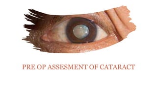
Pre-Op Assessment of Cataract
- 1. PRE OP ASSESMENT OF CATARACT
- 2. OBJECTIVES To find visual prognosis To exclude ocular or systemic focus of infection To find out the fitness status of patient for surgery
- 3. PRE-OPERATIVE ASSESSMENT OCULAR HISTORY SYSTEMIC HISTORY OCULAR EXAMINATION OCULAR INVESTIGATIONS SYSTEMIC EXAMINATION LABOROTARY INVESTIGATIONS
- 4. OCULAR HISTORY Ocular Symptoms Like Blurring Of Vision,coloured Halos,diplopia,glare Nuclear Cataract-distant> Near Posterior Subcapsular-near>distant Duration And Progression H/O Of Previous Intraocular Disease H/O Previous Cataract Surgery H/O Intraocular Injuries
- 5. SYSTEMIC HISTORY H/o of all systemic diseases,current medications patient is on.. Medications relevant to eye surgery Antiplatelets and anticoagulants Antihypertensives like diuretics-electrolyte imbalances Long term steroids-delayed healing Drug allergy to sulfonamides and other antibiotics
- 6. OCULAR EXAMINATION Components of Ocular Examination: • Testing of Visual Acuity • External Ocular Examination • Fundus Eamination • Visual field examination
- 7. Testing Of Visual Acuity Visual acuity - distant (Snellen’s chart) and near vision (Jaeger’s chart) Ocular movements must be tested Perception of light must be noted Projection of light rays – Test for the function of peripheral retina. Potential visual acuity tests – Laser inferometry & Potential acuity meter test (PAM)
- 8. External Ocular Examination Examination of the head posture, forehead and facial symmetry Examination of eyebrows - in cases of ptosis and madarosis Examination of the eyelids - position, movements, abnormalities of skin should be noted Examination of Lacrimal apparatus - inspection of lacrimal sac, lacrimal puncta and lacrimal syringing to locate any blockage Examination of eyeball as a whole - position, visual axes, size and movements to be noted Examination of conjunctiva - for any congestion, scarring, discolouration Examination of sclera - for any inflammation, traumatic perforations, etc. Examination of cornea - size, curvature, surface, sheen, transparency, vascularization, sensations, endotheial status (guttata); Specular microscopy examination for endothelial cell count and morphology.
- 9. Examination of anterior chamber for the depth and its constituents; and Intraocular Pressure (IOP) - in cases of glaucoma and hypotony Examination of iris and pupil - Light reactions and RAPD (Relative Afferent Pupilary Defect) should be checked
- 10. Examination of Lens Cataractous lens should be evaluated for morphology and maturity of cataract and for grade of nuclear sclerosis (important for phacoemulsification) Other signs to be particularly looked for are: pseudoexfoliation, pigment over anterior lens capsule and anterior chamber depth. Size of lens nucleus and grading of nuclear sclerosis should be determined for planning size of incision and type of surgery Nuclear cataract are harder and need more power with phacoemulsification
- 11. Fundus Examination Retinal and optic nerve function must be assessed preop as if it is defective operation becomes valueless. Pathology such as Retinal Detachment can adversely affect visual outcome. Structure to be assesed during fundus examination: Media, Optic disc, Macula, retinal blood vessels and genral background Hence, a thorough fundus evaluation is important.
- 12. Visual Field Examination Perimetry is the technique used to asses the visual field of a patient Common types of defects in visual fields are: Altitudinal field defects, Enlargements of blind spots, Constriction of peripheral fields, bitemporal hemianopia, binasal field defects, etc.
- 13. Ophthalmic investigations Retinal/Macular Function tests Two light discrimination test Entopic visualization Projection of Light
- 14. Maddox rod test Colour perception test
- 15. Entoptic visualization Ultrasonic investigation by B-scan technique
- 16. INTRAOCULAR PRESSURE Should be in each case by APPLANTATION TONOMETRY. Management needed in case of inc. IOP
- 17. REFRACTIVE ERROR Its critical to obtain patients pre-operative refractive status in order to guide IOL implant selection. BIOMETRY facilitate calculation of lens power likely to result in desired post op refractory outcome. It involves 1.Keratometry 2.A SCAN AXIAL LENGTH-curvature of anterior corneal surface calculation by interferometry apparatus. Use SRK formula (Sanders, Retlaff & Kraff) P = A – 2.5L – 0.9K P : Power of IOL L : Axial length (mm) K : Average keratometric reading (Avg. corneal curvature) A : Constant specific to the lens implant to be used
- 18. LAB INVESTIGATIONS NORMAL-RBS,ECG,SCREENING,BP XRAY,URINE RFT,APTT,PT INR-in patients with individual risk factors or planned for general anesthesia, Preop-antibiotic eye drops QID-3 DAYS PRIOR TO SURGERY ANTIANXIETY DRUGS if patient is apprehensive Preparing eye-cutting lashes Asked to take a normal meal,normal sleep, normal bath,continue systemic medications .
- 19. INFORMED WRITTEN CONSENT Patient should give full informed written consent before cataract surgery. 1 in 1000-achieves very little or no sight 1 in 10000-lose eye completely Mild complications-periocular echymosis,raised IOP,mild iridocyclitis, wound leak. Moderate-posterior capsular rupture, zonular dehiscence, corneal decompensation,CME,RD(1%) SEVERE ENDOPTHALMITIS(0.1%) SUPRACHOROIDAL HEMORRHAGE
- 20. THANK YOU