This document summarizes research characterizing Thioflavin T (ThT) and its fluorescent derivatives using high performance liquid chromatography (HPLC). The author developed HPLC methods to detect and separate ThT from multiple derivatives formed during storage. Absorbance and fluorescence profiles of the compounds were recorded. At least three major derivatives and several minor ones were found. A mobile phase of 70% acetonitrile and 30% water provided optimal separation of the derivatives. However, ThT and one derivative degraded more and interacted poorly with the column at higher water contents. Future work may identify the derivatives using mass spectrometry and study their binding properties.
Rapid Quantification of Perfluorinated Compounds in Drinking and Surface Wate...
Poster
1. Acknowledgements
The author would like to thank the Department of Chemistry at Penn State
University for providing the facilities and instrumentation for this research, Dr. Mark
Maroncelli for suggesting the research topic and supplying sample compound as well
as Dr. Dan Sykes for his invaluable knowledge and guidance throughout this project.
Materials and Methods
HPLC parameters
Conclusions
Introduction
Thioflavin T (ThT) is a benzothiazole dye that experiences enhanced fluorescence
upon binding to amyloid fibrils and is commonly used to stain and detect amyloid
fibrils.4 Amyloid fibrils or misfolded proteins are the cause of many diseases such as
Alzheimer’s disease, transmissible spongiform encephalopathy, Parkinson’s disease,
and systemic amyloidosis.1
However, because it contains a positive charge, ThT cannot cross the blood brain
barrier easily, which limits its efficiency to detect amyloid fibrils in the brain.2 As a
result, many researches have looked into developing uncharged derivatives of ThT
which have shown to be 600-fold more lipophilic and possess higher affinity3 to fibrils
than the original charged compound.
Previous work has shown that ThT forms several derivatives during storage and
the amount, chemistry, and photophysical properties of these derivatives are not fully
understood.1 The goal of this research is to develop an analytical method for the
detection and separation of ThT and its derivative and also to characterize their
fluorescent properties. Future applications can involve identification of ThT
derivatives, purification and isolation of ThT and targeted derivatives as well as study
the mechanisms by which ThT and its derivatives bind with amyloid fibrils.
Results and Discussion
References
1. Hsu, J. C.-C.; Chen, E. H.-L.; Snoeberger, R. C.; Luh, F. Y.; Lim, T.-S.; Hsu, C.-P.;
Chen, R. P.-Y. Thioflavin T And Its Photoirradiative Derivatives: Exploring Their
Spectroscopic Properties in the Absence and Presence of Amyloid Fibrils. The Journal
of Physical Chemistry B J. Phys. Chem. B. 2013, 3459–3468.
2. Alí-Torres, J.; Rimola, A.; Rodríguez, C.; Rodríguez-Santiago, L.; Sodupe, M.
Insights on the Binding of Thioflavin Derivative Markers to Amyloid-Like Fibril
Models from Quantum Chemical Calculations. The Journal of Physical Chemistry B.
2013 117 (22), 6674-6680.
3. Klunk, WE.; Wang, Y.; Huang GF.; Debnath, ML.; Holt, DP.; Mathis, CA.
Uncharged thioflavin-T derivatives bind to amyloid-beta protein with high affinity and
readily enter the brain. Life Sci. 2001, 69(13),1471-84.
4. Khurana, R.; Coleman, C.; Ionescu-Zanetti, C.; Carter, S. A.; Krishna, V.; Grover,
R. K.; Roy, R.; Singh, S. Mechanism Of Thioflavin T Binding to Amyloid Fibrils.
Journal of Structural Biology. 229–238.
Fluorescence study of Thioflavin T (ThT) and its derivatives
using High Performance Liquid Chromatography
Trang Vu, Dr. Dan Sykes
Department of Chemistry, The Pennsylvania State University, University Park, PA 16802
Abstract
High performance liquid chromatography (HPLC) methods using several
solvent combinations of water and acetonitrile have been developed to detect and
separate Thioflavin T (ThT) and its derivatives. The absorbance profiles at 412 nm and
fluorescence emission profiles at 450 nm of ThT and its derivatives were recorded. In
this study, we found that during storage, ThT degraded into multiple derivatives. There
are at least three major derivatives and several minor ones that need higher water
content mobile phase for optimal separation. Nevertheless, at higher water content,
ThT and one of its derivatives exhibit rapid degradation and poor interaction with the
stationary phase.
Sample preparation
1 mg of ThT was added to a 25-ml volumetric flask,
diluted with water and shaken until all solid dissolved
then nanopure water was filled to the mark to make a 40
ppm solution. 2 ml of this solution was transferred to a
shorty vial for HPLC analysis. All solutions were
wrapped with aluminum foil and stored in the fridge
after use. ThT sample was provided by Dr. Maroncelli
from the Department of Chemistry at Penn State
University.
Instrument: Shimadzu UFLC XR
Column: Restek Pinnacle DB C18 5µm 150 x 4.6mm
Total flow rate: 1ml/min
Mobile phase: Isocratic
Mobile
phase
% acetonitrile % water Run time
1 90 % 10 % 20 minutes
2 80 % 20 % 30 minutes
3 70% 30% 40 minutes
Figure 1. The UV-Vis
absorbance profile of pure ThT.
The absorbance peak showed up
at 412 nm, which was expected
because ThT appears as a yellow
color, indicating that it absorbs
its complementary color, violet,
which has a wavelength of about
400 nm.
Thioflavin T
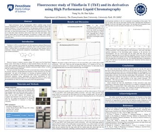
Figure 3. Fluorescence profile of ThT and its derivatives at 450 nm when excited at 370 nm and
412 nm using HPLC with a mobile phase of 90% acetonitrile : 10% water. There were four single
peaks which were circled and one that looked like an overlap of multiple peaks. The last peak at
13.687 minutes corresponds to the pure ThT conformation, indicating that the rest of the peaks are
ThT derivatives.
Figure 2. Absorbance profile of ThT mixture at 412 nm using HPLC with a mobile phase of 90%
acetonitrile : 10% water. There was only one peak that eluted at 13.57 minutes at 412 nm. This
confirmed that the peak at 13.57 minutes was the pure ThT conformation. No other peaks showed
up suggested that none of its derivatives absorbed at this wavelength.
Figure 4. Fluorescence profile of ThT and its derivatives at 450 nm when excited at
370 nm using HPLC with a mobile phase of 70% acetonitrile : 30% water. This show
optimum separation of the fast-eluting peaks compared to the previous two
chromatograms. The two circled peaks and the peaks right after that got great
separation, suggesting they are two different compounds. However the last two peaks
came out at a much later retention time with broader appearances, which was cut off
from the graph due to insufficient run time. This indicated high degree of degradation
of pure ThT and one of its derivatives and their poor interaction with the column at
high water content.
conformational similarity in the two compounds corresponding to those peaks. The
last two peaks, blue and red, had longer retention times, indicating their likelihood to
degrade in solvents with higher water content. The last peak still corresponds to pure
ThT since there was only one peak in the pink curve at 25.308 minutes.
Figure 4. Fluorescence profile of ThT and its derivatives at 450 nm when excited at 370 nm and
412 nm using HPLC with a mobile phase of 80% acetonitrile : 20% water. More peaks started to
appear when the polarity of the mobile phase increased and there was a shift of peaks to the right
when the water content was higher. The four single peaks were still there, but there were more
peaks that got separated from the huge peaks and two smaller peaks that eluted right after the
yellow and green circled peaks, indicating a coelution using the first mobile phase and a
18.410
9.5169.158
5.632
5.268
2.172
The study showed that only pure ThT absorbed at 412 nm but ThT and its
derivatives showed fluorescence emission at 450 nm when excited at 350 nm. There
were three major derivatives that were consistent throughout all chromatograms in all
methods and multiple minor derivatives of which the number of peaks differ from one
mobile phase to the next. Out of the three mobile phase, 90% acetonitrile : 10% water
was the best one to separate ThT and one of its derivative which had poor results and
lengthy retention times in other methods as the water content increased. 70%
acetonitrile : 30% water proved to be the best one to isolate the rest of the derivatives
that are more polar and elute faster.
Future research can look into identification of these derivatives using mass
spectrometry to determine whether the derivatives are charged or uncharged and their
binding affinity to amyloid fibrils. Other mobile phases can be attempted to provide
optimal chromatography for all peaks. Also, looking at absorption and fluorescence of
ThT at different wavelengths is another thing we want to study.
UV-Vis absorbance profile of ThT
HPLC Absorbance profile of ThT at 412 nm