2. INTRODUCTION
Pleural effusion, a collection of fluid in the pleural space, is
rarely a primary disease process but is usually secondary to
other diseases
The pleural space normally contains only about 10-20 ml of
serous fluid
Pleural fluid normally seeps continually into the pleural
space from the capillaries lining the parietal pleura and is
reabsorbed by the visceral pleural capillaries and lymphatic
system
Any condition that interferes with either secretion or
drainage of this fluid leads to pleural effusion
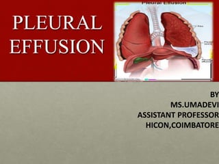
3. What Is Pleural Effusion?
Pleural effusion, sometimes referred to as
“water on the lungs,” is the build-up of
excess fluid between the layers of
the pleura outside the lungs. The pleura are thin
membranes that line the lungs and the inside of
the chest cavity and act to lubricate and
facilitate breathing
4. DEFINITION
Pleural effusion is excess fluid that
accumulates between the two pleural layers,
the fluid-filled space that surrounds the
lungs. Excessive amounts of such fluid can
impair breathing by limiting the expansion of
the lungs during ventilation.
6. Classification
•Can be unilateral or bilateral and classified
A)Based on site
Apical
Interlobar
Sub-pulmonic
Mediastinal
B)Based on mechanism and type of pleural fluid
Transudative (alteration in hydrostatic and oncotic pressure)
Exudative (alteration in pleural permeability)
7. c) Based on mechanism and type of pleural fluid
formed
Pyogenic
Chylous
Haemothorax
Pseudochylous
Hydrothorax
8.
9.
10.
11.
12. Pathogenesis
• Increased vascular permeability allows migration of inflammatory
cells (neutrophils, lymphocytes, and eosinophils) into the pleural
space.
• The process is mediated by a number of cytokines such as
interleukin IL-1, IL-6, IL-8, tumour necrosis factor (TNF)-alpha and
platelet activating factor released by mesothelial cells lining the
pleural space. The result is the exudative stage of a pleural
effusion. This progresses to the fibro-purulent stage due to
increased fluid accumulation and bacterial invasion across the
damaged epithelium.
• Neutrophil migration occurs as well as activation of the
coagulation cascade leading to pro-coagulant activity and
decreased fibrinolysis. Deposition of fibrin in the pleural space
then leads to septation or loculation. The pleural fluid pH and
glucose level falls while LDH levels increase.
21. Clinical features
Many patients have no symptoms due to the effusion when
effusion is small.
Pleuritic chest pain is the usual symptom of pleural
inflammation.
Irritation of the pleural surfaces may also result in a dry,
nonproductive cough.
With larger effusions, dyspnea results from lung
compression.
22. Common symptoms
•chest pain
•dry cough
•fever
•difficulty breathing when lying down
•shortness of breath
•difficulty taking deep breaths
•persistent hiccups
•difficulty with physical activity
23. Physical examination
Inspection:
Absent or diminished movements of affected side
Fullness of chest with bulging intercostal spaces
Palpation:
Diminished breath sounds over the site of the effusion
Decreased or absent tactile fremitus
Percussion:
Stony dullness to percussion
Auscultation:
Absence of breath sounds over the effusion
Vocal resonance absent
Signs of pneumonia like bronchial breathing, crackles etc.
24. Investigations
Total and differential leucocyte counts
• Acute phase reactants-white cell count, total neutrophil
count, CRP, ESR, pro-calcitonin distinguish bacterial from
viral causes
Radiological examination
• X-ray chest PAview done in erect position-a total of
300mL of fluid is needed to diagnose pleural effusion
clinically and radiologically
• Even 50mL of fluid can be demonstrated radiologically in
lateral decubitus
25. Findings
• Obliteration of cardiophrenic and costophrenic angles
• Loculated effusions
• Subpulmonic effusion-collection of fluid below the
diaphragm will lead to elevation of diaphragm, confirmed
by X-ray in lateral decubitus
• Lateral decubitus on side of effusion will show a shift in
the fluid level
• Tracheal and mediastinal shifts are seen in massive
effusion
26.
27. Ultrasonogram
Useful in differentiating between loculated pleural effusion and tumour
CT Scan
Helpful if the effusion is minimal or loculated
Pleural fluid aspiration (Thoracocentesis)
Diagnostic: Helps to differentiate between exudates and transudates
Therapeutic: Massive collection or rapid collection of pleural fluid
Severe respiratory distress
Suspected empyema
Massive mediastinal shift
30. LIGHTS CRITERIA
An accurate diagnosis of the cause of the effusion,
transudate versus exudate, relies on a comparison
of the chemistries in the pleural fluid to those in the
blood, using Light's criteria.
According to Light's criteria (Light, et al. 1972), a
pleural effusion is likely exudative if at least one of
the following exists:
31. LIGHT’S CRITERIA:
• Atleast one of the following criteria should be
satisfied to identify exudates:
Pleural fluid to serum total protein ratio- more than
0.5
Pleural fluid to serum LDH ratio- more than 0.6
Pleural fluid LDH- more than two-third of serum LDH
None of these criteria should be satisfied in a
transudative effusion
32. Roth’s criteria
• If serum-pleural fluid albumin gradient
is more than 1.2 it is transudate, else
exudate.
33. Pleural Fluid Biochemistry
• pH
• Glucose
• Lactate dehydrogenase(LDH)
• Sodium, potassium and calcium conc
• Amylase
• Adenosine deaminase
• Ratio of protein in pleural fluid to serum
• Ratio of LDH values in pleural fluid to serum
34. PLEURAL FLUID CYTOLOGY
WBC Count
Predominant cell type(neutrophil, lymphocytes, eosinophils, red
blood cells)
Lymphocytosis- if >50% leucocytosis then suspect TB
Malignant cells
PLEURAL FLUID MICROBIOLOGY
Gram stain
Acid fast for AFB
Pleural fluid Culture
AFB Culture
PCR for TB
35. Pleural Biopsy
• Can be done at maximum dullness on percussion or
at a maximum thickening of pleura. Abram’s pleural
biopsy needle is used for biopsy
• Most helpful in evaluating for TB
• Limited utility for CA (40-50% positive)
Repeat cytology x 3
• Sarcoid, fungal: might be helpful
39. MANAGEMENT
GOAL of treatment is to:
1. Remove the fluid
2. Prevent fluid from building up again
3. Determine and treat the cause of the
fluid buildup
40. Management
SUPPORTIVE TREATMENT
• Oxygen is necessary if SpO2 <92%
• Fluid therapy if child dehydrated or unable/unwilling
in drinking water
• Initiate IV antibiotics
• Analgesics and antipyretics
• Chest radiography & U/S
41. REMOVAL OF FLUID
Removing the fluid (thoracentesis) may
be done if there is a lot of fluid and it is
causing chest pressure, shortness of
breath, or a low oxygen level.
Removing the fluid allows the lung to
expand, making breathing easier.
42. Medical
• Treat the cause
Pneumonia- initial blind antibiotic treatment
A) Following community acquired pneumonia
• Cefuroxime
• Co-amoxiclav
• Penicillin & flucloxacillin
• Amoxicillin & flucloxaxillin
• Clindamycin
B) Hospital acquired pneumonia
• Broader spectrum antibiotics that cover aerobic gram negative rods
43. • Tuberculosis- Category I treatment
2HRZE+4HRE
Prednisolone 1-2mg/kg orally 4-6weeks promotes
rapid absorption of the pleural fluid and prevents
fibrosis
• Congestive cardiac failure- treat with diuretics and
other anti-failure medications
44. Surgical
• Pleural fluid aspiration is done by using a wide bore
needle. If the fluid is thick and cannot be drained by a
needle, an intercostal drainage(under water seal) at the
most dependant part should be done.
• Indications
Empyema
Presence of causative organisms in the fluid
Pleural fluid glucose <50mg/dL
Pleural fluid pH <7.0
45. Pleural effusions that cannot be managed through drainage
or pleural sclerosis may require surgical treatment.
The two types of surgery include:
1.Video-assisted thoracoscopic surgery (VATS)
A minimally-invasive approach that is completed through 1
to 3 small (approximately ½ -inch) incisions in the chest.
Also known as thoracoscopic surgery, this procedure is
effective in managing pleural effusions that are difficult to
drain or recur due to malignancy. Sterile talc or an antibiotic
may be inserted at the time of surgery to prevent the
recurrence of fluid build-up.
46. 2.A thoracThoracotomy (Also referred to as traditional,
“open” thoracic surgery)
otomy is performed through a 6- to 8-inch incision in the
chest and is recommended for pleural effusions when
infection is present.
A thoracotomy is performed to remove all of the fibrous
tissue and aids in evacuating the infection from the pleural
space.
Patients will require chest tubes for 2 days to 2 weeks
after surgery to continue draining fluid.
47. • Complications
• Pleural shock
• Introduction of infection
• Pneumothorax
• Pulmonary embolism
• Air embolism
• Acute pulmonary edema
• Injury to neovascular bundles
• Hydropneumothorax