
Recommended
More Related Content
What's hot
What's hot (20)
Similar to Immunity, antigen and antibody- Introduction
Similar to Immunity, antigen and antibody- Introduction (20)
Recently uploaded
Recently uploaded (20)
Immunity, antigen and antibody- Introduction
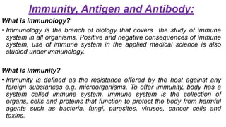
- 1. Immunity, Antigen and Antibody: What is immunology? • Immunology is the branch of biology that covers the study of immune system in all organisms. Positive and negative consequences of immune system, use of immune system in the applied medical science is also studied under immunology. What is immunity? • Immunity is defined as the resistance offered by the host against any foreign substances e.g. microorganisms. To offer immunity, body has a system called immune system. Immune system is the collection of organs, cells and proteins that function to protect the body from harmful agents such as bacteria, fungi, parasites, viruses, cancer cells and toxins.
- 2. Types of immunity: Immunity can be broadly classified into two classes: 1. innate (by birth) immunity and 2. Adaptive (acquired) immunity.
- 3. Innate immunity Adaptive / acquired immunity It is the resistance to infection that an individual possesses from birth Resistance to infection that an individual acquires during lifetime Immune response occurs in minutes Immune response occurs in days This is nonspecific immunity This is specific immunity Immunological memory is absent Immunological memory is present Innate immunity is provided by: Anatomical barriers- skin Physiological barriers-mucous present in respiratory tract, gastrointestinal tract, conjunctional secretion, genitourinary tract, body temperature Phagocytes- neutrophils, macrophages, monocytes Natural killer cells, mast cell, dendritic cell Fever, inflammatory response, normal bacterial flora Acquired immunity is provided by: T cells (T-Lymphocytes) B cells (B-Lymphocytes) Innate immunity depends on: Species Race Individual (age, hormonal level, nutrition) Acquired immunity are of following types: Active (natural or artificial) Natural active- immunity gained due to natural infection Artificial active- immunity gained due to vaccination Passive (natural or artificial) Natural passive- immunity passively transferred from mother to foetus through placenta & milk Artificial passive- immunity transferred to a recipient by parental administration of antibodies
- 4. Cells & organs of the immune system: Organs of immune system: The organs of the immune system are called lymphoid organs Lymphoid organs are the organs where lymphocytes develop, congregate. These organs include the bone marrow, thymus, lymph node, spleen & various other clusters of lymphoid tissue. There are two different types of lymphoid organs in body: Primary lymphoid organs & secondary organs
- 5. Primary lymphoid organ: • Also called central lymphoid organ • It is where immature lymphocytes develop • Organs where differentiation, proliferation and maturation of stem cells into immune competent cells take place • Include thymus and bone marrow
- 6. Thymus: • a bilateral organ located in the mediastinum • attains its peak development during youth Function : the progenitor T cells formed during hematopoiesis in bone marrow enter into the thymus. Here they multilply, differentiate and get matured. Finally from thymus, matured T cells enter into circulation and protect the body from infection Bone marrow: • Bone marrow is the soft tissue in the hollow shafts of the flat bones Function: all the cells of the immune system are initially derived from the bone marrow hrough a process called hematopoiesis Immature B cells proliferate & differentiate with in the bone marrow Responsible for the production of important immune system cells like B cells, granulocytes, natural killer cells and immature thymocytes. Also produces platelets and RBC
- 7. Secondary lymphoid organs: • Also called peripheral lymphoid organs • They maintain mature naïve lymphocytes and initiate an adaptive immune response • The peripheral lymphoid organs are the sites of lymphocyte activation by antigens • Secondary lymphoid organs are: Spleen, lymph nodes, tonsils, appendix, payer’s patches
- 8. Spleen: The spleen is a large, ovoid filtering organ situated in the left abdminal cavity. This organ is composed of T-cell, B-cell, natural killer cells, macrophages, dendritic cells & red blood cells. It is a production site of antibodies and activated lymphocytes. Spleen filters blood and traps blood borne antigen and thus respond to systemic infection Lymph nodes: Bean-shaped, encapsulated structures distributed throughout the body along the course of lymphatic vessel. They are made up of mostly B-cells, T-cells, macrophages and dendritic cells. They act as immunologic filters and drain the lymph from most of the body tissues and filter out the antigens present in them, before allowing the lymph to return to circulation
- 9. Tonsils: Two masses of soft glandular tissue on either side or the back of the mouth Function: Traps bacteria and viruses from inhaled air Appendix: Thin, dead-end tube measuring about three-to-four inches in length and hangs from the cecum Functions: Help tell the lymphocytes exactly where they have to head over to attack infection and it also enhances the massive intestines defences to a range of drugs and foods Peyer’s patches: The nodules of lymphatic cells that combine to make patches or bundles and appear generally only within the lowest part of intestine (ileum) Functions: Detect antigens such as bacteria and toxins and mobilize highly specialized white blood cells termed B-cells to produce an antibody
- 11. Cells of immune system: The response to pathogens is managed by the complex interactions and activities of the large number of diverse cell types involved in the immune response The innate immune response is the first line of defense and occurs soon after pathogen exposure. It is carried out by phagocytic cells such as neutrophils and macrophages, cytotoxic natural killer (NK) cells, and granulocytes The subsequent adaptive immune response includes antigen-specific defense mechanisms and may take days to develop. Cell types with critical roles in adaptive immunity are antigen-presenting cells including macrophages and dendritic cells. Antigen-dependent stimulation of various cell types including T cell subsets, B cells, and macrophages
- 12. Defense Mechanism: It is the way in which the body protects itself from invasion of pathogens The body has developed defense mechanisms to control and to cope with the constant attack of microorganisms The body has three lines of defense 1. First line of defense: Physical Barriers and chemical barrier 2. Second line of defense: Defensive Cells & Proteins, Inflammation, and Fever 3. Third line of defense: Humoral and cell mediated response
- 13. The First Line of Defense: •These are a combination of physical and chemical barriers that prevent all types of foreign agents from penetrating the outer layer of the body •No specific foreign agent is targeted at this level
- 14. Physical Barriers: • Skin • Cells filled with keratin, making skin impenetrable, waterproof, and resistant to disruptive toxins and most invaders • Dead cells are shed and replaced (1 million every 40 min), taking microbes with them • Mucous Membranes • The inner surfaces of the body are guarded by mucous membranes that line the respiratory, digestive, urinary, and reproductive systems and protect the internal lining • But, mucous membranes are more vulnerable than skin • Hair in the nose act as a coarse filter
- 15. Chemical barrier: • Sweat produced by glands in the skin wash away microbes and their acidity slows bacterial growth. •Mucous membranes produce sticky mucous that traps many microbes • Saliva and tears contain an enzyme called lysozyme that kills bacteria by rupturing their cell walls • Cerumen (ear wax) – produced in the ear canal and protects the canal by trapping dirt and dust particles
- 17. The Second Line of Defense: Defensive Cells • If a pathogen penetrates the first line of defense, these cells play a role in inhibiting or destroying the pathogen before it harms the body. They are non-specific and react to the presence of any foreign organism or substance • Phagocytes • Engulf pathogens, damaged tissue, or dead cells • Neutrophils • Macrophages • Eosinophils • Discharge destructive enzymes to destroy pathogens too big for phagocytes (e.g., parasitic worms) • Natural Killer Cells • Seek out abnormal cells (e.g., cancer cells)
- 18. Defensive Proteins • Interferon Protein • A virus enters a cell • The infected cell produces interferon • The interferon binds with other cells that become infected with a virus, and protects it by stimulating the cell to produce antiviral proteins that prevent the virus from making copies of itself • The interferon attracts and stimulates natural killer cells and macrophages to kill cells infected with the virus
- 19. Complement System -Destruction of pathogen (Cell lysis) -Enhancement of phagocytosis (Opsonization) -Stimulation of inflammation -Chemotaxis - attracting macrophages and neutrophils
- 20. Inflammation When body tissues are injured or damaged, a series of events called the inflammatory response occurs Redness: caused by increased blood flow to the damaged area Heat: increased blood flow elevates the temperature in the area of injury, increasing metabolic rate of the body cells Swelling: histamine makes capillaries more permeable than usual Pain: causes person to protect the area and prevent additional injury
- 21. Fever • A fever is an abnormally high body temperature caused by pyrogens (chemicals that set the “thermostat” in the brain to a higher set point) • A mild or moderate fever helps the body fight bacterial infections by slowing the growth of bacteria and stimulating body defense responses
- 22. The Third Line of Defense: • When the first two line of defense of the body can not prevent the infection, the immune system acts to eliminate the infectious agent and prevent the body from infection • Specific resistance is a third line of defense • Forms the immune response and targets specific pathogens • Cells of third line of defense or specific immunity are called lymphocytes • Types of lymphocytes: B- lymphocytes: produce specific proteins called antibodies, which are produced against specific antigens T-lymphocytes: target pathogens directly • Third line of defense works by two mechanisms: Antibody mediated & cell mediated immune response
- 23. • Antibody mediated immune response: This type of defense is provided by B lymphocyte cells that produce antibody against the non-self substance, i.e. antigen When B cells interact with an antigen, they are differentiated into antibody producing plasma cells, which produce and release specific antibodies into the blood circulation These secreted antibodies bind to the antigen specifically and facilitate its clearance from the body by various mechanisms: Antitoxin (antibody against toxin) inactivates toxin Neutralizing antibodies coat viruses, coated viruses cannot penetrate cells in the body Antibodies that combine with antigen on the surface of bacteria attract macrophages and are phagocytosed
- 24. •Cell mediated immune response: The principle role of cell mediated immune response is to detect and eliminate cells that harbor intracellular pathogens The defense is provided by both antigen specific and non-specific cells Antigen specific cells include CD8 cytotoxic T lymphocytes and cytokine secreting CD4 Th cells Non-specific cells include NK cells, macrophages, neutrophils and eosinophils
- 25. Antigen: Definition: antigen is a substance which when introduced parentally into the body stimulates the production of an antibody with which it reacts specifically and in an observable manner. •Example: Foreign protein, Nucleic acids, Large CHO, Some lipids, Pollen grains, microorganisms Epitope or antigenic determinant: An epitope, also known as antigenic determinant, is the part of an antigen that is recognized by the immune system, specifically by antibodies, Bcells, or Tcells. An antigen may have several epitopes. Each epitope is recognised by a different antibody
- 26. Characteristics of antigens: • Foreignness: antigen must be a foreign substance to elicit an immune response. Molecules recognised as “self” are not immunogenic • Molecular size: larger molecules are highly antigenic. Substance having molecular weight less than 10,000Dalton are either non- antigenic or weakly antigenic • Chemical nature: antigens are mainly proteins or polysaccharides; lipids & nucleic acids are less antigenic on their own but do so when combined with proteins • Susceptibility to tissue enzymes: substance that can be metabolised & are able to be digested by the action of tissue enzyme behave as antigen
- 27. Types of antigen 1. Based on the immunogenicity: • Complete antigen-A complete antigen can induce antibody formation and produce a specific and observable reaction with the antibody so produced. They are high molecular weight proteins but some are polysaccharides also • Incomplete antigen or Hapten- haptens are substances that are incapable of inducing antibody formation by themselves but can react specifically with antibodies. Hapten becomes immunogenic (capable of inducing antibodies) on combining with a larger carrier molecule e.g. serum protein such as albumin, globulin or polypeptide.
- 28. 2. Based on origin: • Exogenous antigen: exogenous antigens are antigens, that have entered the body from the outside for example by inhalation, ingestion or injection. • Endogenous antigen: endogenous antigens are antigens that have been generated within previously normal cells as a result of normal cell metabolism or because of viral or intracellular bacterial infection • Autoantigen: an auto antigen is usually a normal protein or complex of proteins (and sometime DNA or RNA) that is recognised by the immune system of patients suffering from autoimmune diseases. These antigens, under normal conditions, should not be the target of the immune system, but due to genetic or environmental factors, the normal immunological tolerance for such an antigen is lost
- 29. Antibody: •Definition: antibody or immunoglobulin is a specialised glycoprotein produced from activated B lymphocytes (plasma cells) in response to an antigen and react specifically with that antigen. Chemically antibodies are globulins and hence are also called immunoglobulins.
- 30. Structure of antibody: • Antibody molecules are globular proteins. They are Y-shaped structures, made up of two identical heavy (H) and two identical light (L) polypeptide chains. The longer chains are called heavy (H) chains and the shorter ones are called light (L) chains • Each L- chain is attached to H-chain by a disulphide (S-S) bond and the two symmetrical H-chains are held together by one to five S-S bonds, depending upon the type of immunoglobulin • Light chain consists polypeptides of around 22,000 dalton and heavy chain consists larger polypeptide of around 50,000 dalton or more.
- 31. • There are five types of immunoglobulin, based on the structure of constant region of heavy chains, denoted by Greek letters – gamma (), mu (µ), alpha (), delta (δ) & epsilon (ε). • The light chains exist in two forms- lambda (λ) and kappa (κ). Each immunoglobulin molecule shows either two kappa or two lambda light chains but they are never found in combination. • All chains of antibody have two ends- an amino terminal end (NH3) and a carboxy terminal end (COOH) • An antibody is made up of a variable region and a constant region • Variable region is the region that changes to various structure depending on differences in antigen. It is the part near amino terminal end of both light and heavy chain. Variable region represents the antigen binding site of antibody. • Constant region is the remaining structure in antibody towards the carboxy terminal end. • Hinge region is the junction formed between constant regions of heavy chains of IgG, IgA and IgD. It is absent in IgE & IgM. This region is rich in proline & cysteine amino acids. The hinge region is quite flexible, thus helps the antibody in reaching towards the antigen
- 33. Classes of immunoglobulins / antibodies: On the basis of nature of heavy chain, immunoglobulins are classified as below: • Immunoglobulin G (IgG) – contains gamma () chain • Immunoglobulin A (IgA) – contains alpha () chain • Immunoglobulin M (IgM) – contains mu (µ) chain • Immunoglobulin D (IgD) – contains delta (δ) chain • Immunoglobulin E (IgE) – contains epsilon (ε) chain
- 34. •Immunoglobulin G: IgG molecule consists of two heavy and two light chains linked by disulphide bonds. IgG has two identical antigen binding sites so that they are called divalent. IgG appears generally two weeks after infection but persist for long period of time. IgG is the predominant antibody in secondary immune response. It is the only immunoglobulin that passes through placenta and provides natural passive immunity to newborn •Immunoglobulin A: it is the major immunoglobulin that appears in the sero-mucous secretions such as milk, saliva, tears, nasal fluids, colostrum & secretions of respiratory, intestinal and genital tracts. It protects the exposed mucous membrane against attack by microorganisms
- 35. • Immunoglobulin M: it consists of 10 heavy chains and 10 light chains. IgM appears early in response to infection and its presence in serum indicates recent infection. It cannot cross placenta, hence presence of IgM in the serum of newborn indicates intrauterine infection. IgM remains largely to blood stream and offers protection against bacteraemia. Its deficiency may lead to septicaemia. • Immunoglobulin D: it is structurally similar to IgG. They occur on the surface of unstimulated B-lymphocytes and serve as recognition receptors for antigen. Combination of cell membrane bound IgD with corresponding antigen leads to specific stimulation of the B cell to produce antibody. • Immunoglobulin E: these antibodies are also called regains and inactivated by heat at 56oC for 1 hour. It does not pass the placenta. It is mostly extravascular in distribution and found in insignificant amount in normal serum but found higher amount in allergy and intestinal parasitic infection.