
Recommended
Recommended
More Related Content
Similar to Health Cardiorespiratory sx 2.1.ppt
Similar to Health Cardiorespiratory sx 2.1.ppt (20)
Recently uploaded
Recently uploaded (20)
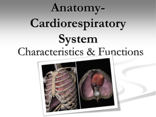
Health Cardiorespiratory sx 2.1.ppt
- 2. Anatomy-Cardiorespiratory System Terminology Fitness-the ability of the heart, blood vessels, lungs, and muscles to work together to perform a physical activity. Cardiorespiratory endurance-the ability of the heart, lungs, and blood vessels to distribute nutrients and oxygen to and remove wastes from the body’s tissues. Cardiology-the study of the heart and the body’s network of vessels and their functions.
- 3. Terminology continued. . . Oxygenated High oxygen level Deoxygenated Low oxygen level
- 4. The Heart: Location and Function Location: The medial cavity of the thorax Flanked on both sides by the lungs Protected by the thoracic cage Slightly tilted off the midline at the apex Function: To pump blood throughout the body Blood carries oxygen and nutrients to the cells Blood carries carbon dioxide and waste products away from the cells
- 5. Anatomy-Cardiovascular System Heart Heart: Function: to pump blood throughout the body Blood carries oxygen and nutrients to the cells Blood carries carbon dioxide and waste products away from the cells
- 6. Anatomy-Cardiovascular System Heart • Divided into 4 chambers • R and L atrium (upper chambers) • R and L ventricles (lower chambers) • Septum-thick vertical wall divides left and right chambers. https://youtu.be/kvfJafl4Es0
- 7. The Heart Heart valves separate each chamber and prevent a backflow of the blood Tricuspid valve (right A- V valve) Bicuspid (mitral) valve- (left A-V valve) Pulmonary valve Aortic valve https://www.sciencelearn.org.nz/videos/1608-blood-flow-through-the-heart
- 8. How does the blood flow through the Heart? Blood enters the heart through two large veins, the inferior and superior vena cava, emptying oxygen-poor blood from the body into the right atrium of the heart. As the atrium contracts, blood flows from your right atrium into your right ventricle through the open tricuspid valve. When the ventricle is full, the tricuspid valve shuts. This prevents blood from flowing backward into the atria while the ventricle contracts. As the ventricle contracts, blood leaves the heart through the pulmonic valve, into the pulmonary artery and to the lungs, where it is oxygenated and then returns to the left atrium through the pulmonary veins. Right Side of Heart
- 9. The Heart Left side of the Heart
- 10. How blood flows to the lungs? Once blood travels through the pulmonic valve, it enters your lungs. This is called the pulmonary circulation. From pulmonic valve, blood travels to the pulmonary artery to tiny capillary vessels in the lungs. Carbon dioxide leaves the body when you exhale. Once the blood is purified and oxygenated, it travels back to the left atrium through the pulmonary veins.
- 11. Blood Flow
- 12. Blood Vessels Blood vessels Elastic tubes Carry blood Arteries Arterioles Capillaries Venules Veins Arteries Veins
- 13. Anatomy-Cardiovascular System Arteries Arteries Muscular, thick, elastic blood vessels Carry oxygenated blood away from the heart to body cells, tissue & organs Except for pulmonary artery artery
- 14. Anatomy-Cardiovascular System Arterioles Arterioles Small terminal branches of arteries Connect arteries to capillaries arteriole artery
- 15. Anatomy-Cardiovascular System Capillaries Capillaries Very small blood vessels Connect arterioles to venules arteriole artery capillary
- 16. Anatomy-Cardiovascular System Venules Venules Small veins Connect capillaries to larger veins arteriole artery capillary venule
- 17. Anatomy-Cardiovascular System Veins Veins Thin walled blood vessels Carry deoxygenated blood to the heart from body cells, tissue & organs Except for pulmonary vein arteriole artery capillary venule vein
- 18. Direction blood pumps through the heart
- 19. Blood Pressure Systole-Contraction reading • Deoxygenated blood moves from Rt ventricle through the Pulmonary valve up into the pulmonary trunk. • Oxygenated blood moves from Lt Ventricle through the Aortic valve. Diastole-Relaxation reading • Deoxygenated blood moves through the tricuspid valve from the atrium. • Oxygenated blood moves from Lt atrium through the Mitral valve to the Lt ventricle https://youtu.be/4YNdp3pRjig
- 20. Movement of Blood Heart Rate: the speed of the heartbeat measured by the number of contractions of the heart per minute. Pulse: Occurs in time with heartbeat. Can be measured and detected at radial arteries(wrist) and carotid arteries(neck). Stroke Volume: The volume of blood ejected from ventricles with each contraction. Increase fitness level =decreases resting heart rate
- 21. Pneumology The study of the respiratory system- structure, functions, disorders, and diseases.
- 22. The Lungs-Primary Organs Of Respiratory System Location: On either side of the heart Consist of a total of five lobes Right lung has 3 lobes Left lung has 2 lobes Functions: Exchange carbon dioxide for oxygen Help to remove heat from the body
- 23. Respiratory Tract Pathway of air: Air enters through mouth or nose Air is warmed, moistened, and filtered Passes into the pharynx Next through the glottis, the larynx, and into the trachea (voice box). The trachea is a cartilage ringed tube
- 24. Gas Exchange within the Lungs Trachea divides into two branches Called bronchi (plural) Enters the thoracic cavity Division of bronchi occur within the lungs called bronchioles Bronchioles divide and end in small sac-like structures called alveoli Alveoli microscopic sacs surrounded by capillaries. This is the site of gas exchange for the body.
- 25. Mechanics of Breathing Thoracic cavity Airtight chamber Moveable floor called diaphragm Diaphragm-muscle dividing thoracic cavity and abdominal cavity Inhalation causes the diaphragm to contract and move down
- 26. Mechanics of Breathing Inhalation Lifts the ribcage and sternum upwards and outward Enlargement of thoracic area reduces pressure on the lungs and air rushes in the equalize pressure. Exhalation Diaphragm, rib and chest muscles relax reducing size of cavity Pressure on the surface increases and air rushes in from lungs to equalize pressure
- 28. Lung Capacity Normal breath measures approximately ½ liter of air. Total lung capacity approximately 6 liters. Vital capacity is about 4 ½ liters-which is exhale and inhale as much air as one can. The lungs are never completely without air. Increase volume (deep breathing) increases oxygen intake. During exercise deep breathing decreases the stress on the heart and lungs and improves endurance while delaying fatigue.