Excretory SYSTEM.pptx
•Download as PPTX, PDF•
0 likes•9 views

Human Excretory System Grade 11

Recommended
More Related Content
Similar to Excretory SYSTEM.pptx
Similar to Excretory SYSTEM.pptx (20)
Recently uploaded
Recently uploaded (20)
Excretory SYSTEM.pptx
- 1. E X C R E T O R Y S Y S T E M . • Life of every organism depends on certain basic processes. Excretion is one among them. Different organisms follow different modes of excretion. In complex organisms including humans, there is a specialized system for excretion called human excretory system. • Excretion is a process in which metabolic waste is eliminated from an organism. In vertebrates this is primarily carried out by the lungs, kidneys, and skin. This is in contrast with secretion, where the substance may have specific tasks after leaving the cell. Excretion is an essential process in all forms of life
- 2. L E S S O N O B J E C T I V E S . At the end of the lesson, learners will be able to : • Explain the purpose of the kidneys, bladder, and urethra. • Describe the excretory system. • Demonstrate an understanding of the path of excretory system
- 3. E X C R E T O R Y I N VA R I O U S O R G A N S Excretory organs include: The lungs. The kidney or bladder. The liver. The alimentary canal (gut) and The skin.
- 4. T H E L U N G S The lungs are part of the respiratory system, but they are also important organs of excretion. They are responsible for the excretion of gaseous wastes from the body. The main waste gas excreted by the lungs is carbon dioxide, which is a waste product of in cells throughout the body. Carbon dioxide is diffused from the blood into the air in the tiny air sacs called in the lungs). By expelling carbon dioxide from the blood, the lungs help maintain acid-base . In fact, it is the pH of blood that controls the rate of breathing. Water vapor is also picked up from the lungs and other organs of the respiratory tract as the exhaled air passes over their moist linings, and the water vapor is excreted along with the carbon dioxide. Trace levels of some other waste gases are exhaled, as well.
- 5. T H E K I D N E Y S & B L A D D E R . The paired kidneys are often considered the main organs of excretion. The primary function of the kidneys is the elimination of excess water and wastes from the bloodstream by the production of the liquid waste known as . The main structural and functional units of the kidneys are tiny structures called nephrons. filter materials out of the blood, return to the blood what is needed, and excrete the rest as urine. As shown in the Figure , the kidneys are organs of the urinary system, which also includes the ureters, bladder, and urethra — organs that transport, store, and eliminate urine, respectively.
- 6. T H E L I V E R . • The liver (shown in the Figure) has numerous major functions, including secreting bile for digestion of lipids, synthesizing many proteins and other compounds, storing glycogen and other substances, and secreting endocrine hormones. In addition to all of these functions, the liver is a very important organ of excretion. The liver breaks down many substances in the blood, including toxins. For example, the liver transforms ammonia — a poisonous by-product of protein catabolism — into urea, which is filtered from the blood by the kidneys and excreted in urine. The liver also excretes in its bile the protein bilirubin, a byproduct of hemoglobin catabolism that forms when red blood cells die. Bile travels to the small intestine and is then excreted in feces by the large intestine.
- 7. T H E A L I M E N TA R Y C A N A L ( G U T ) • True metabolic wastes are excreted by means of the flow of bile from the liver into the intestine. The destruction of cells in animals produces bile pigments—residues of hemoglobin and other pigments—which may be considered to be the principal metabolic wastes eliminated via the alimentary canal
- 8. T H E S K I N . The is part of the integumentary system, but it also plays a role in excretion through the production of by sweat glands in the dermis. Although the main role of sweat production is to cool the body and maintain temperature , sweating also eliminates excess water and salts, as well as a small amount of urea. When sweating is copious, as in the Figure , ingestion of salts and water may be helpful to maintain
- 9. T H E U R I N A R Y S Y S T E M . • Consist of paired kidneys, renal artery, renal vein, ureter, bladder and urethra. • Each kidney is supplied with blood by a renal artery (impure, oxygenated blood) and drained by a renal vein (pure, deoxygenated blood). • Urine exits each kidney through a duct called the ureter. • Both urethers drain into a common urinary bladder, and urine is expelled through a urethra
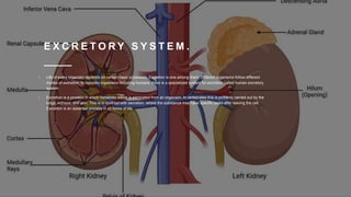
- 10. S T R U C T U R E O F K I D N E Y.
- 11. C O N T I N U AT I O N O F S T R U C T U R E T H E K I D N E Y. • Microscopically each kidney is composed of over 1 million tiny tubules called nephrons. • The nephrons of the kidneys produce urine.
- 12. T H E N E P H R O N • The nephrons of the kidneys produce urine. • Some nephrons are located primarily in the renal cortex, but others dip down into the renal medulla. • The nephron, the functional unit of the vertebrate kidney, consists of a single long tubule and a ball of capillaries called the glomerulus.
- 13. N E P H R O N • https://study.com/learn/lesson/nephron-parts-function-location.html
- 14. T H E F U N C T I O N O F T H E K I D N E Y I N U R I N A R Y F O R M AT I O N . An average person produces between 1 - 2 liters of urine daily. Urine production requires 3 distinct processes: • Glomerular filtration at Malpighian body (Glomerulus & Capsule of Bowman.) • Tubular reabsorption at convoluted tubules. • Tubular secretion at convoluted tubules.
- 15. U R I N A R Y F O R M AT I O N .
- 16. G L O M A R U L A R F I LT R AT I O N . • Blood enters kidney via renal artery (impure / oxygenated). • This branches many times eventually forming many afferent arterioles, each of which delivers blood to an individual kidney nephron. • The diameter of afferent (incoming) arteriole is greater than diameter of efferent arteriole ( • Pressure of blood inside glomerulus is increased due to difference in diameter of incoming & out-going arterioles.
- 17. G L O M E R U L A R F I LT R AT I O N C O N T … • Increased blood pressure helps to force following components of blood out of glomerular capillaries into cavity of capsule of Bowman: • Most of the water; • Most/all of the salts; • Most/all of the glucose; • Most/all of the urea.
- 18. T U B U L A R R E A B S O R P T I O N . Only about 1% of glomerular filtrate leaves body (rest - useful substances) is reabsorbed into the blood . Tubular reabsorption & occurs via 3 mechanisms. They are: Osmosis Diffusion Active Transport
- 19. T U B U L A R R E A B S O R T I O N C O N T. . Reabsorption varies according to body's needs enabling body to retain most of its nutrients. Processes of tubular reabsorption occur in following order: Proximal convoluted tubule (PCT). Loop of Henle Distal convoluted tubule (DCT
- 20. T U B U L A R R E A B S O R P T I O N : P R O X I M A L C O N V U L A T E D T U B U L E . Most of volume of filtrate solution is reabsorbed in proximal convoluted tubule. This includes some water and most/all of glucose. Solutes are selectively moved from glomerular filtrate to BLOOD plasma by active transport. Rest of the glucose, amino acids, & high amounts of ions, are reabsorbed again later.
- 21. T U B U L A R R E A B S O R P T I O N : L O O P O F H E N L E . • Water, urea, & salts contained within ascending limb of Henle eventually pass into distal convoluted tubule (DCT). • DCT reacts to amount of anti-diuretic hormone (ADH) in blood: • The more ADH is present in blood, the more water is re-absorbed into it. • Because presence of ADH in blood causes cells of DCT to become more permeable to water, therefore they allow more water to pass from tubular fluid back into blood. This results in more concentrated urine.
- 22. Tubular reabsorption IN the loop of henle. If level of ADH in blood is reduced then cells in DCT becomes less permeable to water • less water is able to pass from tubular fluid back into blood - which results in less concentrated urine.
- 23. T U B U L A R S E C R E T I O N . • Involves substances being added to tubular fluid. • Removes excessive quantities of certain dissolved substances from body. • Maintains blood at a normal healthy pH (pH 7.35 to pH 7.45) • Substances that are secreted into tubular fluid (for removal from body) include: • Potassium ions (K+), • Hydrogen ions (H+), • Ammonium ions (NH4 +), • creatinine, • urea, • some hormones, and • some drugs (e.g. penicillin).
- 24. K I D N E Y A N D H O M E O S TA S I S • Homeostasis the property of a system in which variables are regulated so that internal conditions remain stable. • The kidneys are organs of homeostasis for 4 main reasons: • Excrete metabolic waste such as urea (primary metabolic waste of humans). • Maintain the water salt balance in the body. • Maintain the acid-base (pH) balance in the body. • Secrete hormones.
- 25. K I D N E Y A N D H E M E O S TA S I S . • Through hormone ADH,water is kept constant. (Refer to reabsorption in DCT) • Salt is reabsorbed through hormone Aldosterone by means of a Sodium pump mechanism. • Aldosterone is a hormone that • increases the reabsorption of sodium (Na) & water & • release (secretion) of potassium (K) in kidneys.