
Recommended
More Related Content
Similar to Classification , function of HORMONES in our body.pdf
Similar to Classification , function of HORMONES in our body.pdf (20)
Recently uploaded
Recently uploaded (20)
Classification , function of HORMONES in our body.pdf
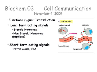
- 1. Biochem 03 Cell Communication November 4, 2009 •Function: Signal Transduction • Long term acting signals –Steroid Hormones –Non Steroid Hormones (peptides) • Short term acting signals –Nitric oxide, NO
- 2. Endocrine System • Small molecules are released from these glands into the bloodstream where they travel to a distant site and change the pace or architecture of the target tissue. • Hypothalamus in the brain is the mission control center
- 3. Endocrine System Anterior Pituitary Most of these are PEPTIDE HORMONES Posterior Pituitary
- 4. Hormones enter cells through different methods depending on their chemical nature Steroid hormones Peptide hormones
- 5. Steroid Hormones • are small molecules • all exhibit lots of chemical similarity • all are fundamentally non-polar, hydrophobic
- 6. Steroid Synthesis • derived from cholesterol • grouped by the receptors to which they bind: – glucocorticoids – mineralocorticoids – androgens – estrogens – progestagens – Vitamin D a sixth closely related hormone system with homologous receptors
- 7. Action of Steroids – bind to receptors – complex migrates to the nucleus- binds to DNA – binding to DNA affects transcription – the pattern of gene expression is changed – the time scale for this event is slow (minutes to days)
- 8. Nuclear Receptor Subfamily Yellow shows the highly variable activation domain Blue shows the highly conserved DNA binding domain- ~66 amino acids Red shows the hormone binding domain estrogen receptor glucocorticoid receptor
- 10. Estrogen Receptor Hormone binding domain Estradiol binds in a deep cleft of a binding site in this mostly helical 240 residue domain of ER. Somehow this gets communicated to DNA binding domain Estrogen
- 11. ER Hormone binding domain Structure 1A52 http://www.pdb.org/pd b/explore.do?structure Id=1A52
- 12. ER Hormone binding domain Structure 1A52 Hydrogen Bonds Ring A: Oxygen ___________ ___________ ___________ Ring D Oxygen ___________
- 13. ER Hormone binding domain Structure 1A52 Hydrophobic Int. @3.7 Å Ring A: Ring B: Now change distance to 3.8 Å and see what other interactions you see
- 14. Steroid Receptors DNA binding domain • Zn finger proteins are the 2nd largest class of proteins • Genes for > 700 different Zn fingers in the human genome (antibodies largest) • DNA binding domain of a classic Zinc finger Zif268 shown left • the red helix interacts with the DNA…..How?
- 15. • Structural motif is highly versatile (one type of structure seems to be used in 700 different ways.) • # of Zn fingers in one protein ranges from 1 to 37 • Extended DNA sequences can be specifically recognized • Recognition happens through complementarity between amino acid side groups on helix (blue below) and base pair sequence of DNA Steroid Receptors DNA binding domain
- 16. Common Motifs in Steroid Receptors • The DNA binding domain of the receptor binds to the hormone response element on the DNA • Changes of only two base pairs within each palindromic unit on the DNA switches the recognition from GR to ER What the DNA looks like...
- 17. •Estrogen Receptor’s DNA binding domain also a zinc cluster protein. •Shown here is the DNA binding domain similar to glucocorticoid receptor Estrogen Receptor * DNA binding domain
- 18. ER DNA binding domain Structure 1hcq http://www.pdb.org/ pdb/explore.do?stru ctureId=1HCQ
- 19. Do other types of molecules bind to this nuclear receptor subfamily? • Non-Natural non-steroidal ligands • Environmental Estrogens – Phytoestrogens from plants but remember plants don’t have cholesterol so must be non-steroidal pathways to derivatives. – Xenoestrogens - DDT is the most potent estrogenic mimic known, must stronger than estrogen itself in inducing proliferative cell growth, bisphenol A, THC, • see handout of xenoestrogens • see estrogen mimics under external links, signal transduction folder.
- 20. Steroid Synthesis • Steroid biosynthesis starts with the fatty acid squalene, whose carbon backbone is shown here • Synthesis occurs in the gonads and in the adrenal glands SQUALENE CHOLESTEROL
- 21. Glucocorticoid Receptor dimeric protein (blue and yellow balls above right) Each stabilized by a pair of zinc clusters (small purple balls) Recognition helix fits snugly into major groove of DNA, which widens 2 A in the process. Iglu.pdb from Sigler et al, Nature 1991