SlideShare a Scribd company logo
1 of 54
Download to read offline
Chairperson: Dr. Ravi D
Moderator: Dr. Balaji NK
Presenter: Dr. Shreya Sinha
â–Ş Introduction
â–Ş Premalignant lesions Vs premalignant conditions
â–Ş Progression of oral cancer
â–Ş Rate of malignant transformation
â–Ş Leukoplakia
â–Ş Erythroplakia
â–Ş Oral Submucous fibrosis
â–Ş Oral Lichen planus
â–Ş Candidiasis
â–Ş Actinic chelitis
â–Ş Screening protocol
â–Ş Others
â–Ş These are lesions and conditions that are not malignant but have the
potential to become malignant.
â–Ş Stratified squamous epithelium lines the majority of the oral cavity, and
aberrancy in the epithelium gives rise to premalignant lesions.
â–Ş Previously, various terms like Pre-cancerous, Pre-malignant, Intra epithelial
dysplasia, Intra epithelial neoplasia were used.
▪ Recently, a more precise phrase, “Oral potentially malignant disorders” has
been adopted by the WHO collaborating center.
Pre-malignant lesions
â–Ş A morphologically altered tissue in
which malignancy is more likely to
occur than its apparently normal
counterpart.
â–Ş Leukoplakia
â–Ş Erythroplakia
â–Ş Submucous fibrosis
â–Ş Lichen planus
â–Ş Actinic chelitis
Pre-malignant conditions
â–Ş A generalised state associated with a
significantly increased risk of
malignancy.
â–Ş Plummer Vinson syndrome
â–Ş Syphilis
â–Ş The most feared complicationof oral premalignant disorders is progression to carcinoma.
â–Ş A recent meta-analysis revealed that the rate of malignant transformation of all oral
premalignant disorders over a period of 5 years combined was around 7.9%. In contrast,
individual transformation rates were:
â–Ş lichen planusis around 1.4%,
â–Ş leukoplakia 9.5%,
â–Ş oral submucousfibrosis 5.2%,
â–Ş erythroplakia 33.1%.
â–Ş It refers to a white patch in the mucosa that does not rub off and cannot be
clinically identified as another entity.
â–Ş It is a diagnosis of exclusion.
â–Ş Risk factors include: chronic smoking, tobacco abuse, areca nut and betel
nut chewing, alcohol abuse, chronic trauma (friction induced hyperkeratosis),
chronic sun exposure, sanguinaria (herbal root extract in some mouthwashes
and toothpaste), male sex, people in fourth decade of life, etc.
â–Ş Clinical forms are:
1. Homogenous leukoplakia: There is a smooth or wrinkled white patch, well
demarcated, has a consistent pattern throughout and is less often associated
with malignancy.
2. Nodular (Speckled) leukoplakia: There are nodular white patches with
erythematous base.
3. Erosive leukoplakia (Erythroleukoplakia) presents white patches which has
erosions and fissures and is interspersed with red patches (erythroplakia).
4. Proliferative verrucous leukoplakia: This uncommon variant of leukoplakia
is multifocal and persistent and occurs more often in women. A thin flat white
patch progresses to leathery thickened and papillary to verrucous appearance.
homogenous homogenous speckled
erythroleukoplakia proliferative
verrucous
â–Ş Histopathology: It ranges from hyperkeratosis and acanthosis to dysplasia
(disordered cell growth and architectural distortion) or carcinoma in situ to
invasive squamous cell carcinoma. The dysplasia is traditionally graded as mild,
moderate, or severe.
▪ Potential for malignant change: The chances of malignant change are from 1–
17.5% , which varies according to the site, type and duration of the lesion and
age of the patient.
â–Ş Risk factors associated with malignant transformation of oral leukoplakia are:
1. Female sex
2. Long standing leukoplakia
3. Leukoplakia in non smokers
4. Leukoplakia over tongue or floor of mouth
5. Size >200 mm2
6. Non homogenous type of leukoplakia
7. Leukoplakia with presence of epithelial dysplasia
A clinical staging system (OL system) for oral leukoplakia on the lines of TNM
staging was recommended by WHO in 2005, taking the size (L) and histopathological
features (P) into consideration.
â–Ş Management:
1. Biopsy: An incisional biopsy (multiple biopsies in extensive lesions) must be taken
from suspicious areas (such as erythematous, granular, ulcerated and indurated)
to know the grades of dysplasia and rule out malignancy.
2. Medical management
a) L-ascorbic acid (vitamin C)
b) a-tocoferol (vitamin E)
c) Retinoic acid (vitamin A)
d) Vitamin A derivative, isotretinoin and 13-cis retinoic acid: 28,500 IU per day.
e) Beta carotene : 150,000 IU of beta carotene twice per week for 6 months.
f) Bleomycin: topical bleomycin has been used in the treatment of oral leukoplakia in
dosage of 0.5% per day for 12-15 days or 1% per day for 14 days. It prevents the
progression though dysplasia to carcinoma. Bleomycin is a glycopeptide antibiotic
that acts by binding to guanosine-cytosine rich portion of the DNA via association
of the S tripeptide and by partial intercalation of the bithiazole rings.
â–Ş Surgical management includes:
1. Scalpel excision/ stripping
2. Electrocautery
3. Cryotherapy
4. CO2 laser therapy
1. Scalpel excision:
• Area is outlined using few (1-2) mm of normal tissue. It is incised with scalpel
and patch is undermined by scalpel or blunt dissection to a depth of 2-4 mm.
This allows the leukoplakia to be removed in one piece. The mucosal defect if
small is closed primarily or it is covered by transported local mucosal flaps. Larger
defects are grafted with split thickness skin graft.
2. Electrocautery ( Fulguration):
• This requires local or general anaesthesia. Here, multiple areas of lesion are
pierced with electrocautery and left to heal. The healing process is slow and
painful.
3. Cryotherapy :
• It is a method of superfreezing the tissue in order to destroy it.
• It is done using a cotton swab that has been dipped in liquid nitrogen or a probe
that has liquid nitrogen flowing through it. The technique involves freezing the
mucosa with cryoprobe for 1.5-2 minutes, then waiting for 2 minutes, followed by
further freezing for 1.5-2 minutes. Thicker lesions may require 2-3 minutes
freezing.
4. CO2 Laser therapy:
• CO2 laser beam wavelength – 10.6 micro meter
• Well absorbed by water and hence by soft tissues
• The absorbed energy causes vaporisation of the intra and
extracellular fluid and destruction of cell membrane. The cell debris
are released and burned in the laser beam, depositing a carbonised
layer on the tissue surfaces.
• Two techniques are used to remove the leukoplakia using CO2 laser:
a) Excision- to excise a patch, the laser is used to cut around the
margins, which can be held in tissue forceps while the laser
undermines the leukoplakic patch.
b) Vaporisation- it is done by moving the laser beam back and forth
across the surface of the lesion. It has the risk of leaving small bits
of abnormal tissue which are deep under thickly keratinised
tissue.
Surgical excision of lesions using laser offers several advantages over scalpel
excision which includes
â–Ş bloodless surgical and postsurgical events;
â–Ş the ability to precisely coagulate, vaporise, or cut tissue;
â–Ş minimal swelling and scarring;
â–Ş reduction of surgical time, postsurgical pain with high patient acceptance.
â–Ş Differentia diagnosis:
Linea alba buccalis Leukoedema
Fordyce granules Traumatic keratosis
Geographic tongue
â–Ş It is a fiery red patch that cannot be clinically or pathologically classified as
another entity.
â–Ş Also known as Erythroplasia of Queyart.
â–Ş These lesions may have a flat, macular, velvety appearance and may be speckled
with white spots representing foci of keratosis.
â–Ş It is much less common than leukoplakia, but has a much greater probability
(91%) of showing signs of dysplasia or malignancy at the time of diagnosis.
â–Ş The rate of malignant transformation over a period of 5 years was found to be
33.1%
â–Ş Differential diagnosis:
1. Erythematous (atrophic) candidiasis
2. Kaposi’s sarcoma
3. Contact stomatitis
4. Vascular malformation
5. Squamous cell carcinoma
6. Geographic tongue/ erythema migrans
â–Ş Management:
• It is the same as that for Invasive carcinoma or Carcinoma in-situ and
includes surgery, radiation and cauterisation.
• Surgical excision if proven dysplastic /malignant.
â–Ş It is an insidious painless oral cavity disease, which is characterized by juxta
epithelial inflammatory reaction, followed by fibro-elastic change of lamina
propria with epithelial atrophy, leading to stiffness of oral mucosa and
causing trismus and inability to eat.
â–Ş It appears as white fibrotic bands extending from retromolar trigone to soft
palate, buccal mucosa, tongue and pharynx.
â–Ş It is common in Indian population. Incidence is 0.2-0.5 %.
â–Ş Etiology:
1. Prolonged local irritation due to paan, betel nut, tobacco, smoking of cigarette
or bidis, excessive amount of spices and chillies in food.
2. Dietary deficiency of iron, vitamin B-complex and vitamin A .
3. Cell mediated immune process: Some consider it a cell mediated immune
reaction to arecoline.
4. Racial: Disease usually affects Indians or people of Indian origin living abroad.
â–Ş Pathogenesis:
oral mucosa
Betel quid habit
Constant irritation
Chronic inflammation
Activated T cell and macrophages at the site
Increase in cytokines- IL6, TNF, IFN-a
Increse in growth factors- TGF-B
Increased susceptibility due to
deficiency of iron & vitamin B12
cytokines
Increase collagen
production
Decrease collagen
degradation
Increased collagen
(insoluble form – Cross
linking of insoluble collagen)
Fibrosis
Oral submucous fibrosis
▪ The malignant transformation has been observed in 3–7.6% of cases.
â–Ş Initial stage-
• Burning sensation on eating spicy
food
• Blisters or vesicles on the palate
• Ulceration or recurrent stomatitis
• Excessive salivation
• Dryness of mouth
â–Ş Later stage-
• Difficulty in opening mouth
• Inability to whistle or blow
• Difficulty swallowing
• Referred pain to the ear
• Change in voice tone due to vocal
cord involvement
• Deafness due to occlusuion of
eustachian tube
â–Ş Clinical features:
â–Ş Clinical and functional staging:
âť‘ Clinical staging:
Stage 1- faucial bands only
Stage 2- faucial and buccal bands
Stage 3- faucial, buccal and labial bands
âť‘Functional staging:
A- mouth opening >= 20 mm
B- mouth opening 11-19 mm
C- mouth opening <= 10 mm
â–Ş Management:
1. Local steroids/hyluronidase: Topical injection of steroids, which may be
combined with hyluronidase, into the area of fibrous bands (injection
dexamethasone 4 mg and hylase 1500 IU in one ml, intraoral submucosal
biweekly at different sites for 8–10 weeks). This brings significant improvement
in symptoms and relieves trismus. Steroids suppress inflammatory
reactions, thereby preventing fibrosis by decreasing fibroblastic proliferation
and deposition of collagen.
2. Use of Placentrix 2ml solution at interval of 3 days.
3. Avoidance of irritant factors
4. Systemic immunomodulators:
• Levamisole 150mg for 3 weeks orally
• Dapsone 75mg OD for 90 days orally
5. Vitamins and minerals: Treatment of existent anemia or vitamin
deficiencies. Vitamin A, zinc and antioxidants therapy has shown some
beneficial effect. They are effective in controlling the burning sensation and
ulceration in OSMF
6. Physiotherapy (jaw opening exercises)
â–Ş Surgical management:
Fibrotomy (scalpel, electrocautery, laser)
Coronoidectomy and Temporalis myotomy
Extraction of 3rd molars
reconstruction
â–Ş Grafts and flaps tried for reconstruction in the
management of oral submucous fibrosis:
• Bilateral tongue flap
• Nasolabial flaps (most common)
• Island palatal mucoperiosteal flap
• Bilateral radial forearm free flap
• Buccal pad of fat graft
• Temporalis fascia graft
• Split skin graft
â–Ş It is a mucocutaneous immunologic disorder that occurs due to T-cell
lymphocytic reaction to surface epithelial antigen.
â–Ş Oral lesions may present in various forms such as reticular, plaque, atrophic,
erosive and bullous. Concomitant presence of various forms is not uncommon.
â–Ş Premalignant nature of this lesion is still controversial.
â–Ş In cases of erosive lichen planus or atrophic lichen planus, there is risk of
malignant change.
â–Ş Risk factors include:
Autoimmunity, genetic background, dentures, infectious agents like
Hepatitis C virus, bowel diseases like Chron’s and Ulcerative colitis, stress,
diabetes and hypertension.
â–Ş Pathogenesis
unknown antigen
antigen presenting cells
CD 4 lymphocytes
Interferon gamma produced
Basal cell degeneration with sub-epithelial
lymphocytic infiltration
â–Ş Treatment:
1. Corticosteroids: Topical, intralesional injections and systemic.
2. Other alternatives: immunomodulatory drugs like Hydroxychloroquine,
azathioprine and retinoids for at least 3-6 months.
3. Surgical: Resection of the lesion, lasers and cryotherapy.
â–Ş Infection caused by Candida albicans has two forms: thrush and chronic
hypertrophic candidiasis.
â–Ş Certain strains of Candida have been shown to produce Nitrosamines
(chemical carcinogen), hence these lesions carry a risk of malignant
transformation.
â–Ş Risk factors:
1. Systemic: Diabetes, antibiotics, age and immunosuppression.
2. Local: Corticosteroids, xerostomia, heavy smoking and dentures.
â–Ş Clinical features: Symptoms range from none to burning, dysgeusia,
sensitivity and generalized discomfort. Odynophagia indicates involvement of
pharynx.
Acute Pseudomembranous
Candidiasis (Thrush)
Chronic Hyperplastic
Candidiasis
â–Ş Diagnosis: Microscopic evaluation of lesion smears-
1. Potassium hydroxide preparation to demonstrate hyphae
2. Periodic acid Schiff (PAS) stain
3. Culture on Sabouraud’s corn meal or Potato agar medium
4. Biopsy with PAS, Gomori’s methenamine silver(GMS), or other fungal stains of
microscopic sections.
â–Ş Treatment:
1. Topical therapy-
• Nystatin oral suspension (100,000 units/mL); rinse 5 ml and swallow 4 times
per day
• Clotrimazole solution 1% ; rinse 5 mL and swallow 4 times per day
2. Systemic therapy-
• Fluconazole 100mg, 2 tablets on day 1, followed by 1 tablet on days 2-7, and
1 tablet every other day for days 8-21
• Ketoconazole 200mg, 1 tablet per day for 21 days
â–Ş It is a premalignant lesion that affects the vermilion border of the lower lip.
â–Ş It usually results due to chronic sun exposure or UV radiation exposure.
â–Ş In early stage, redness and swelling of the lips is found alongwith
desquamation of epithelium.
â–Ş As the disease progresses, erosions and scrub covered ulcers may develop.
â–Ş Treatment includes:
1. Topical: 5-fluorouracil, Imiquimod, trichloroacetic acid, etc.
2. Laser treatment
3. Vermilionectomy or surgical excision
4. Phototherapy
â–Ş Some other lesions are:
1. Palatal lesions in reverse smokers
2. Hereditary conditions like Dyskeratosis congenita and Epidermolysis
bullosa.
3. In the year 2007, the WHO and the Cancer meetings also decided to
include the Oral manifestations of Graft Versus Host Disease and
Lichenoid lesions like Lichen pemphigoid, Oral lichen planus, Atypical
lichenoid stomatitis, lichenoid dysplasia, etc. under potentially
malignant disorders of oral cavity.
â–Ş It is recommended that all adult patients should be screened annually even if
medical and dental histories elicit no risk factors. Known at risk patients
should be screened every 6 months.
1. Biopsy protocol
• Apart from incisional and excisional biopsy used for definitive diagnosis of
oral cancers, Brush biopsy is another technique that can be used as a
preliminary diagnostic tool.
• It uses a stiff brush to obtain a full thickness sampling of epithelial cells for
examination in patients with mucosal lesions.
• It is a simple, relatively inexpensive, high sensitive and less invasive to
perform, but the existence of false positives have been pointed out in this
method.
• Studies have suggested that reduced nuclear size and increased cytoplasm
size are useful early indicators of malignant transformation, and thus
exfoliative cytology is of value for monitoring clinically suspected lesions.
2. Toluidine blue staining
• Vital staining with toluidine blue (as a 1% rinse or application) has been
suggested as a means of surveillance in patients at risk of developing oral
cancer, and for those who have had a confirmed neoplasm on other parts of
aerodigestive tract.
• It is most useful in secondary care for delineating the extent of lesions and for
surveillance of patients at risk of recurrent disease.
3. Autofluorescence
• This phenomenon is based on the interaction of various fluorescent tissue
compounds (fluorophores) that occur naturally in the body. When excited by an
appropriate light stimulus, these compounds emit visible fluorescent light in the
violet to green region of the spectrum.
• VELscope is a portable device comprising of light source and a viewing hand piece.
The technology is based on direct visualisation of tissue fluorescence.
• Both keratinised and nonkeratinized squamous epithelium with a normal
submucosa show a typical homogenous, pale green fluorescence. Sharply
circumscribed areas of decreased auto fluorescence might indicate areas of
musosal abnormalities that should be monitored or investigated by tissue biopsy.
• Microlux is a handheld device that used light emitting diodes( LEDs) as the
elimination source. Prior to exposing the mucosa to light, the patient rinses for 30-
60 seconds with 1% acetic acid. Upon illumination, abnormal tissue will appear
white( aceto-white).
4. Salivary biomarkers
• Abnormal DNA, RNA and protein molecules released by the malignant cells can be
easily obtained from the saliva.
• It includes :
a) Non organic compounds- Na, Ca, F, Mg
b) Peptides- Defensin 1
c) Proteins- IL-1, IL-6, IL-8, TNF-a, Endothelin-1
d) DNAs- p53 gene codon 63
e) mRNAs- IL-1B, H3F3A, etc
f) Oxidative stress related molecules- glutathione, peroxidase
g) Metabolites- lactic acid, valine, etc
h)Glycosylation related molecules- sialic acid, a-L-fucosidase, etc
Linea alba buccalis leukoedema Fordyce granules
Traumatic keratosis
Geographic tongue
Oral Potentially Malignant Disorders

More Related Content

What's hot

Osteoradionecrosis
OsteoradionecrosisOsteoradionecrosis
OsteoradionecrosisMohammad Naved
 
Oral squamous cell carcinoma
Oral squamous cell carcinomaOral squamous cell carcinoma
Oral squamous cell carcinomaDr.Satheesh Kumar.K
 
Delayed Eruption
Delayed EruptionDelayed Eruption
Delayed EruptionCing Sian Dal
 
Diagnostic aids in oral cancer
Diagnostic aids in oral cancerDiagnostic aids in oral cancer
Diagnostic aids in oral cancerVIGNESH PRABHU.T
 
Biomarkers for oral cancer
Biomarkers for oral cancerBiomarkers for oral cancer
Biomarkers for oral cancerSakshi Saxena
 
Non odontogenic tumors
Non odontogenic tumorsNon odontogenic tumors
Non odontogenic tumorsMohammed Rhael
 
Granulomatous diseases of the head &amp; neck
Granulomatous diseases of the head &amp; neckGranulomatous diseases of the head &amp; neck
Granulomatous diseases of the head &amp; neckMammootty Ik
 
lichen planus and lichenoid reaction 4 .ppt
lichen planus and lichenoid reaction 4 .pptlichen planus and lichenoid reaction 4 .ppt
lichen planus and lichenoid reaction 4 .pptmadhusudhan reddy
 
Salivary gland pathoology 1
Salivary gland pathoology 1Salivary gland pathoology 1
Salivary gland pathoology 1IAU Dent
 
Case of Radicular Cyst
Case of Radicular CystCase of Radicular Cyst
Case of Radicular CystDr Bhavik Miyani
 
Principles Of Radiographic Interpretation
Principles Of Radiographic InterpretationPrinciples Of Radiographic Interpretation
Principles Of Radiographic InterpretationDrJamilAlossaimi
 
Aetiology and management of trismus
Aetiology and management of trismusAetiology and management of trismus
Aetiology and management of trismusHope Inegbenosun
 
DIFFERENTIAL DIAGNOSIS FOR PERIAPICAL RADIOLUCENCY.pptx
DIFFERENTIAL DIAGNOSIS FOR PERIAPICAL RADIOLUCENCY.pptxDIFFERENTIAL DIAGNOSIS FOR PERIAPICAL RADIOLUCENCY.pptx
DIFFERENTIAL DIAGNOSIS FOR PERIAPICAL RADIOLUCENCY.pptxPooja461465
 
Non odontogenic cysts or fissural cysts
Non odontogenic cysts or fissural cystsNon odontogenic cysts or fissural cysts
Non odontogenic cysts or fissural cystsmadhusudhan reddy
 
Fibro Osseous Lesions
Fibro Osseous LesionsFibro Osseous Lesions
Fibro Osseous LesionsSanchit Goyal
 

What's hot (20)

Osteoradionecrosis
OsteoradionecrosisOsteoradionecrosis
Osteoradionecrosis
 
Oral squamous cell carcinoma
Oral squamous cell carcinomaOral squamous cell carcinoma
Oral squamous cell carcinoma
 
Delayed Eruption
Delayed EruptionDelayed Eruption
Delayed Eruption
 
Ameloblastoma
AmeloblastomaAmeloblastoma
Ameloblastoma
 
Diagnostic aids in oral cancer
Diagnostic aids in oral cancerDiagnostic aids in oral cancer
Diagnostic aids in oral cancer
 
Biomarkers for oral cancer
Biomarkers for oral cancerBiomarkers for oral cancer
Biomarkers for oral cancer
 
Non odontogenic tumors
Non odontogenic tumorsNon odontogenic tumors
Non odontogenic tumors
 
Granulomatous diseases of the head &amp; neck
Granulomatous diseases of the head &amp; neckGranulomatous diseases of the head &amp; neck
Granulomatous diseases of the head &amp; neck
 
lichen planus and lichenoid reaction 4 .ppt
lichen planus and lichenoid reaction 4 .pptlichen planus and lichenoid reaction 4 .ppt
lichen planus and lichenoid reaction 4 .ppt
 
Salivary gland pathoology 1
Salivary gland pathoology 1Salivary gland pathoology 1
Salivary gland pathoology 1
 
COMPOUND ODONTOMA
COMPOUND ODONTOMA COMPOUND ODONTOMA
COMPOUND ODONTOMA
 
Case of Radicular Cyst
Case of Radicular CystCase of Radicular Cyst
Case of Radicular Cyst
 
Principles Of Radiographic Interpretation
Principles Of Radiographic InterpretationPrinciples Of Radiographic Interpretation
Principles Of Radiographic Interpretation
 
Aetiology and management of trismus
Aetiology and management of trismusAetiology and management of trismus
Aetiology and management of trismus
 
pigmented lesions
pigmented lesionspigmented lesions
pigmented lesions
 
White lesions
White lesionsWhite lesions
White lesions
 
DIFFERENTIAL DIAGNOSIS FOR PERIAPICAL RADIOLUCENCY.pptx
DIFFERENTIAL DIAGNOSIS FOR PERIAPICAL RADIOLUCENCY.pptxDIFFERENTIAL DIAGNOSIS FOR PERIAPICAL RADIOLUCENCY.pptx
DIFFERENTIAL DIAGNOSIS FOR PERIAPICAL RADIOLUCENCY.pptx
 
Non odontogenic cysts or fissural cysts
Non odontogenic cysts or fissural cystsNon odontogenic cysts or fissural cysts
Non odontogenic cysts or fissural cysts
 
Furcation involvement
Furcation involvementFurcation involvement
Furcation involvement
 
Fibro Osseous Lesions
Fibro Osseous LesionsFibro Osseous Lesions
Fibro Osseous Lesions
 

Similar to Oral Potentially Malignant Disorders

Precancerous lesions of oral cavity
Precancerous lesions of oral cavityPrecancerous lesions of oral cavity
Precancerous lesions of oral cavityDr. Bibina George
 
Premalignant lesions and biopsy
Premalignant lesions and biopsyPremalignant lesions and biopsy
Premalignant lesions and biopsySujay Patil
 
Premalignant lesions and conditions
Premalignant lesions and conditionsPremalignant lesions and conditions
Premalignant lesions and conditionsakshay shete
 
Oral premalignancy
Oral premalignancyOral premalignancy
Oral premalignancyEdward Kaliisa
 
Oral Pathology and Oesophagus
Oral Pathology and OesophagusOral Pathology and Oesophagus
Oral Pathology and OesophagusEvith Pereira
 
Oral premalignant lesions
Oral premalignant lesionsOral premalignant lesions
Oral premalignant lesionsMO'men AbuDaif
 
premalignant lesions& conditions.pptx
premalignant lesions& conditions.pptxpremalignant lesions& conditions.pptx
premalignant lesions& conditions.pptxDrNonithaS
 
Oral precancerous lesions
Oral precancerous lesionsOral precancerous lesions
Oral precancerous lesionsMehul Shinde
 
oralprecancerouslesions-150430102957-conversion-gate02.pdf
oralprecancerouslesions-150430102957-conversion-gate02.pdforalprecancerouslesions-150430102957-conversion-gate02.pdf
oralprecancerouslesions-150430102957-conversion-gate02.pdfkelnia
 
Premalignant conditions omr
Premalignant conditions omrPremalignant conditions omr
Premalignant conditions omrcyriacjohn
 
ORAL EPITHELIAL DYSPLASIA: recognition, grading, and clinical significance.pptx
ORAL EPITHELIAL DYSPLASIA: recognition, grading, and clinical significance.pptxORAL EPITHELIAL DYSPLASIA: recognition, grading, and clinical significance.pptx
ORAL EPITHELIAL DYSPLASIA: recognition, grading, and clinical significance.pptxKunal Jha
 
leukoplakia.pptx
leukoplakia.pptxleukoplakia.pptx
leukoplakia.pptxMaharijNoor
 
Leukoplakia
LeukoplakiaLeukoplakia
LeukoplakiaAnnu Mj
 
Red and White lesions Part 1
Red and White lesions Part 1Red and White lesions Part 1
Red and White lesions Part 1Arun Panwar
 
Premalignant lesions
Premalignant lesionsPremalignant lesions
Premalignant lesionsMoola Reddy
 
Oral lesions with dyskeratosis
Oral lesions with dyskeratosisOral lesions with dyskeratosis
Oral lesions with dyskeratosisYousef Abosaif
 
Pre cancerous lesions & conditions
Pre cancerous lesions & conditionsPre cancerous lesions & conditions
Pre cancerous lesions & conditionsDr. Anindya Chakrabarty
 
VITAL STAINING
VITAL STAININGVITAL STAINING
VITAL STAININGishita1994
 

Similar to Oral Potentially Malignant Disorders (20)

Precancerous lesions of oral cavity
Precancerous lesions of oral cavityPrecancerous lesions of oral cavity
Precancerous lesions of oral cavity
 
Premalignant lesions and biopsy
Premalignant lesions and biopsyPremalignant lesions and biopsy
Premalignant lesions and biopsy
 
Premalignant lesions and conditions
Premalignant lesions and conditionsPremalignant lesions and conditions
Premalignant lesions and conditions
 
Oral premalignancy
Oral premalignancyOral premalignancy
Oral premalignancy
 
Oral Pathology and Oesophagus
Oral Pathology and OesophagusOral Pathology and Oesophagus
Oral Pathology and Oesophagus
 
Oral premalignant lesions
Oral premalignant lesionsOral premalignant lesions
Oral premalignant lesions
 
premalignant lesions& conditions.pptx
premalignant lesions& conditions.pptxpremalignant lesions& conditions.pptx
premalignant lesions& conditions.pptx
 
Oral mucosal lesions
Oral mucosal lesionsOral mucosal lesions
Oral mucosal lesions
 
Oral precancerous lesions
Oral precancerous lesionsOral precancerous lesions
Oral precancerous lesions
 
oralprecancerouslesions-150430102957-conversion-gate02.pdf
oralprecancerouslesions-150430102957-conversion-gate02.pdforalprecancerouslesions-150430102957-conversion-gate02.pdf
oralprecancerouslesions-150430102957-conversion-gate02.pdf
 
Premalignant conditions omr
Premalignant conditions omrPremalignant conditions omr
Premalignant conditions omr
 
ORAL EPITHELIAL DYSPLASIA: recognition, grading, and clinical significance.pptx
ORAL EPITHELIAL DYSPLASIA: recognition, grading, and clinical significance.pptxORAL EPITHELIAL DYSPLASIA: recognition, grading, and clinical significance.pptx
ORAL EPITHELIAL DYSPLASIA: recognition, grading, and clinical significance.pptx
 
leukoplakia.pptx
leukoplakia.pptxleukoplakia.pptx
leukoplakia.pptx
 
Leukoplakia
LeukoplakiaLeukoplakia
Leukoplakia
 
Red and White lesions Part 1
Red and White lesions Part 1Red and White lesions Part 1
Red and White lesions Part 1
 
Premalignant condition
Premalignant conditionPremalignant condition
Premalignant condition
 
Premalignant lesions
Premalignant lesionsPremalignant lesions
Premalignant lesions
 
Oral lesions with dyskeratosis
Oral lesions with dyskeratosisOral lesions with dyskeratosis
Oral lesions with dyskeratosis
 
Pre cancerous lesions & conditions
Pre cancerous lesions & conditionsPre cancerous lesions & conditions
Pre cancerous lesions & conditions
 
VITAL STAINING
VITAL STAININGVITAL STAINING
VITAL STAINING
 

Recently uploaded

Sonagachi Call Girls Services 9907093804 @24x7 High Class Babes Here Call Now
Sonagachi Call Girls Services 9907093804 @24x7 High Class Babes Here Call NowSonagachi Call Girls Services 9907093804 @24x7 High Class Babes Here Call Now
Sonagachi Call Girls Services 9907093804 @24x7 High Class Babes Here Call NowRiya Pathan
 
Vip Call Girls Anna Salai Chennai 👉 8250192130 ❣️💯 Top Class Girls Available
Vip Call Girls Anna Salai Chennai 👉 8250192130 ❣️💯 Top Class Girls AvailableVip Call Girls Anna Salai Chennai 👉 8250192130 ❣️💯 Top Class Girls Available
Vip Call Girls Anna Salai Chennai 👉 8250192130 ❣️💯 Top Class Girls AvailableNehru place Escorts
 
Call Girl Lucknow Mallika 7001305949 Independent Escort Service Lucknow
Call Girl Lucknow Mallika 7001305949 Independent Escort Service LucknowCall Girl Lucknow Mallika 7001305949 Independent Escort Service Lucknow
Call Girl Lucknow Mallika 7001305949 Independent Escort Service Lucknownarwatsonia7
 
Ahmedabad Call Girls CG Road 🔝9907093804 Short 1500 💋 Night 6000
Ahmedabad Call Girls CG Road 🔝9907093804  Short 1500  💋 Night 6000Ahmedabad Call Girls CG Road 🔝9907093804  Short 1500  💋 Night 6000
Ahmedabad Call Girls CG Road 🔝9907093804 Short 1500 💋 Night 6000aliya bhat
 
Call Girls Electronic City Just Call 7001305949 Top Class Call Girl Service A...
Call Girls Electronic City Just Call 7001305949 Top Class Call Girl Service A...Call Girls Electronic City Just Call 7001305949 Top Class Call Girl Service A...
Call Girls Electronic City Just Call 7001305949 Top Class Call Girl Service A...narwatsonia7
 
Hemostasis Physiology and Clinical correlations by Dr Faiza.pdf
Hemostasis Physiology and Clinical correlations by Dr Faiza.pdfHemostasis Physiology and Clinical correlations by Dr Faiza.pdf
Hemostasis Physiology and Clinical correlations by Dr Faiza.pdfMedicoseAcademics
 
Call Girls Kanakapura Road Just Call 7001305949 Top Class Call Girl Service A...
Call Girls Kanakapura Road Just Call 7001305949 Top Class Call Girl Service A...Call Girls Kanakapura Road Just Call 7001305949 Top Class Call Girl Service A...
Call Girls Kanakapura Road Just Call 7001305949 Top Class Call Girl Service A...narwatsonia7
 
Call Girls Jayanagar Just Call 7001305949 Top Class Call Girl Service Available
Call Girls Jayanagar Just Call 7001305949 Top Class Call Girl Service AvailableCall Girls Jayanagar Just Call 7001305949 Top Class Call Girl Service Available
Call Girls Jayanagar Just Call 7001305949 Top Class Call Girl Service Availablenarwatsonia7
 
Call Girls Thane Just Call 9910780858 Get High Class Call Girls Service
Call Girls Thane Just Call 9910780858 Get High Class Call Girls ServiceCall Girls Thane Just Call 9910780858 Get High Class Call Girls Service
Call Girls Thane Just Call 9910780858 Get High Class Call Girls Servicesonalikaur4
 
Call Girls Service Chennai Jiya 7001305949 Independent Escort Service Chennai
Call Girls Service Chennai Jiya 7001305949 Independent Escort Service ChennaiCall Girls Service Chennai Jiya 7001305949 Independent Escort Service Chennai
Call Girls Service Chennai Jiya 7001305949 Independent Escort Service ChennaiNehru place Escorts
 
Kolkata Call Girls Services 9907093804 @24x7 High Class Babes Here Call Now
Kolkata Call Girls Services 9907093804 @24x7 High Class Babes Here Call NowKolkata Call Girls Services 9907093804 @24x7 High Class Babes Here Call Now
Kolkata Call Girls Services 9907093804 @24x7 High Class Babes Here Call NowNehru place Escorts
 
Call Girls Frazer Town Just Call 7001305949 Top Class Call Girl Service Avail...
Call Girls Frazer Town Just Call 7001305949 Top Class Call Girl Service Avail...Call Girls Frazer Town Just Call 7001305949 Top Class Call Girl Service Avail...
Call Girls Frazer Town Just Call 7001305949 Top Class Call Girl Service Avail...narwatsonia7
 
Asthma Review - GINA guidelines summary 2024
Asthma Review - GINA guidelines summary 2024Asthma Review - GINA guidelines summary 2024
Asthma Review - GINA guidelines summary 2024Gabriel Guevara MD
 
Call Girl Bangalore Nandini 7001305949 Independent Escort Service Bangalore
Call Girl Bangalore Nandini 7001305949 Independent Escort Service BangaloreCall Girl Bangalore Nandini 7001305949 Independent Escort Service Bangalore
Call Girl Bangalore Nandini 7001305949 Independent Escort Service Bangalorenarwatsonia7
 
Russian Call Girls in Pune Riya 9907093804 Short 1500 Night 6000 Best call gi...
Russian Call Girls in Pune Riya 9907093804 Short 1500 Night 6000 Best call gi...Russian Call Girls in Pune Riya 9907093804 Short 1500 Night 6000 Best call gi...
Russian Call Girls in Pune Riya 9907093804 Short 1500 Night 6000 Best call gi...Miss joya
 
Call Girls Budhwar Peth 7001305949 All Area Service COD available Any Time
Call Girls Budhwar Peth 7001305949 All Area Service COD available Any TimeCall Girls Budhwar Peth 7001305949 All Area Service COD available Any Time
Call Girls Budhwar Peth 7001305949 All Area Service COD available Any Timevijaych2041
 
Call Girls Service in Bommanahalli - 7001305949 with real photos and phone nu...
Call Girls Service in Bommanahalli - 7001305949 with real photos and phone nu...Call Girls Service in Bommanahalli - 7001305949 with real photos and phone nu...
Call Girls Service in Bommanahalli - 7001305949 with real photos and phone nu...narwatsonia7
 
Bangalore Call Girls Marathahalli đź“ž 9907093804 High Profile Service 100% Safe
Bangalore Call Girls Marathahalli đź“ž 9907093804 High Profile Service 100% SafeBangalore Call Girls Marathahalli đź“ž 9907093804 High Profile Service 100% Safe
Bangalore Call Girls Marathahalli đź“ž 9907093804 High Profile Service 100% Safenarwatsonia7
 
Call Girl Service Bidadi - For 7001305949 Cheap & Best with original Photos
Call Girl Service Bidadi - For 7001305949 Cheap & Best with original PhotosCall Girl Service Bidadi - For 7001305949 Cheap & Best with original Photos
Call Girl Service Bidadi - For 7001305949 Cheap & Best with original Photosnarwatsonia7
 

Recently uploaded (20)

Sonagachi Call Girls Services 9907093804 @24x7 High Class Babes Here Call Now
Sonagachi Call Girls Services 9907093804 @24x7 High Class Babes Here Call NowSonagachi Call Girls Services 9907093804 @24x7 High Class Babes Here Call Now
Sonagachi Call Girls Services 9907093804 @24x7 High Class Babes Here Call Now
 
Vip Call Girls Anna Salai Chennai 👉 8250192130 ❣️💯 Top Class Girls Available
Vip Call Girls Anna Salai Chennai 👉 8250192130 ❣️💯 Top Class Girls AvailableVip Call Girls Anna Salai Chennai 👉 8250192130 ❣️💯 Top Class Girls Available
Vip Call Girls Anna Salai Chennai 👉 8250192130 ❣️💯 Top Class Girls Available
 
Call Girl Lucknow Mallika 7001305949 Independent Escort Service Lucknow
Call Girl Lucknow Mallika 7001305949 Independent Escort Service LucknowCall Girl Lucknow Mallika 7001305949 Independent Escort Service Lucknow
Call Girl Lucknow Mallika 7001305949 Independent Escort Service Lucknow
 
Ahmedabad Call Girls CG Road 🔝9907093804 Short 1500 💋 Night 6000
Ahmedabad Call Girls CG Road 🔝9907093804  Short 1500  💋 Night 6000Ahmedabad Call Girls CG Road 🔝9907093804  Short 1500  💋 Night 6000
Ahmedabad Call Girls CG Road 🔝9907093804 Short 1500 💋 Night 6000
 
Call Girls Electronic City Just Call 7001305949 Top Class Call Girl Service A...
Call Girls Electronic City Just Call 7001305949 Top Class Call Girl Service A...Call Girls Electronic City Just Call 7001305949 Top Class Call Girl Service A...
Call Girls Electronic City Just Call 7001305949 Top Class Call Girl Service A...
 
Hemostasis Physiology and Clinical correlations by Dr Faiza.pdf
Hemostasis Physiology and Clinical correlations by Dr Faiza.pdfHemostasis Physiology and Clinical correlations by Dr Faiza.pdf
Hemostasis Physiology and Clinical correlations by Dr Faiza.pdf
 
Call Girls Kanakapura Road Just Call 7001305949 Top Class Call Girl Service A...
Call Girls Kanakapura Road Just Call 7001305949 Top Class Call Girl Service A...Call Girls Kanakapura Road Just Call 7001305949 Top Class Call Girl Service A...
Call Girls Kanakapura Road Just Call 7001305949 Top Class Call Girl Service A...
 
Call Girls Jayanagar Just Call 7001305949 Top Class Call Girl Service Available
Call Girls Jayanagar Just Call 7001305949 Top Class Call Girl Service AvailableCall Girls Jayanagar Just Call 7001305949 Top Class Call Girl Service Available
Call Girls Jayanagar Just Call 7001305949 Top Class Call Girl Service Available
 
Call Girls Thane Just Call 9910780858 Get High Class Call Girls Service
Call Girls Thane Just Call 9910780858 Get High Class Call Girls ServiceCall Girls Thane Just Call 9910780858 Get High Class Call Girls Service
Call Girls Thane Just Call 9910780858 Get High Class Call Girls Service
 
Call Girls Service Chennai Jiya 7001305949 Independent Escort Service Chennai
Call Girls Service Chennai Jiya 7001305949 Independent Escort Service ChennaiCall Girls Service Chennai Jiya 7001305949 Independent Escort Service Chennai
Call Girls Service Chennai Jiya 7001305949 Independent Escort Service Chennai
 
Kolkata Call Girls Services 9907093804 @24x7 High Class Babes Here Call Now
Kolkata Call Girls Services 9907093804 @24x7 High Class Babes Here Call NowKolkata Call Girls Services 9907093804 @24x7 High Class Babes Here Call Now
Kolkata Call Girls Services 9907093804 @24x7 High Class Babes Here Call Now
 
Call Girls Frazer Town Just Call 7001305949 Top Class Call Girl Service Avail...
Call Girls Frazer Town Just Call 7001305949 Top Class Call Girl Service Avail...Call Girls Frazer Town Just Call 7001305949 Top Class Call Girl Service Avail...
Call Girls Frazer Town Just Call 7001305949 Top Class Call Girl Service Avail...
 
Asthma Review - GINA guidelines summary 2024
Asthma Review - GINA guidelines summary 2024Asthma Review - GINA guidelines summary 2024
Asthma Review - GINA guidelines summary 2024
 
Call Girl Bangalore Nandini 7001305949 Independent Escort Service Bangalore
Call Girl Bangalore Nandini 7001305949 Independent Escort Service BangaloreCall Girl Bangalore Nandini 7001305949 Independent Escort Service Bangalore
Call Girl Bangalore Nandini 7001305949 Independent Escort Service Bangalore
 
Russian Call Girls in Pune Riya 9907093804 Short 1500 Night 6000 Best call gi...
Russian Call Girls in Pune Riya 9907093804 Short 1500 Night 6000 Best call gi...Russian Call Girls in Pune Riya 9907093804 Short 1500 Night 6000 Best call gi...
Russian Call Girls in Pune Riya 9907093804 Short 1500 Night 6000 Best call gi...
 
Call Girls Budhwar Peth 7001305949 All Area Service COD available Any Time
Call Girls Budhwar Peth 7001305949 All Area Service COD available Any TimeCall Girls Budhwar Peth 7001305949 All Area Service COD available Any Time
Call Girls Budhwar Peth 7001305949 All Area Service COD available Any Time
 
Call Girls Service in Bommanahalli - 7001305949 with real photos and phone nu...
Call Girls Service in Bommanahalli - 7001305949 with real photos and phone nu...Call Girls Service in Bommanahalli - 7001305949 with real photos and phone nu...
Call Girls Service in Bommanahalli - 7001305949 with real photos and phone nu...
 
Russian Call Girls in Delhi Tanvi ➡️ 9711199012 💋📞 Independent Escort Service...
Russian Call Girls in Delhi Tanvi ➡️ 9711199012 💋📞 Independent Escort Service...Russian Call Girls in Delhi Tanvi ➡️ 9711199012 💋📞 Independent Escort Service...
Russian Call Girls in Delhi Tanvi ➡️ 9711199012 💋📞 Independent Escort Service...
 
Bangalore Call Girls Marathahalli đź“ž 9907093804 High Profile Service 100% Safe
Bangalore Call Girls Marathahalli đź“ž 9907093804 High Profile Service 100% SafeBangalore Call Girls Marathahalli đź“ž 9907093804 High Profile Service 100% Safe
Bangalore Call Girls Marathahalli đź“ž 9907093804 High Profile Service 100% Safe
 
Call Girl Service Bidadi - For 7001305949 Cheap & Best with original Photos
Call Girl Service Bidadi - For 7001305949 Cheap & Best with original PhotosCall Girl Service Bidadi - For 7001305949 Cheap & Best with original Photos
Call Girl Service Bidadi - For 7001305949 Cheap & Best with original Photos
 

Oral Potentially Malignant Disorders

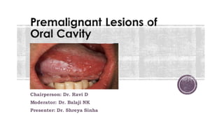
  • 1. Chairperson: Dr. Ravi D Moderator: Dr. Balaji NK Presenter: Dr. Shreya Sinha
  • 2. â–Ş Introduction â–Ş Premalignant lesions Vs premalignant conditions â–Ş Progression of oral cancer â–Ş Rate of malignant transformation â–Ş Leukoplakia â–Ş Erythroplakia â–Ş Oral Submucous fibrosis â–Ş Oral Lichen planus â–Ş Candidiasis â–Ş Actinic chelitis â–Ş Screening protocol â–Ş Others
  • 3. â–Ş These are lesions and conditions that are not malignant but have the potential to become malignant. â–Ş Stratified squamous epithelium lines the majority of the oral cavity, and aberrancy in the epithelium gives rise to premalignant lesions. â–Ş Previously, various terms like Pre-cancerous, Pre-malignant, Intra epithelial dysplasia, Intra epithelial neoplasia were used. â–Ş Recently, a more precise phrase, “Oral potentially malignant disorders” has been adopted by the WHO collaborating center.
  • 4. Pre-malignant lesions â–Ş A morphologically altered tissue in which malignancy is more likely to occur than its apparently normal counterpart. â–Ş Leukoplakia â–Ş Erythroplakia â–Ş Submucous fibrosis â–Ş Lichen planus â–Ş Actinic chelitis Pre-malignant conditions â–Ş A generalised state associated with a significantly increased risk of malignancy. â–Ş Plummer Vinson syndrome â–Ş Syphilis
  • 5.
  • 6.
  • 7. â–Ş The most feared complicationof oral premalignant disorders is progression to carcinoma. â–Ş A recent meta-analysis revealed that the rate of malignant transformation of all oral premalignant disorders over a period of 5 years combined was around 7.9%. In contrast, individual transformation rates were: â–Ş lichen planusis around 1.4%, â–Ş leukoplakia 9.5%, â–Ş oral submucousfibrosis 5.2%, â–Ş erythroplakia 33.1%.
  • 8. â–Ş It refers to a white patch in the mucosa that does not rub off and cannot be clinically identified as another entity. â–Ş It is a diagnosis of exclusion. â–Ş Risk factors include: chronic smoking, tobacco abuse, areca nut and betel nut chewing, alcohol abuse, chronic trauma (friction induced hyperkeratosis), chronic sun exposure, sanguinaria (herbal root extract in some mouthwashes and toothpaste), male sex, people in fourth decade of life, etc.
  • 9. â–Ş Clinical forms are: 1. Homogenous leukoplakia: There is a smooth or wrinkled white patch, well demarcated, has a consistent pattern throughout and is less often associated with malignancy. 2. Nodular (Speckled) leukoplakia: There are nodular white patches with erythematous base. 3. Erosive leukoplakia (Erythroleukoplakia) presents white patches which has erosions and fissures and is interspersed with red patches (erythroplakia). 4. Proliferative verrucous leukoplakia: This uncommon variant of leukoplakia is multifocal and persistent and occurs more often in women. A thin flat white patch progresses to leathery thickened and papillary to verrucous appearance.
  • 11. â–Ş Histopathology: It ranges from hyperkeratosis and acanthosis to dysplasia (disordered cell growth and architectural distortion) or carcinoma in situ to invasive squamous cell carcinoma. The dysplasia is traditionally graded as mild, moderate, or severe. â–Ş Potential for malignant change: The chances of malignant change are from 1– 17.5% , which varies according to the site, type and duration of the lesion and age of the patient.
  • 12. â–Ş Risk factors associated with malignant transformation of oral leukoplakia are: 1. Female sex 2. Long standing leukoplakia 3. Leukoplakia in non smokers 4. Leukoplakia over tongue or floor of mouth 5. Size >200 mm2 6. Non homogenous type of leukoplakia 7. Leukoplakia with presence of epithelial dysplasia
  • 13. A clinical staging system (OL system) for oral leukoplakia on the lines of TNM staging was recommended by WHO in 2005, taking the size (L) and histopathological features (P) into consideration.
  • 14. â–Ş Management: 1. Biopsy: An incisional biopsy (multiple biopsies in extensive lesions) must be taken from suspicious areas (such as erythematous, granular, ulcerated and indurated) to know the grades of dysplasia and rule out malignancy. 2. Medical management a) L-ascorbic acid (vitamin C) b) a-tocoferol (vitamin E) c) Retinoic acid (vitamin A) d) Vitamin A derivative, isotretinoin and 13-cis retinoic acid: 28,500 IU per day. e) Beta carotene : 150,000 IU of beta carotene twice per week for 6 months. f) Bleomycin: topical bleomycin has been used in the treatment of oral leukoplakia in dosage of 0.5% per day for 12-15 days or 1% per day for 14 days. It prevents the progression though dysplasia to carcinoma. Bleomycin is a glycopeptide antibiotic that acts by binding to guanosine-cytosine rich portion of the DNA via association of the S tripeptide and by partial intercalation of the bithiazole rings.
  • 15. â–Ş Surgical management includes: 1. Scalpel excision/ stripping 2. Electrocautery 3. Cryotherapy 4. CO2 laser therapy 1. Scalpel excision: • Area is outlined using few (1-2) mm of normal tissue. It is incised with scalpel and patch is undermined by scalpel or blunt dissection to a depth of 2-4 mm. This allows the leukoplakia to be removed in one piece. The mucosal defect if small is closed primarily or it is covered by transported local mucosal flaps. Larger defects are grafted with split thickness skin graft.
  • 16. 2. Electrocautery ( Fulguration): • This requires local or general anaesthesia. Here, multiple areas of lesion are pierced with electrocautery and left to heal. The healing process is slow and painful. 3. Cryotherapy : • It is a method of superfreezing the tissue in order to destroy it. • It is done using a cotton swab that has been dipped in liquid nitrogen or a probe that has liquid nitrogen flowing through it. The technique involves freezing the mucosa with cryoprobe for 1.5-2 minutes, then waiting for 2 minutes, followed by further freezing for 1.5-2 minutes. Thicker lesions may require 2-3 minutes freezing.
  • 17. 4. CO2 Laser therapy: • CO2 laser beam wavelength – 10.6 micro meter • Well absorbed by water and hence by soft tissues • The absorbed energy causes vaporisation of the intra and extracellular fluid and destruction of cell membrane. The cell debris are released and burned in the laser beam, depositing a carbonised layer on the tissue surfaces. • Two techniques are used to remove the leukoplakia using CO2 laser: a) Excision- to excise a patch, the laser is used to cut around the margins, which can be held in tissue forceps while the laser undermines the leukoplakic patch. b) Vaporisation- it is done by moving the laser beam back and forth across the surface of the lesion. It has the risk of leaving small bits of abnormal tissue which are deep under thickly keratinised tissue.
  • 18. Surgical excision of lesions using laser offers several advantages over scalpel excision which includes â–Ş bloodless surgical and postsurgical events; â–Ş the ability to precisely coagulate, vaporise, or cut tissue; â–Ş minimal swelling and scarring; â–Ş reduction of surgical time, postsurgical pain with high patient acceptance.
  • 19. â–Ş Differentia diagnosis: Linea alba buccalis Leukoedema Fordyce granules Traumatic keratosis
  • 21. â–Ş It is a fiery red patch that cannot be clinically or pathologically classified as another entity. â–Ş Also known as Erythroplasia of Queyart. â–Ş These lesions may have a flat, macular, velvety appearance and may be speckled with white spots representing foci of keratosis. â–Ş It is much less common than leukoplakia, but has a much greater probability (91%) of showing signs of dysplasia or malignancy at the time of diagnosis. â–Ş The rate of malignant transformation over a period of 5 years was found to be 33.1%
  • 22.
  • 23. â–Ş Differential diagnosis: 1. Erythematous (atrophic) candidiasis 2. Kaposi’s sarcoma 3. Contact stomatitis 4. Vascular malformation 5. Squamous cell carcinoma 6. Geographic tongue/ erythema migrans â–Ş Management: • It is the same as that for Invasive carcinoma or Carcinoma in-situ and includes surgery, radiation and cauterisation. • Surgical excision if proven dysplastic /malignant.
  • 24. â–Ş It is an insidious painless oral cavity disease, which is characterized by juxta epithelial inflammatory reaction, followed by fibro-elastic change of lamina propria with epithelial atrophy, leading to stiffness of oral mucosa and causing trismus and inability to eat. â–Ş It appears as white fibrotic bands extending from retromolar trigone to soft palate, buccal mucosa, tongue and pharynx. â–Ş It is common in Indian population. Incidence is 0.2-0.5 %.
  • 25. â–Ş Etiology: 1. Prolonged local irritation due to paan, betel nut, tobacco, smoking of cigarette or bidis, excessive amount of spices and chillies in food. 2. Dietary deficiency of iron, vitamin B-complex and vitamin A . 3. Cell mediated immune process: Some consider it a cell mediated immune reaction to arecoline. 4. Racial: Disease usually affects Indians or people of Indian origin living abroad.
  • 26. â–Ş Pathogenesis: oral mucosa Betel quid habit Constant irritation Chronic inflammation Activated T cell and macrophages at the site Increase in cytokines- IL6, TNF, IFN-a Increse in growth factors- TGF-B Increased susceptibility due to deficiency of iron & vitamin B12
  • 27. cytokines Increase collagen production Decrease collagen degradation Increased collagen (insoluble form – Cross linking of insoluble collagen) Fibrosis Oral submucous fibrosis
  • 28.
  • 29. â–Ş The malignant transformation has been observed in 3–7.6% of cases.
  • 30. â–Ş Initial stage- • Burning sensation on eating spicy food • Blisters or vesicles on the palate • Ulceration or recurrent stomatitis • Excessive salivation • Dryness of mouth â–Ş Later stage- • Difficulty in opening mouth • Inability to whistle or blow • Difficulty swallowing • Referred pain to the ear • Change in voice tone due to vocal cord involvement • Deafness due to occlusuion of eustachian tube â–Ş Clinical features:
  • 31. â–Ş Clinical and functional staging: âť‘ Clinical staging: Stage 1- faucial bands only Stage 2- faucial and buccal bands Stage 3- faucial, buccal and labial bands âť‘Functional staging: A- mouth opening >= 20 mm B- mouth opening 11-19 mm C- mouth opening <= 10 mm
  • 32. â–Ş Management: 1. Local steroids/hyluronidase: Topical injection of steroids, which may be combined with hyluronidase, into the area of fibrous bands (injection dexamethasone 4 mg and hylase 1500 IU in one ml, intraoral submucosal biweekly at different sites for 8–10 weeks). This brings significant improvement in symptoms and relieves trismus. Steroids suppress inflammatory reactions, thereby preventing fibrosis by decreasing fibroblastic proliferation and deposition of collagen. 2. Use of Placentrix 2ml solution at interval of 3 days.
  • 33. 3. Avoidance of irritant factors 4. Systemic immunomodulators: • Levamisole 150mg for 3 weeks orally • Dapsone 75mg OD for 90 days orally 5. Vitamins and minerals: Treatment of existent anemia or vitamin deficiencies. Vitamin A, zinc and antioxidants therapy has shown some beneficial effect. They are effective in controlling the burning sensation and ulceration in OSMF 6. Physiotherapy (jaw opening exercises)
  • 34. â–Ş Surgical management: Fibrotomy (scalpel, electrocautery, laser) Coronoidectomy and Temporalis myotomy Extraction of 3rd molars reconstruction
  • 35. â–Ş Grafts and flaps tried for reconstruction in the management of oral submucous fibrosis: • Bilateral tongue flap • Nasolabial flaps (most common) • Island palatal mucoperiosteal flap • Bilateral radial forearm free flap • Buccal pad of fat graft • Temporalis fascia graft • Split skin graft
  • 36. â–Ş It is a mucocutaneous immunologic disorder that occurs due to T-cell lymphocytic reaction to surface epithelial antigen. â–Ş Oral lesions may present in various forms such as reticular, plaque, atrophic, erosive and bullous. Concomitant presence of various forms is not uncommon. â–Ş Premalignant nature of this lesion is still controversial. â–Ş In cases of erosive lichen planus or atrophic lichen planus, there is risk of malignant change.
  • 37. â–Ş Risk factors include: Autoimmunity, genetic background, dentures, infectious agents like Hepatitis C virus, bowel diseases like Chron’s and Ulcerative colitis, stress, diabetes and hypertension. â–Ş Pathogenesis unknown antigen antigen presenting cells CD 4 lymphocytes Interferon gamma produced Basal cell degeneration with sub-epithelial lymphocytic infiltration
  • 38. â–Ş Treatment: 1. Corticosteroids: Topical, intralesional injections and systemic. 2. Other alternatives: immunomodulatory drugs like Hydroxychloroquine, azathioprine and retinoids for at least 3-6 months. 3. Surgical: Resection of the lesion, lasers and cryotherapy.
  • 39. â–Ş Infection caused by Candida albicans has two forms: thrush and chronic hypertrophic candidiasis. â–Ş Certain strains of Candida have been shown to produce Nitrosamines (chemical carcinogen), hence these lesions carry a risk of malignant transformation. â–Ş Risk factors: 1. Systemic: Diabetes, antibiotics, age and immunosuppression. 2. Local: Corticosteroids, xerostomia, heavy smoking and dentures. â–Ş Clinical features: Symptoms range from none to burning, dysgeusia, sensitivity and generalized discomfort. Odynophagia indicates involvement of pharynx.
  • 41. â–Ş Diagnosis: Microscopic evaluation of lesion smears- 1. Potassium hydroxide preparation to demonstrate hyphae 2. Periodic acid Schiff (PAS) stain 3. Culture on Sabouraud’s corn meal or Potato agar medium 4. Biopsy with PAS, Gomori’s methenamine silver(GMS), or other fungal stains of microscopic sections. â–Ş Treatment: 1. Topical therapy- • Nystatin oral suspension (100,000 units/mL); rinse 5 ml and swallow 4 times per day • Clotrimazole solution 1% ; rinse 5 mL and swallow 4 times per day
  • 42. 2. Systemic therapy- • Fluconazole 100mg, 2 tablets on day 1, followed by 1 tablet on days 2-7, and 1 tablet every other day for days 8-21 • Ketoconazole 200mg, 1 tablet per day for 21 days
  • 43. â–Ş It is a premalignant lesion that affects the vermilion border of the lower lip. â–Ş It usually results due to chronic sun exposure or UV radiation exposure. â–Ş In early stage, redness and swelling of the lips is found alongwith desquamation of epithelium. â–Ş As the disease progresses, erosions and scrub covered ulcers may develop.
  • 44.
  • 45. â–Ş Treatment includes: 1. Topical: 5-fluorouracil, Imiquimod, trichloroacetic acid, etc. 2. Laser treatment 3. Vermilionectomy or surgical excision 4. Phototherapy
  • 46. â–Ş Some other lesions are: 1. Palatal lesions in reverse smokers 2. Hereditary conditions like Dyskeratosis congenita and Epidermolysis bullosa. 3. In the year 2007, the WHO and the Cancer meetings also decided to include the Oral manifestations of Graft Versus Host Disease and Lichenoid lesions like Lichen pemphigoid, Oral lichen planus, Atypical lichenoid stomatitis, lichenoid dysplasia, etc. under potentially malignant disorders of oral cavity.
  • 47. â–Ş It is recommended that all adult patients should be screened annually even if medical and dental histories elicit no risk factors. Known at risk patients should be screened every 6 months. 1. Biopsy protocol • Apart from incisional and excisional biopsy used for definitive diagnosis of oral cancers, Brush biopsy is another technique that can be used as a preliminary diagnostic tool. • It uses a stiff brush to obtain a full thickness sampling of epithelial cells for examination in patients with mucosal lesions. • It is a simple, relatively inexpensive, high sensitive and less invasive to perform, but the existence of false positives have been pointed out in this method.
  • 48. • Studies have suggested that reduced nuclear size and increased cytoplasm size are useful early indicators of malignant transformation, and thus exfoliative cytology is of value for monitoring clinically suspected lesions. 2. Toluidine blue staining • Vital staining with toluidine blue (as a 1% rinse or application) has been suggested as a means of surveillance in patients at risk of developing oral cancer, and for those who have had a confirmed neoplasm on other parts of aerodigestive tract. • It is most useful in secondary care for delineating the extent of lesions and for surveillance of patients at risk of recurrent disease.
  • 49. 3. Autofluorescence • This phenomenon is based on the interaction of various fluorescent tissue compounds (fluorophores) that occur naturally in the body. When excited by an appropriate light stimulus, these compounds emit visible fluorescent light in the violet to green region of the spectrum. • VELscope is a portable device comprising of light source and a viewing hand piece. The technology is based on direct visualisation of tissue fluorescence. • Both keratinised and nonkeratinized squamous epithelium with a normal submucosa show a typical homogenous, pale green fluorescence. Sharply circumscribed areas of decreased auto fluorescence might indicate areas of musosal abnormalities that should be monitored or investigated by tissue biopsy.
  • 50. • Microlux is a handheld device that used light emitting diodes( LEDs) as the elimination source. Prior to exposing the mucosa to light, the patient rinses for 30- 60 seconds with 1% acetic acid. Upon illumination, abnormal tissue will appear white( aceto-white). 4. Salivary biomarkers • Abnormal DNA, RNA and protein molecules released by the malignant cells can be easily obtained from the saliva. • It includes : a) Non organic compounds- Na, Ca, F, Mg b) Peptides- Defensin 1 c) Proteins- IL-1, IL-6, IL-8, TNF-a, Endothelin-1
  • 51. d) DNAs- p53 gene codon 63 e) mRNAs- IL-1B, H3F3A, etc f) Oxidative stress related molecules- glutathione, peroxidase g) Metabolites- lactic acid, valine, etc h)Glycosylation related molecules- sialic acid, a-L-fucosidase, etc
  • 52. Linea alba buccalis leukoedema Fordyce granules