Apoptosis Post Microwave Ablation of the Liver: Does it Change with Power?
poster NEPG oct 14 A
1. Developing biomarkers for
autoimmune related
psychiatric illness
Introduction
In anti-NMDA receptor autoimmune encephalitis, a potentially fatal, but
treatable disease, autoantibodies bind and internalize the NMDA receptor. This
condition usually includes psychiatric (hallucinations, anxiety) and
neurological (seizures, autonomic instability) symptoms, but the presence of
these antibodies has been found in some psychiatric patients in absence of the
neurological symptoms. The antibodies can be detected in serum and
cerebrospinal fluid with a immunofluorescence cell based assay using HEK
cells expressing NR1 subunits of the NMDA receptor.
NMDA receptors play an important role in the generation of gamma
oscillations, which are found disrupted in psychiatric illness, such as
schizophrenia. Ketamine, an NMDAR antagonist, causes a reduction in the
power of gamma oscillations in vitro in the medial entorhinal cortex (mEC).
We evaluate a novel electrophysiology assay attempting to detect the
presence of the antibodies, and study their effect on gamma oscillations,
which might also give us an insight of the role of the antibodies in the
pathology.
This is part of a larger study, investigating the prevalence of these antibodies
in patients with psychosis or catatonia, and the correlation between results
with the cell based assay and the electrophysiology assay. The effect of serum
from healthy individuals on this assay had not previously been studied.
Aims
• To evaluate the effect of serum from psychiatric patients on
gamma oscillations in vitro.
• Compare the effects between those patients with confirmed
presence of anti-NMDAR antibodies, and those without, as
well as against healthy controls.
Methods
a)
b)
• Patient recruitment and testing on
the cell based assay was performed
as part of a larger study; for this
MRes study we focused on healthy
controls, the positive patient, from
whom 2 samples were available, and
three patients with a negative result
in the cell based assay.
• Electrophysiology assay: Rat brain
slices were obtained and perfused
with aCSF (artificial cerebrospinal
fluid), electrodes were inserted in
the mEC (figure 2.)
• Gamma oscillations were induced
with kainic acid. Once these were
stable, 10 µl of serum was added to
50 mL of circulating aCSF and left for
an hour, followed by an hour wash
out.
• Power spectrums were obtained and
analysed for power, peak frequency
and peak amplitude (See figure 3).
• Power spectrums were obtained and
analysed for power, peak frequency
and peak amplitude (See figure 2).
Results
Peak
amplitude
Peak Frequency
Power
Power Spectrum
Conclusions
Serum of healthy individuals has no effect on gamma oscillations
Correlation of results of negative patients among electrophysiology assay
and CBA
Though the positive patient’s serum had an effect on gamma oscillation,
the effect of the anti-NMDAR AB cannot be established with our results, further
research is required.
Psychiatric Patient
Recruitment
(EIP or catatonia)
Blood Sample
Oxford- cell based assay *
Positive for anti-NMDAR:
n=1
Ncl-Electrophysiology
assay
Healthy
controls n=5
Blood Sample
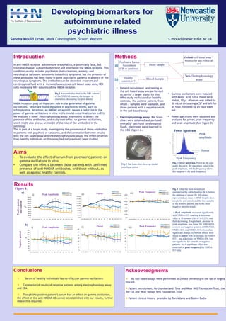
Fig.3 Power spectrum, Power as the area
under the curve, the maximum value is the
peak amplitude, and the frequency where
this happens is the peak frequency
Fig 4 . Data has been normalized
considering the stable baseline (KA) before
the addition of serum (S). All values
represented are mean ± SEM. Graphs show
results for a) Controls and the two samples
of the positive patient, and b) the three
negative patients tested.
i) Peak amplitude increased significantly
with NMDA 033, reaching a maximum
value at 30 minutes (S6) of 161.23%, and
then decreasing. A significant, decrease in
peak amplitude was found for NMDA 036
controls and negative patients (NMDA 011,
NMDA 012, and NMDA 013) showed no
significant change. ii) Similar effects were
found in power with an increase for NMDA
033 , and a decrease for NMDA 036, but
not significant for controls or negative
patients. iii) A significant effect was
observed in peak frequency for NMDA
033 only.
Acknowledgments
All cell based assays were performed at Oxford University in the lab of Angela
Vincent.
Patient recruitment: Northumberland Tyne and Wear NHS Foundation Trust, the
Tee Esk and Wear Valleys NHS Foundation Trust
Patient clinical history provided by Tom Adams and Roshni Budia
0.00
20.00
40.00
60.00
80.00
100.00
120.00
140.00
160.00
180.00
200.00
KA1KA2KA3 S1 S2 S3 S4 S5 S6 S7 S8 S9 S10 S11 S12
Peak Amplitude
Control
NMDA 033
NMDA 036
*
i)
*
***
* *
**
*
* ** *
**
0.00
20.00
40.00
60.00
80.00
100.00
120.00
140.00
160.00
180.00
KA1KA2KA3 S1 S2 S3 S4 S5 S6 S7 S8 S9 S10 S11 S12
Power
Control
NMDA 033
NMDA 036
***
ii)
0.00
20.00
40.00
60.00
80.00
100.00
120.00
140.00
KA1KA2KA3 S1 S2 S3 S4 S5 S6 S7 S8 S9 S10 S11 S12
Peak Frequency
Control
NMDA 033
NMDA 036iii)
0
20
40
60
80
100
120
140
160
180
KA1 KA2 KA3 S1 S2 S3 S4 S5 S6 S7 S8 S9 S10 S11 S12
Peak Amplitude
NMDA 011
NMDA 012
NMDA 013
0
20
40
60
80
100
120
140
KA1KA2KA3 S1 S2 S3 S4 S5 S6 S7 S8 S9 S10 S11 S12
Power
NMDA 011
NMDA 012
NMDA 013
0
20
40
60
80
100
120
140
KA1 KA2 KA3 S1 S2 S3 S4 S5 S6 S7 S8 S9 S10 S11 S12
Peak Frequency
NMDA 011
NMDA 012
NMDA 013i)
b)
ii) iii)
a)
Figure 4.
%
%
Sandra Mould Urías, Mark Cunningham, Stuart Watson s.mould@newcastle.ac.uk
NR1
Positivepatient
andcontrolsNegativepatient
Fig 2. Rat brain slice showing medial
entorhinal cortex
Fig 1 Autoantibodies bind to the NR1 subunit
of the NMDAR, causing the receptor to
internalize, decreasing receptor density