1. RESULTS
Low and medium doses of chronic intranasal oxytocin (OT) reduce
quantity of OT cells in PVN of male prairie vole
Caleigh D. Guoynes, Catherine Yun, David Patron, Louiza Livschitz, Griffin Downing, Crystal Vardakis,
Chathurika Peiris, Allison Perkeybile & Karen L. Bales
ABSTRACT: For voles given chronic intranasal OT, behavioral assays have shown that low and
medium of doses of OT caused adult male prairie voles to lose partner preference and low doses
caused adult female prairie voles to attack pups more during alloparental care tests. Our goal was to
determine if low, medium, and high doses of chronic intranasal OT change the number of cells
containing OT in the PVN and SON in prairie voles. This study used non-behavior tested animals to
establish a baseline for OT in brains of voles given one of three OT doses or saline. Using oxytocin
immunohistochemistry, we found that voles given the low and medium doses of OT had
significantly fewer cells (F=5.55, p=.0274 and F=3.00, p=.0966, respectively) in the PVN. There were
no significant changes in the SON and no changes in OT plasma levels across groups. These results
suggest that the decreased number of cells containing OT in the PVN largely contributed to the
observed behavioral abnormalities.
BACKGROUND & INTRODUCTION
•Oxytocin (OT) is a naturally occurring neuropeptide that
has a significant impact on the formation and
maintenance of social bonds.
•Our study aims to confirm the short-term benefits of
intranasal OT and explore possible long-term side
effects.
•The PVN contains parvocellular cells that make OT
available to the brain. The SON contains both
parvocellular and magnocellular cells, making OT
available to both the brain and body.
•Our previous study indicates that low and medium
doses of OT cause long-term behavioral deficits, so we
predict those doses will cause a decrease in cell number
in the brain (Fig. 1).
•Quantifying OT antigens may make it possible to
understand how various doses of chronic OT affect brain
development and the consequent adult social behavior.
•Clinical researchers are using intranasal OT for patients
with autism, schizophrenia, and post-traumatic stress in
conjunct with behavioral therapy to maximize patient
benefit.
METHODS
At post-natal day (PND) 21 through PND 42 (sexual maturity) voles were given daily treatments between 8 a.m.
and 12 p.m. Each vole was given either a low (0.08 IU/kg), medium (0.8 IU/kg), or high (8.0 IU/kg) dose of OT or
saline. Days 42-50 served as a washout period and PND 50-55, brains were removed. After all brains were sliced,
immunohistochemistry was performed using anti-rabbit primary and secondary antibodies, and all cells in PVN
and SON were counted by C.D.G. The assay for the blood plasma was performed by K.L.B. and T.W. Below, Fig. 2
shows the timeline of these events.
ACKNOWLEDGMENTS
Thanks to Julie
Vanwesterhuyzen, Meredith
Lee, Fontaine Ma, Caryn
Covella, and John Helmy.
Thanks to Allison Perkeybile
and Tamara Weinstein for
their help with assays. Special
thanks to Dr. Karen Bales for
designing the project and
giving me this incredible
opportunity. This work was
supported NIH grant
HD071998 to K. Bales.
DISCUSSION
•There was a significant decrease in cell number in the PVN of both
low and medium dose treated animals.
•It follows that these animals were not producing as much of their
own OT as control animals.
•There were no significant changes in the SON, so this suggests the
behavioral abnormalities observed in the previous study were
mediated by PVN fibers extending into the brain rather than SON
fibers extending to the posterior pituitary.
•A previous autoradiography study showed the medium dose in
males had significantly higher OT receptor binding in the posterior
cingulate cortex (F=5.85, p=0.0421) and lower binding in the BNST
(F=4.62, p=0.0842). There were no significant changes with the low
dose in males.
•This suggests that the abnormal social behaviors in the low dose
were caused by a decrease in OT cells and the abnormal social
behaviors in the medium dose were caused by a combination of
changes in receptors and a decrease in OT cells.
•We have plans to quantify the data on female brains and run a
vasopressin assay to look for AVP expression in these cells.
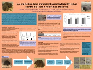
Figure 5
Males given the low dose had significantly fewer cells producing OT in the PVN when compared to
saline controls. Two-tailed probability shows F=5.55, p=0.0274. For male prairie voles given the medium
dose, the one-tailed probability is significant (p=0.0483).
**significant for two-tailed probability, *significant for one-tailed probability
•Stained brains were sorted into one of seven levels of PVN (Fig. 2) and
averaged for total cell counts throughout the various levels of PVN (Fig. 4).
Same methods were used for quantification of SON.
•There were fewer OT-expressing cells in the PVN of voles given low and
medium doses of OT. Two-tailed probabilities were significant for voles given
the low dose (p=0.0274) and showed a trend medium doses (p=0.0965).
(Fig. 5)
•The one-tailed probability for the medium dose is significant (p=0.0483) as
seen in Figure 5.
Figure 4
The averaged number of cells expressing OT in the PVN. Standard errors (STE) were
used the calculate the variability of cell number in the PVN between different test
subjects.
Dosage Low Medium High Saline
Cell number ± STE 37.97 ± 4.82 40.29 ± 4.81 44.67 ± 4.64 46.71 ± 1.25
CA B
ED
G
F
PND 50-55
(adulthood): brain
removal
Immunohistochemistry
assay for OT
Brains sorted from
anterior to posterior
and stained cells
counted
PND 21-42 (juvenile
period): daily intranasal
OT treatments
PND 42-50 (adulthood):
washout period, no
treatments
Juvenile prairie vole Adult prairie vole
PVN from anterior to posterior:
above, pictures A -G show how the shape of the PVN
changes significantly from anterior to posterior. In this
study, we averaged the PVN and SON cells for the sum of
all brain areas. However, we plan to examine possible
changes in PVN density throughout the different areas.
Figure 3
40 micron slice with PVN, PVN fibers, and SON
labeled. These areas were stained using a rabbit OT
primary and secondary antibody with DAB staining.
Slices were mounted within one week of the assay.
PVN fibers
SON
PVN
Figure 1
Results from the behavioral assay (as published in Biological
Psychiatry). Short term, OT increased social behavior for all
doses. Long-term, low and medium doses caused deficits in
social bond while high and saline produced typical social
bonds.
0
10
20
30
40
50
60
Low dose vs. saline Medium dose vs.
saline
High dose vs. saline
AveragenumberofcellsinPVN
Effects of OT on cell number in PVN of males
dose
saline** *
Figure 2
Timeline of study events.
•The PVN and SON were stained and then quantified as shown in Figure 3.
•There was a trend showing that the medium dose had fewer cells (average of 8.988 ) in the
SON compared to the saline control (average of 13.532). However, the one-tailed probability
of the medium dose versus saline is p=0.05.
•There were no significant changes in the SON for the low or high doses compared to the
saline dose.
•No significant differences were found in OT plasma levels across groups.